

Multi-cell type human liver microtissues for hepatotoxicity testing
Open accessThe DOI matches its Crossref record; 12 of the 12 references it cites were found in a registry or an index.
Abstract
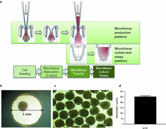
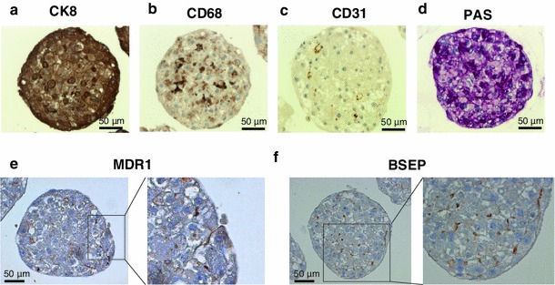
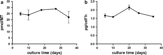
Current 2-dimensional hepatic model systems often fail to predict chemically induced hepatotoxicity due to the loss of a hepatocyte-specific phenotype in culture. For more predictive in vitro models, hepatocytes have to be maintained in a 3-dimensional environment that allows for polarization and cell-cell contacts. Preferably, the model will reflect an in vivo-like multi-cell type environment necessary for liver-like responses. Here, we report the characterization of a multi-cell type microtissue model, generated from primary human hepatocytes and liver-derived non-parenchymal cells. Liver microtissues were stable and functional for 5 weeks in culture enabling, for example, long-term toxicity testing of acetaminophen and diclofenac. In addition, Kupffer cells were responsive to inflammatory stimuli such as LPS demonstrating the possibility to detect inflammation-mediated toxicity as exemplified by the drug trovafloxacin. Herewith, we present a novel 3D liver model for routine testing in 96-well format capable of reducing the risk of unwanted toxic effects in the clinic.
Overview
The claims and statistics of this paper haven’t been checked yet.
Free: it reads the open-access text and checks the first 5 claims that cite a source we can check. Create a free account to check every claim.
Integrity signals
Registry
Cites
Statistics
Figures

Verification
Claims the paper makes
The claims and statistics of this paper haven’t been checked yet.
Free: it reads the open-access text and checks the first 5 claims that cite a source we can check. Create a free account to check every claim.
Claims other papers make about this one
Each is checked against this paper’s own text: does it say what the citing paper says it does?
The source backs part of the claim, not all of it — or backs it for a different population, condition or comparison.
In the citing paper
“Three-dimensional (3D) spheroid hepatic cultures have emerged as promising tools to assess mechanisms of hepatotoxicity, as they demonstrate enhanced liver phenotype, metabolic activity, and stability in culture not attainable with conventional two-dimensional hepatic models.”
From Utility of spherical human liver microtissues for prediction of clinical drug-induced liver injury (2017) · cites this paper as Messner et al. 2013
Experimental — accuracy not yet measuredThe source backs part of the claim, not all of it — or backs it for a different population, condition or comparison.
In the citing paper
“3D spheroid models are reported to produce more accurate assessment of acute and possibly also chronic drug-induced hepatotoxicity than traditional 2D culture models (Messner et al. 2013) and to be capable of detecting compounds with cholestatic liability (Hendriks et al. 2016).”
From Utility of spherical human liver microtissues for prediction of clinical drug-induced liver injury (2017) · cites this paper as Messner et al. 2013
Experimental — accuracy not yet measuredThe source backs part of the claim, not all of it — or backs it for a different population, condition or comparison.
In the citing paper
“Moreover, 3D multicellular liver spheroids require low cell numbers (e.g., 500–5000 cells), express relevant transporters, maintain functionality over 28 days in culture, and can be produced in a 96-well format (Messner et al. 2013), which make them amenable to higher throughput long-term repeat-dose testing in early discovery.”
From Utility of spherical human liver microtissues for prediction of clinical drug-induced liver injury (2017) · cites this paper as Messner et al. 2013
Experimental — accuracy not yet measured
Self-check
Percentages, sums and reported differences re-computed from the paper’s own numbers.
The self-check needs the paper’s full text.
Citations
Cites
Concordance of the toxicity of pharmaceuticals in humans and in animals
Olson, H., Betton, G., Robinson, D., Thomas, K., Monro, A., Kolaja, G., Lilly, P., Sanders, J., Sipes, G., Bracken, W., et al. (2000) · Regulatory Toxicology and Pharmacology · doi:10.1006/rtph.2000.1399 · Cited by 2,050 · cited 1×
“Here, we report the characterization of a multi-cell type microtissue model, generated from primary human hepatocytes and liverderived non-parenchymal cells.”
Toxicology for the twenty-first century
Hartung, T. (2009) · Nature · doi:10.1038/460208a · Cited by 661
THE ROLE OF METABOLIC ACTIVATION IN DRUG-INDUCED HEPATOTOXICITY
Park, B. K., Kitteringham, N. R., Maggs, J. L., Pirmohamed, M., & Williams, D. P. (2005) · Annual Review of Pharmacology and Toxicology · doi:10.1146/annurev.pharmtox.45.120403.100058 · Cited by 453
Berthiaume, F., Moghe, P. V., Toner, M., & Yarmush, M. L. (1996) · The FASEB Journal · doi:10.1096/fasebj.10.13.8940293 · Cited by 401
Organotypic liver culture models: meeting current challenges in toxicity testing
LeCluyse, E. L., Witek, R. P., Andersen, M. E., & Powers, M. J. (2012) · Critical Reviews in Toxicology · doi:10.3109/10408444.2012.682115 · Cited by 363
Microscale tissue engineering using gravity-enforced cell assembly
Kelm, J. M., & Fussenegger, M. (2004) · Trends in Biotechnology · doi:10.1016/j.tibtech.2004.02.002 · Cited by 297
Self-citationDiclofenac Toxicity to Hepatocytes: A Role for Drug Metabolism in Cell Toxicity
Bort, R., Ponsoda, X., Jover, R., Gómez-Lechón, M. J., & Castell, J. V. (1999) · The Journal of Pharmacology and Experimental Therapeutics · doi:10.1016/s0022-3565(24)37925-x · Cited by 236
Fey, S. J., & Wrzesinski, K. (2012) · Toxicological Sciences · doi:10.1093/toxsci/kfs122 · Cited by 188
Shaw, P. J., Hopfensperger, M. J., Ganey, P. E., & Roth, R. A. (2007) · Toxicological Sciences · doi:10.1093/toxsci/kfm218 · Cited by 159
Shaw, P. J., Ganey, P. E., & Roth, R. A. (2010) · Toxicological Sciences · doi:10.1093/toxsci/kfq168 · Cited by 123
Drug-induced liver injury: a summary of recent advances
Stine, J. G., & Lewis, J. H. (2011) · Expert Opinion on Drug Metabolism & Toxicology · doi:10.1517/17425255.2011.577415 · Cited by 53
Liguori, M. J., Ditewig, A. C., Maddox, J. F., Luyendyk, J. P., Lehman‐McKeeman, L. D., Nelson, D. M., Bhaskaran, V. M., Waring, J. F., Ganey, P. E., Roth, R. A., et al. (2010) · International Journal of Molecular Sciences · doi:10.3390/ijms11114697 · Cited by 22
Cited by
Bruderer, R., Bernhardt, O. M., Gandhi, T., Miladinović, S. M., Cheng, L.-Y., Messner, S., Ehrenberger, T., Zanotelli, V., Butscheid, Y., Escher, C., et al. (2015) · Molecular & Cellular Proteomics · doi:10.1074/mcp.m114.044305 · Cited by 1,238
Self-citationOrgans-on-chips at the frontiers of drug discovery
Esch, E. W., Bahinski, A., & Huh, D. (2015) · Nature Reviews Drug Discovery · doi:10.1038/nrd4539 · Cited by 1,185
Bell, C. C., Hendriks, D. F. G., Moro, S. M. L., Ellis, E., Walsh, J., Renblom, A., Fredriksson Puigvert, L., Dankers, A. C. A., Jacobs, F., Snoeys, J., et al. (2016) · Scientific Reports · doi:10.1038/srep25187 · Cited by 739
Organoid-on-a-chip and body-on-a-chip systems for drug screening and disease modeling
Skardal, A., Shupe, T., & Atala, A. (2016) · Drug Discovery Today · doi:10.1016/j.drudis.2016.07.003 · Cited by 512
“The second traditional type of model system, in vitro 2D cultures, fails to recapitulate the 3D microenvironment, and often function, of in vivo tissues [3,11].”
BackgroundA microfluidic culture model of the human reproductive tract and 28-day menstrual cycle
Xiao, S., Coppeta, J. R., Rogers, H. B., Isenberg, B. C., Zhu, J., Olalekan, S. A., McKinnon, K. E., Dokic, D., Rashedi, A. S., Haisenleder, D. J., et al. (2017) · Nature Communications · doi:10.1038/ncomms14584 · Cited by 479
Metadata
- Authors
- Messner, S., Agarkova, I., Moritz, W., & Kelm, J. M.
- Corresponding author
- Kelm, J. M.
- Venue
- Archives of Toxicology
- Published
- 2012-11-11
- Volume
- 87
- Issue
- 1
- Pages
- 209-213
- ISSN
- 0340-5761 (ISSN-L)
- 1432-0738
- PMC
- PMC3535351
- OpenAlex
- W2079277029
- Cited by
- 319
- References
- 12
- Citations by year
- 2026: 6 · 2025: 19 · 2024: 17 · 2023: 13 · 2022: 17 · 2021: 32 · 2020: 30 · 2019: 26 · 2018: 29 · 2017: 29 · 2016: 44 · 2015: 24 · 2014: 26 · 2013: 6 · 2012: 1
- FWCI
- 20.61
- Type
- journal article
- Language
- en
- Open access
- hybrid
- doi.org
- Licence
- cc-by
- Locations
- Archives of Toxicology (publishedVersion, cc-by)
- Europe PMC (PubMed Central) (publishedVersion, cc-by)
- PubMed Central (publishedVersion, cc-by)
- PubMed (publishedVersion)
- Funders
- European Commission (296257)
- Topics
- Life Sciences › Pharmacology, Toxicology and Pharmaceutics › Pharmacology › Drug-Induced Hepatotoxicity and Protection
- Health Sciences › Medicine › Hepatology › Liver physiology and pathology
- Health Sciences › Veterinary › Small Animals › Animal testing and alternatives
- Keywords
- hepatotoxicity testing, 3D liver model, human hepatocytes, non-parenchymal liver cells, Kupffer cells, acetaminophen toxicity, trovafloxacin, lipopolysaccharide, 96-well format, in vitro toxicity prediction
- MeSH terms
- Humans, Kupffer Cells, Lipopolysaccharides, Naphthyridines, Toxicity Tests, Hepatocytes, Fluoroquinolones, Tissue Culture Techniques
About these checks
Registry, retraction, Cites and statistics results are computed, not judged, with one exception: a printed reference that no index lists is read once by a model before it counts as a citation error. Claims are extracted by a model and kept only when their quote appears verbatim in the text; each cited source is then checked, with a model’s help, for whether it says what the claim relies on. Those verdicts can be wrong — read the claim’s quote and the cited source before relying on one.
Statistics and arithmetic are recomputed, not judged.
Confidence bands are shown only for fields calibrated on our annotated test set. None is calibrated yet, so no band is shown.
What counts as a claim ›How we measure confidence ›How we check this ›
Are you an author, or do you see a problem with this page? Request a review
Data sources
Bibliographic data: Semantic Scholar, OpenAlex, Crossref.
Contains information from the Semantic Scholar Academic Graph Datasets, made available under the ODC Attribution License (ODC-BY 1.0).
Semantic Scholar’s data is described in: Kinney, R., et al. (2023). The Semantic Scholar Open Data Platform. arXiv:2301.10140.
