

The DOI matches its Crossref record; 67 of the 67 references it cites were found in a registry or an index.
Dual modification of Alzheimer’s disease PHF-tau protein by lysine methylation and ubiquitylation: a mass spectrometry approach
Open accessAbstract
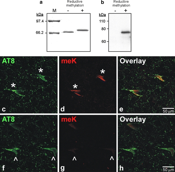
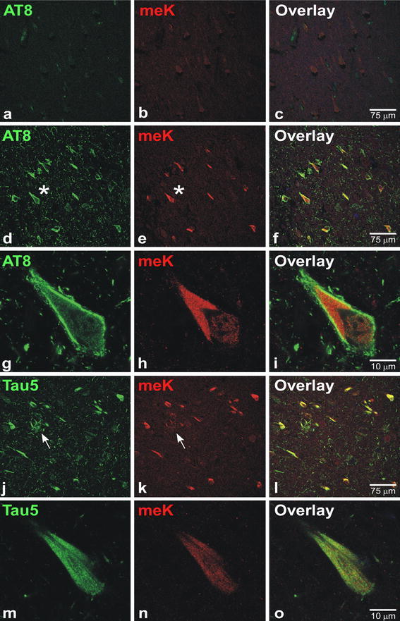
In sporadic Alzheimer's disease (AD), neurofibrillary lesion formation is preceded by extensive post-translational modification of the microtubule associated protein tau. To identify the modification signature associated with tau lesion formation at single amino acid resolution, immunopurified paired helical filaments were isolated from AD brain and subjected to nanoflow liquid chromatography-tandem mass spectrometry analysis. The resulting spectra identified monomethylation of lysine residues as a new tau modification. The methyl-lysine was distributed among seven residues located in the projection and microtubule binding repeat regions of tau protein, with one site, K254, being a substrate for a competing lysine modification, ubiquitylation. To characterize methyl lysine content in intact tissue, hippocampal sections prepared from post mortem late-stage AD cases were subjected to double-label confocal fluorescence microscopy using anti-tau and anti-methyl lysine antibodies. Anti-methyl lysine immunoreactivity colocalized with 78 ± 13% of neurofibrillary tangles in these specimens. Together these data provide the first evidence that tau in neurofibrillary lesions is post-translationally modified by lysine methylation.
Overview
The claims and statistics of this paper haven’t been checked yet.
Run the check — free, no account needed. It reads the open-access text and checks the sources it cites.
Integrity signals
Registry
Cites
Statistics
Figures
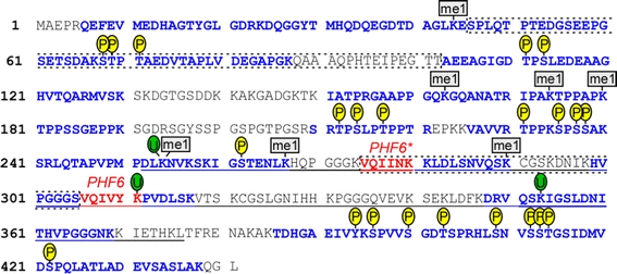
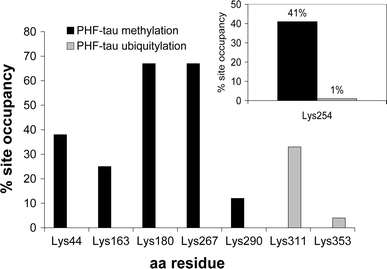
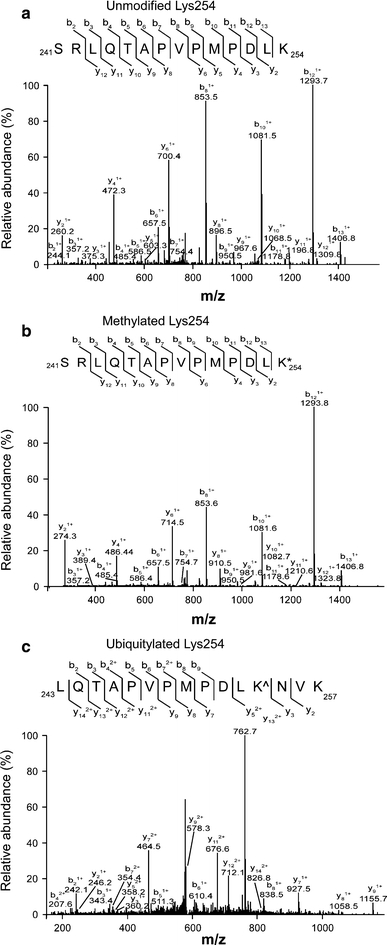
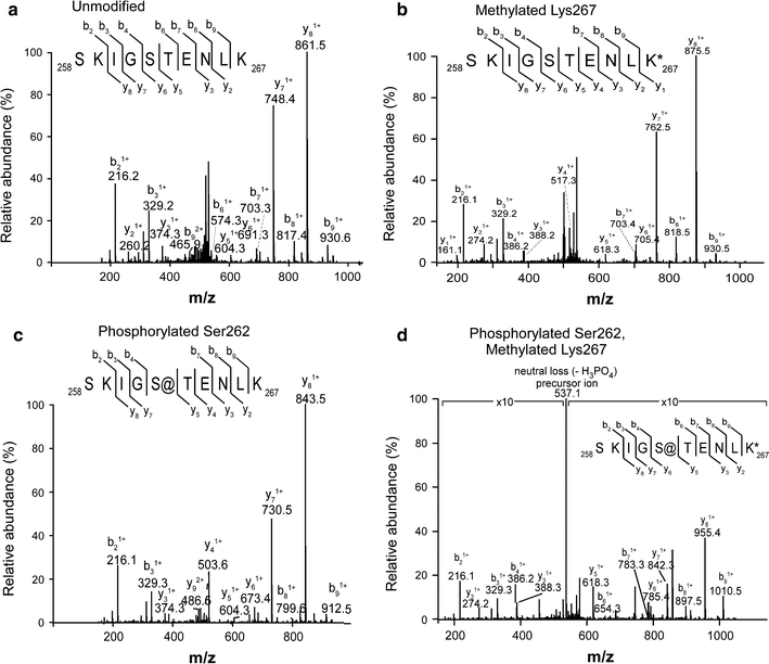
Verification
Claims the paper makes
The claims and statistics of this paper haven’t been checked yet.
Run the check — free, no account needed. It reads the open-access text and checks the sources it cites.
Claims other papers make about this one
Each is checked against this paper’s own text: does it say what the citing paper says it does?
In the citing paper
“a recent study suggests that Lys residues are predominately monomethylated in aging or AD cohorts [174–176].”
From Molecular and cellular mechanisms underlying the pathogenesis of Alzheimer’s disease (2020) · cites this paper as [175]
In the citing paper
“Methylated Lys residues mainly distribute to the projection region and tau microtubule-binding domain [174–176].”
From Molecular and cellular mechanisms underlying the pathogenesis of Alzheimer’s disease (2020) · cites this paper as [175]
Self-check
Percentages, sums and reported differences re-computed from the paper’s own numbers.
The self-check needs the paper’s full text.
Citations
Cites
Neuropathological stageing of Alzheimer-related changes.
Braak, H., & Braak, E. (1991) · Acta Neuropathologica · doi:10.1007/bf00308809 · Cited by 16,123 · cited 1×
“All AD cases satisfied criteria for Braak stages V or VI, whereas none of the control cases met pathological criteria for AD [6].”
Mirra, S. S., Heyman, A., McKeel, D., Sumi, S. M., Crain, B. J., Brownlee, L. M., Vogel, F. S., Hughes, J. P., Belle, G. V., Berg, L., et al. (1991) · Neurology · doi:10.1212/wnl.41.4.479 · Cited by 4,961
Braak, H., Alafuzoff, I., Arzberger, T., Kretzschmar, H., & Del Tredici, K. (2006) · Acta Neuropathologica · doi:10.1007/s00401-006-0127-z · Cited by 3,172
Consortium to Establish a Registry for Alzheimer's Disease (CERAD)
Fillenbaum, G., & Heyman, A. (2002) · doi:10.1002/0470846410.ch42(iii · Cited by 2,859
A Model for Random Sampling and Estimation of Relative Protein Abundance in Shotgun Proteomics
Liu, H., Sadygov, R. G., & Yates, J. R. (2004) · Analytical Chemistry · doi:10.1021/ac0498563 · Cited by 2,533
Buée, L., Bussière, T., Buée‐Scherrer, V., Delacourte, A., & Hof, P. R. (2000) · Brain Research Reviews · doi:10.1016/s0165-0173(00)00019-9 · Cited by 2,142
Profound Loss of Layer II Entorhinal Cortex Neurons Occurs in Very Mild Alzheimer’s Disease
Gómez-Isla, T., Price, J. L., McKeel Jr., D. W., Morris, J. C., Growdon, J. H., & Hyman, B. T. (1996) · The Journal of Neuroscience · doi:10.1523/jneurosci.16-14-04491.1996 · Cited by 1,796 · cited 1×
“Because neurofibrillary lesion density correlates with neurodegeneration [24, 31] and cognitive decline”
BackgroundLarge-scale characterization of HeLa cell nuclear phosphoproteins
Beausoleil, S. A., Jedrychowski, M. P., Schwartz, D., Elias, J. E., Villén, J., Li, J., Cohn, M. A., Cantley, L. C., & Gygi, S. P. (2004) · Proceedings of the National Academy of Sciences · doi:10.1073/pnas.0404720101 · Cited by 1,376
Comparison of Label-free Methods for Quantifying Human Proteins by Shotgun Proteomics
Old, W. M., Meyer-Arendt, K., Aveline-Wolf, L. D., Pierce, K. G., Mendoza, A. M., Sevinsky, J. R., Resing, K. A., & Ahn, N. G. (2005) · Molecular & Cellular Proteomics · doi:10.1074/mcp.m500084-mcp200 · Cited by 1,206
Tangle and neuron numbers, but not amyloid load, predict cognitive status in Alzheimer’s disease
Giannakopoulos, P., Herrmann, F. R., Bussière, T., Bouras, C., Kövari, E., Perl, D. P., Morrison, J. H., Gold, G., & Hof, P. R. (2003) · Neurology · doi:10.1212/01.wnl.0000063311.58879.01 · Cited by 1,118
Cited by
Sorted by citations
Items 101–124 of 124
SUMO inhibits Tau aggregation in Alzheimer’s disease
Chinnathambi, S., & Rangappa, N. (2025) · Advances in protein chemistry and structural biology · doi:10.1016/bs.apcsb.2025.02.001 · Cited by 4
Tau seeding activity in Alzheimer's disease: Characteristics, factors, and applications
Liu, Y., Chen, Y., Chen, Z., Wan, H., & Wu, C. (2025) · Journal of Alzheimer s Disease · doi:10.1177/13872877251378772 · Cited by 4
Post-translational modifications as a regulatory code for tau function in health and disease
Nordbeck, A. J., Sharma, B., Garcia, C. A., & Tseng, J. (2026) · Frontiers in Dementia · doi:10.3389/frdem.2026.1834737 · Cited by 3
Orlandini, M., Bonacini, A., Favero, A., Secchi, A., Lazzarini, L., Verucchi, R., Dalcanale, E., Pedrini, A., Sidoli, S., & Pinalli, R. (2024) · Chemical Science · doi:10.1039/d4sc02076f · Cited by 3
Eissman, J. M., Qiao, M., Kalia, V., Zerlin, M., Reyes‐Dumeyer, D., Piriz, A. L., Dubey, S., Nandakumar, R., Lee, A., Lantigua, R., et al. (2025) · Alzheimer s & Dementia · doi:10.1002/alz.70898 · Cited by 4
Molecular and environmental drivers of tau post-translational modifications and tau pathology
Price, T. M., Tucker, A. E., & Funk, K. E. (2026) · Ageing Research Reviews · doi:10.1016/j.arr.2026.103223 · Cited by 2
Self-citationTau in Health and Neurodegenerative Diseases
Chu, D., & Liu, F. (2021) · IntechOpen eBooks · doi:10.5772/intechopen.101299 · Cited by 2
Quantification of Methylation and Phosphorylation Stoichiometry
Ayoub, C. A., Moore, K. I., & Kuret, J. (2024) · Methods in molecular biology · doi:10.1007/978-1-0716-3629-9_13 · Cited by 1
Self-citationSahu, K. K., Chaudhary, R., Farooqi, M. A., & Kaurav, M. (2024) · Drug Delivery Letters · doi:10.2174/0122103031331346241122152643 · Cited by 1
Xia, P., Feng, D., Qi, B., Guan, L., Sun, Y., & Zou, Y. (2026) · Physical Chemistry Chemical Physics · doi:10.1039/d5cp04873g · Cited by 1
Damdinbazar, D., Bolortuya, B., Oyunbat, N., Ariya, E., Namuun, L., Tuvshinjargal, K., Binderiya, B., Bold, N., Davaakhuu, G., & Oyunsuren, T. (2023) · Proceedings of the Institute of Biology · doi:10.5564/pib.v39i1.3143 · Cited by 1
THE ROLE OF MICROTUBULE-ASSOCIATED PROTEIN TAU IN NEURONAL EXCITABILITY AND EPILEPTOGENESIS
Cloyd, R. (2021) · UKnowledge (University of Kentucky) · doi:10.13023/etd.2021.271
Liquid–liquid phase separation of tau in Alzheimer’s disease
Rajkhowa, S., & Tripathi, T. (2026) · International review of neurobiology · doi:10.1016/bs.irn.2026.01.009
Liu, W., Bai, Y., Qu, W., Zhang, X., Zhou, S., Luo, H., Zhu, M., Li, D., & Fang, X. (2026) · Frontiers in Aging Neuroscience · doi:10.3389/fnagi.2026.1752446
Rôle des modifications post-traductionnelles dans le processus d’agrégation de la protéine Tau
Despres, C. (2017) · theses.fr (ABES) · doi:10.70675/de10f843z8572z4295zb672z763dbf30e2b5
Identification et caractérisation fonctionnelle de nouveaux sites de clivage de la protéine Tau
Derisbourg, M. J. (2015) · theses.fr (ABES) · doi:10.70675/ef5f45b0zf4e8z433fz9beaz8d2cc6de65bc
Yatoui, D. (2022) · theses.fr (ABES) · doi:10.70675/e3620ccez2ea7z43f2za0dcz257a72021b17
Sanchez, M., & Kuret, J. (2026) · Methods in enzymology on CD-ROM/Methods in enzymology · doi:10.1016/bs.mie.2026.05.044
Self-citation
Metadata
- Authors
- Thomas, S. N., Funk, K. E., Wan, Y., Liao, Z., Davies, P., Kuret, J., & Yang, A. J.
- Affiliations
- University of Maryland, Baltimore (US)
- The Ohio State University (US)
- Albert Einstein College of Medicine (US)
- Corresponding author
- Kuret, Jeff
- Yang, Austin J.
- ORCID
- Venue
- Acta Neuropathologica
- Published
- 2011-10-28
- Volume
- 123
- Issue
- 1
- Pages
- 105-117
- ISSN
- 0001-6322 (ISSN-L)
- 1432-0533
- PMC
- PMC3249157
- OpenAlex
- W2155022431
- Cited by
- 123
- References
- 67
- Citations by year
- 2026: 7 · 2025: 12 · 2024: 14 · 2023: 8 · 2022: 10 · 2021: 14 · 2020: 7 · 2019: 9 · 2018: 9 · 2017: 14 · 2016: 5 · 2015: 3 · 2014: 7 · 2013: 1 · 2012: 3
- FWCI
- 2.40
- Type
- journal article
- Language
- en
- Open access
- hybrid
- doi.org
- Licence
- cc-by-nc
- Locations
- Acta Neuropathologica (publishedVersion, cc-by-nc)
- Europe PMC (PubMed Central) (publishedVersion, cc-by-nc)
- PubMed Central (publishedVersion, cc-by-nc)
- PubMed (publishedVersion)
- Funders
- Ohio State University (P30-NS045758)
- University of Rochester
- National Institutes of Health (NS045758)
- National Institute on Aging (AG 25323, R01AG014452, AG 14452, R01AG025323)
- National Institute of Mental Health (MH 59786, R01MH059786)
- National Institute of Neurological Disorders and Stroke (P30NS045758)
- Topics
- Life Sciences › Neuroscience › Cellular and Molecular Neuroscience › Alzheimer's disease research and treatments
- Health Sciences › Medicine › Psychiatry and Mental health › Dementia and Cognitive Impairment Research
- Physical Sciences › Chemistry › Spectroscopy › Advanced Proteomics Techniques and Applications
- Keywords
- Alzheimer's disease, tau protein, lysine methylation, ubiquitination, post-translational modification, paired helical filaments, neurofibrillary tangles, microtubule-binding repeats, hippocampus
- MeSH terms
- Aged, Aged, 80 and over, Alzheimer Disease, Amino Acid Sequence, Female, Humans, Lysine, Male, Methylation, Middle Aged, Molecular Sequence Data, Phosphorylation, Protein Processing, Post-Translational, Neurofibrillary Tangles, tau Proteins, Tandem Mass Spectrometry, Ubiquitination
About these checks
Registry, retraction, Cites and statistics results are computed, not judged, with one exception: a printed reference that no index lists is read once by a model before it counts as a citation error. Claims are extracted by a model and kept only when their quote appears verbatim in the text; each cited source is then checked, with a model’s help, for whether it says what the claim relies on. Those verdicts can be wrong — read the claim’s quote and the cited source before relying on one.
Statistics and arithmetic are recomputed, not judged.
Confidence bands are shown only for fields calibrated on our annotated test set. None is calibrated yet, so no band is shown.
What counts as a claim ›How we measure confidence ›How we check this ›
Are you an author, or do you see a problem with this page? Request a review
