Cited by 143Alzheimer's disease research and treatmentsPrion Diseases and Protein MisfoldingNeuroinflammation and Neurodegeneration Mechanisms

The DOI matches its Crossref record; 76 of the 76 references it cites were found in a registry or an index.

Templated misfolding of Tau by prion-like seeding along neuronal connections impairs neuronal network function and associated behavioral outcomes in Tau transgenic mice

Open access

Abstract

Prion-like seeding and propagation of Tau-pathology have been demonstrated experimentally and may underlie the stereotyped progression of neurodegenerative Tauopathies. However, the involvement of templated misfolding of Tau in neuronal network dysfunction and behavioral outcomes remains to be explored in detail. Here we analyzed the repercussions of prion-like spreading of Tau-pathology via neuronal connections on neuronal network function in TauP301S transgenic mice. Spontaneous and GABA(A)R-antagonist-induced neuronal network activity were affected following templated Tau-misfolding using synthetic preformed Tau fibrils in cultured primary neurons. Electrophysiological analysis in organotypic hippocampal slices of Tau transgenic mice demonstrated impaired synaptic transmission and impaired long-term potentiation following Tau-seed induced Tau-aggregation. Intracerebral injection of Tau-seeds in TauP301S mice, caused prion-like spreading of Tau-pathology through functionally connected neuroanatomical pathways. Electrophysiological analysis revealed impaired synaptic plasticity in hippocampal CA1 region 6 months after Tau-seeding in entorhinal cortex (EC). Furthermore, templated Tau aggregation impaired cognitive function, measured in the object recognition test 6 months post-seeding. In contrast, Tau-seeding in basal ganglia and subsequent spreading through functionally connected neuronal networks involved in motor control, resulted in motoric deficits reflected in clasping and impaired inverted grid hanging, not significantly affected following Tau-seeding in EC. Immunostaining, biochemical and electron microscopic analysis in the different models suggested early pathological forms of Tau, including Tau-oligomers, rather than fully mature neurofibrillary tangles (NFTs) as culprits of neuronal dysfunction. We here demonstrate for the first time using in vitro, ex vivo and in vivo models, that prion-like spreading of Tau-misfolding by Tau seeds, along unique neuronal connections, causes neuronal network dysfunction and associated behavioral dysfunction. Our data highlight the potential relevance of this mechanism in the symptomatic progression in Tauopathies. We furthermore demonstrate that the initial site of Tau-seeding thereby determines the behavioral outcome, potentially underlying the observed heterogeneity in (familial) Tauopathies, including in TauP301 mutants.

Overview

The claims and statistics of this paper haven’t been checked yet.

Run the check — free, no account needed. It reads the open-access text and checks the sources it cites.

A few free checks a day · papers already verified open instantly · results are public

Integrity signals

Registry

Crossref · 10.1007/s00401-015-1413-4

Cites

76 of 76 references verified0 citation errors · 0 not checkable

Statistics

Figures

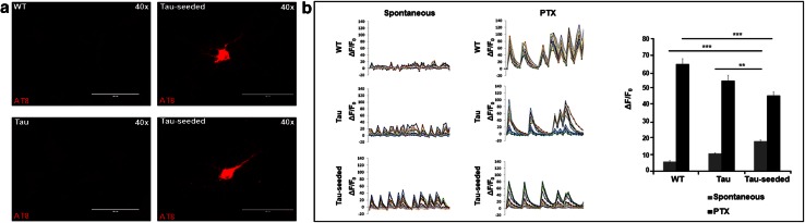
Fig. 1 Tau-seed induced Tau aggregation affected neuronal network activity in primary neurons. a Tau-seeding in primary neurons from TauP301S mice (Tau-seeded) induced somatic accumulation of aggregated Tau, as detected by AT8 staining following stringent extraction to eliminate soluble Tau, in ~5–10 % of the neurons. AT8 staining was not detected in non-seeded TauP301S neurons (Tau) or neurons derived from non-transgenic mice (WT) (scale bar 100 µm). b Calcium oscillations in primary neurons were visualized using Fura-2AM [expressed as background corrected increase in fluorescence divided by the resting fluorescence (ΔF/F 0)] for the indicated conditions. Representative traces of spontaneously oscillating neurons for each condition (WT neurons, Tau-expressing neurons, Tau-seeded Tau-expressing neurons), reveal higher amplitudes, higher synchronicity, and more regularly shaped oscillations in Tau-seeded neurons, compared to Tau and to WT neurons. Application of picrotoxin (PTX) increased amplitudes of oscillations, the synchronicity and the shape of the oscillations. Mean amplitudes were significantly lower in Tau-seeded neurons following PTX administration compared to Tau and WT neurons. Quantitative analysis of amplitudes in the presence and absence of PTX are presented for the different conditions [number of neurons measured per condition (n = 47 (WT); n = 66 (Tau); n = 68 (Tau-seeded); experiments (n = 3); mean ± SEM are presented; **p value <0.01, ***p value <0.001]
Fig. 1 Tau-seed induced Tau aggregation affected neuronal network activity in primary neurons. a Tau-seeding in primary neurons from TauP301S mice (Tau-seeded) induced somatic accumulation of aggregated Tau, as detected by AT8 staining following stringent extraction to eliminate soluble Tau, in ~5–10 % of the neurons. AT8 staining was not detected in non-seeded TauP301S neurons (Tau) or neurons derived from non-transgenic mice (WT) (scale bar 100 µm). b Calcium oscillations in primary neurons were visualized using Fura-2AM [expressed as background corrected increase in fluorescence divided by the resting fluorescence (ΔF/F 0)] for the indicated conditions. Representative traces of spontaneously oscillating neurons for each condition (WT neurons, Tau-expressing neurons, Tau-seeded Tau-expressing neurons), reveal higher amplitudes, higher synchronicity, and more regularly shaped oscillations in Tau-seeded neurons, compared to Tau and to WT neurons. Application of picrotoxin (PTX) increased amplitudes of oscillations, the synchronicity and the shape of the oscillations. Mean amplitudes were significantly lower in Tau-seeded neurons following PTX administration compared to Tau and WT neurons. Quantitative analysis of amplitudes in the presence and absence of PTX are presented for the different conditions [number of neurons measured per condition (n = 47 (WT); n = 66 (Tau); n = 68 (Tau-seeded); experiments (n = 3); mean ± SEM are presented; **p value <0.01, ***p value <0.001]CC BY · PubMed Central
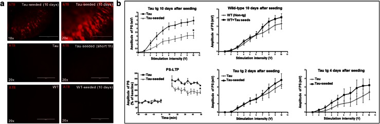
Fig. 2 Synthetic Tau seed induced Tau-aggregation impaired basal synaptic transmission and synaptic plasticity in organotypic hippocampal slices. a Tau-aggregation induced by Tau-seeding at DIV3 and DIV6 in organotypic hippocampal slices derived from P6 heterozygous TauP301S transgenic pups. AT8 staining revealed robust AT8 staining in hippocampal CA1 region 10 days after seeding of TauP301S organotypic cultures [Tau-seeded (10 days)], not detected in neurons of non-seeded TauP301S organotypic cultures (Tau), short application (1 h) of Tau-seeds [Tau-seeded (short (1 h)] on TauP301S organotypic cultures (OC) or organotypic cultures derived from non-transgenic mice (WT) with or without Tau-seeding (10 days) (lower panel) (scale bar ×10—400 µm, ×20—200 µm). b Population spike (PS) measurement in CA1 following stimulation of Schaffer collaterals. Amplitudes of population spikes were significantly decreased 10 days after seeding in Tau-seeded compared to non-seeded TauP301S cultures [n = 15 (Tau); n = 17 (Tau-seeded); mean ± SEM are presented; *p value <0.05]. LTP of the population spike amplitude measured following tetanic stimulation of Schaffer collaterals was significantly impaired 10 days after seeding in Tau-seeded compared to non-seeded slices [n = 3 (Tau); n = 5 (Tau-seeded); mean ± SEM are presented; p value <0.05]. Amplitudes of population spikes were not significantly altered at 2 days [Tau (n = 6), Tau-seeded (n = 9)] or 4 days [Tau (n = 11), Tau-seeded (n = 9)] after Tau-seeding in TauP301S cultures or in non-transgenic (wild-type, WT) cultures 10 days after Tau-seeding [WT (n = 10); WT + Tau-seeds (n = 7)]
Fig. 2 Synthetic Tau seed induced Tau-aggregation impaired basal synaptic transmission and synaptic plasticity in organotypic hippocampal slices. a Tau-aggregation induced by Tau-seeding at DIV3 and DIV6 in organotypic hippocampal slices derived from P6 heterozygous TauP301S transgenic pups. AT8 staining revealed robust AT8 staining in hippocampal CA1 region 10 days after seeding of TauP301S organotypic cultures [Tau-seeded (10 days)], not detected in neurons of non-seeded TauP301S organotypic cultures (Tau), short application (1 h) of Tau-seeds [Tau-seeded (short (1 h)] on TauP301S organotypic cultures (OC) or organotypic cultures derived from non-transgenic mice (WT) with or without Tau-seeding (10 days) (lower panel) (scale bar ×10—400 µm, ×20—200 µm). b Population spike (PS) measurement in CA1 following stimulation of Schaffer collaterals. Amplitudes of population spikes were significantly decreased 10 days after seeding in Tau-seeded compared to non-seeded TauP301S cultures [n = 15 (Tau); n = 17 (Tau-seeded); mean ± SEM are presented; *p value <0.05]. LTP of the population spike amplitude measured following tetanic stimulation of Schaffer collaterals was significantly impaired 10 days after seeding in Tau-seeded compared to non-seeded slices [n = 3 (Tau); n = 5 (Tau-seeded); mean ± SEM are presented; p value <0.05]. Amplitudes of population spikes were not significantly altered at 2 days [Tau (n = 6), Tau-seeded (n = 9)] or 4 days [Tau (n = 11), Tau-seeded (n = 9)] after Tau-seeding in TauP301S cultures or in non-transgenic (wild-type, WT) cultures 10 days after Tau-seeding [WT (n = 10); WT + Tau-seeds (n = 7)]CC BY · PubMed Central
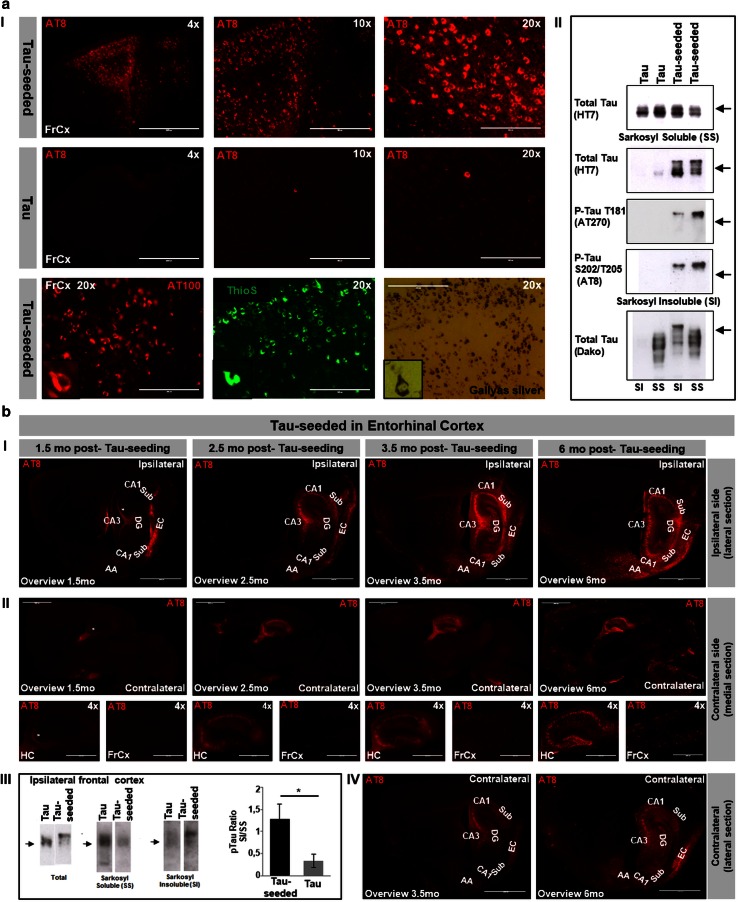
Fig. 3 In vivo aggregation of Tau following injection of Tau-seeds. a Tau-seeding induced Tau-aggregation in vivo. I Intracerebral injection of Tau-seeds in frontal cortex and hippocampus (not shown) at 3 months in TauP301S transgenic mice induced robust Tau-pathology 6 months post-injection, in contrast to buffer only injected TauP301S mice or non-injected TauP301S mice (not shown). Representative AT8 stainings are shown (magnification ×4, ×10, ×20). Staining with AT100, Gallyas silver and ThioS are presented (magnification ×20) (scale bar ×4—1000 µm, ×10—400 µm, ×20—200 µm). II Biochemical analysis revealing Tau-seed induced Tau-aggregation, as demonstrated by immunoblotting of sarkosyl soluble and sarkosyl insoluble fractions of brain extracts. Total levels of sarkosyl soluble Tau were not significantly affected following Tau seeding, but show increased ratio of higher MW to lower MW Tau forms. Marked increase in sarkosyl insoluble Tau following Tau-seeding as revealed by immunoblotting with HT7 (total Tau) antibody, AT8 (P-S202/T205) antibody and with AT270 (P-T181) antibody of sarkosyl insoluble fractions extracted from brains of Tau-seeded mice, 6 months post-injection, compared to non-seeded Tau mice. Sarkosyl insoluble Tau displayed a characteristic shift in mobility compared to sarkosyl insoluble Tau (arrows indicate 68 kDa). b Progressive spreading of Tau-pathology. Longitudinal analysis at different post-injection (p.i.) times (1.5 mo, 2.5 mo, 3.5 mo and 6 mo post-injection). Tau-pathology revealed by AT8 staining spreads progressively from entorhinal cortex (EC) to functionally connected hippocampal regions (CA1, CA3, DG, Sub) and amygdala (AA). At 1.5 mo p.i. NFTs are mainly restricted to EC. Note that weak axonal staining (indicated with asterisk) of mossy fibers and in DG (at 1.5 mo p.i.), does not represent NFTs but axonal staining observed in young TauP301S mice [73]. I Tau-pathology progressively spreads to the contralateral hemisphere. Medial and lateral sagittal sections (cfr Fig. 4a for schematic presentation) of the contralateral hemisphere at different times p.i. are presented (II, IV), demonstrating spreading of Tau-pathology to distant brain regions along neuronal connections (scale bar overviews—2000 µm, ×4—1000 µm). III Biochemical analysis of spreading of Tau-pathology to ipsilateral frontal cortex. Western blotting of total homogenate (frontal cortex) revealed a characteristic mobility shift (AT8 antibody) and sarkosyl insoluble Tau in frontal cortex extracts following Tau seeding in EC while absent in non-seeded Tau mice (AD-2 antibody). Quantitative analysis revealed significant increased ratio of sarkosyl insoluble to sarkosyl soluble Tau in frontal cortex following Tau-seeding in EC (n = 3 per condition; mean ± SEM are presented; *p < 0.05)
Fig. 3 In vivo aggregation of Tau following injection of Tau-seeds. a Tau-seeding induced Tau-aggregation in vivo. I Intracerebral injection of Tau-seeds in frontal cortex and hippocampus (not shown) at 3 months in TauP301S transgenic mice induced robust Tau-pathology 6 months post-injection, in contrast to buffer only injected TauP301S mice or non-injected TauP301S mice (not shown). Representative AT8 stainings are shown (magnification ×4, ×10, ×20). Staining with AT100, Gallyas silver and ThioS are presented (magnification ×20) (scale bar ×4—1000 µm, ×10—400 µm, ×20—200 µm). II Biochemical analysis revealing Tau-seed induced Tau-aggregation, as demonstrated by immunoblotting of sarkosyl soluble and sarkosyl insoluble fractions of brain extracts. Total levels of sarkosyl soluble Tau were not significantly affected following Tau seeding, but show increased ratio of higher MW to lower MW Tau forms. Marked increase in sarkosyl insoluble Tau following Tau-seeding as revealed by immunoblotting with HT7 (total Tau) antibody, AT8 (P-S202/T205) antibody and with AT270 (P-T181) antibody of sarkosyl insoluble fractions extracted from brains of Tau-seeded mice, 6 months post-injection, compared to non-seeded Tau mice. Sarkosyl insoluble Tau displayed a characteristic shift in mobility compared to sarkosyl insoluble Tau (arrows indicate 68 kDa). b Progressive spreading of Tau-pathology. Longitudinal analysis at different post-injection (p.i.) times (1.5 mo, 2.5 mo, 3.5 mo and 6 mo post-injection). Tau-pathology revealed by AT8 staining spreads progressively from entorhinal cortex (EC) to functionally connected hippocampal regions (CA1, CA3, DG, Sub) and amygdala (AA). At 1.5 mo p.i. NFTs are mainly restricted to EC. Note that weak axonal staining (indicated with asterisk) of mossy fibers and in DG (at 1.5 mo p.i.), does not represent NFTs but axonal staining observed in young TauP301S mice [73]. I Tau-pathology progressively spreads to the contralateral hemisphere. Medial and lateral sagittal sections (cfr Fig. 4a for schematic presentation) of the contralateral hemisphere at different times p.i. are presented (II, IV), demonstrating spreading of Tau-pathology to distant brain regions along neuronal connections (scale bar overviews—2000 µm, ×4—1000 µm). III Biochemical analysis of spreading of Tau-pathology to ipsilateral frontal cortex. Western blotting of total homogenate (frontal cortex) revealed a characteristic mobility shift (AT8 antibody) and sarkosyl insoluble Tau in frontal cortex extracts following Tau seeding in EC while absent in non-seeded Tau mice (AD-2 antibody). Quantitative analysis revealed significant increased ratio of sarkosyl insoluble to sarkosyl soluble Tau in frontal cortex following Tau-seeding in EC (n = 3 per condition; mean ± SEM are presented; *p < 0.05)CC BY · PubMed Central
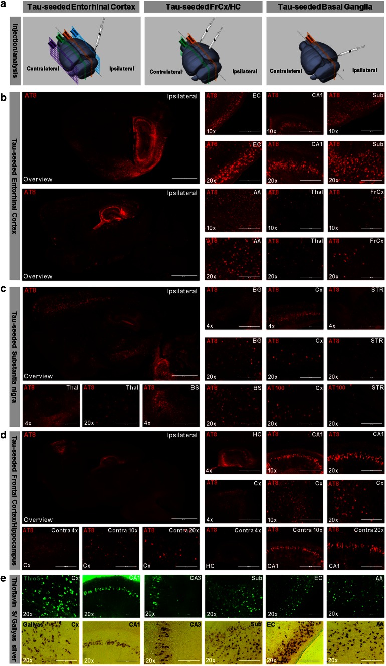
Fig. 4 Seeding and spreading of Tau-aggregation according to connectivity rather than proximity and along disease relevant patterns. Seeding and spreading of Tau-pathology following injection of Tau-PFFs in entorhinal cortex (b), in substantia nigra (c) or in frontal cortex and hippocampus (d), injection sites and sections used for analysis are schematically presented in a (modified after Allen Mouse Brain Atlas, website: http://mouse.brain-map.org/). Distinct patterns of Tau-pathology are observed. b Six months post-injection in entorhinal cortex; AT8 staining is detected in entorhinal cortex (EC), hippocampal Cornu Ammonis (CA1), subiculum (Sub), amygdala (AA), thalamus (Thal) and frontal cortex (FrCx) (right panel). An overview of AT8 staining of a section closely to the medial longitudinal fissure and a more lateral section are presented (left panel). c Six months post-seeding in substantia nigra; AT8 staining is detected in substantia nigra (SN), striatum (STR), thalamus (Thal), brain stem (BS) and motor cortex (Cx). AT8 stained neurons were also detected with AT100 antibody. d Six months post-injection in frontal cortex and hippocampus, Tau-pathology was observed at the contralateral hemisphere in cortical (Cx) and hippocampal (HC; CA1) regions, but less pronounced (marked as contra; lower row). e AT8 stainings used throughout this analysis matched with Gallyas silver and ThioS stainings, as demonstrated in different brain regions (Cx; CA1; CA3, Sub; EC; AA) (scale bar overviews—2000 µm, ×4—1000 µm, ×10—400 µm, ×20—200 µm)
Fig. 4 Seeding and spreading of Tau-aggregation according to connectivity rather than proximity and along disease relevant patterns. Seeding and spreading of Tau-pathology following injection of Tau-PFFs in entorhinal cortex (b), in substantia nigra (c) or in frontal cortex and hippocampus (d), injection sites and sections used for analysis are schematically presented in a (modified after Allen Mouse Brain Atlas, website: http://mouse.brain-map.org/). Distinct patterns of Tau-pathology are observed. b Six months post-injection in entorhinal cortex; AT8 staining is detected in entorhinal cortex (EC), hippocampal Cornu Ammonis (CA1), subiculum (Sub), amygdala (AA), thalamus (Thal) and frontal cortex (FrCx) (right panel). An overview of AT8 staining of a section closely to the medial longitudinal fissure and a more lateral section are presented (left panel). c Six months post-seeding in substantia nigra; AT8 staining is detected in substantia nigra (SN), striatum (STR), thalamus (Thal), brain stem (BS) and motor cortex (Cx). AT8 stained neurons were also detected with AT100 antibody. d Six months post-injection in frontal cortex and hippocampus, Tau-pathology was observed at the contralateral hemisphere in cortical (Cx) and hippocampal (HC; CA1) regions, but less pronounced (marked as contra; lower row). e AT8 stainings used throughout this analysis matched with Gallyas silver and ThioS stainings, as demonstrated in different brain regions (Cx; CA1; CA3, Sub; EC; AA) (scale bar overviews—2000 µm, ×4—1000 µm, ×10—400 µm, ×20—200 µm)CC BY · PubMed Central
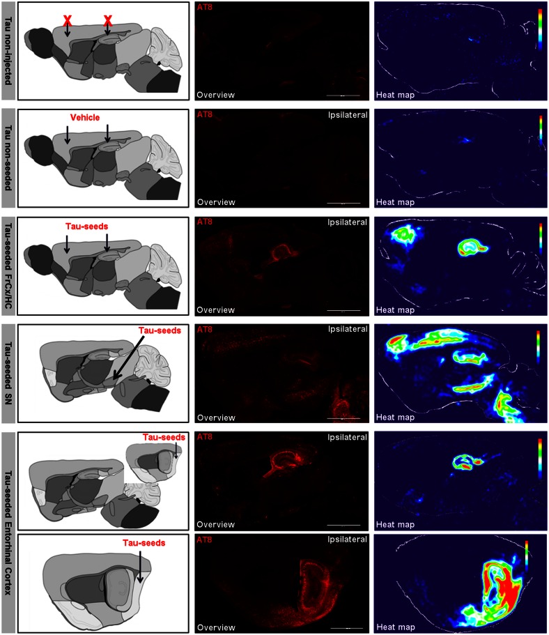
Fig. 5 Averaged heat maps for semi-quantitative analysis of spreading of Tau-pathology according to connectivity rather than proximity and along disease relevant patterns. Heat maps representing color scored averaged images of AT8 staining were generated for different injection paradigms. Heat maps of Tau mice, of Tau mice injected with vehicle, Tau mice injected with Tau-seeds in frontal cortex and hippocampus (FrCx/HC), Tau mice injected with Tau-seeds in substantia nigra (SN) and Tau mice injected with Tau-seeds in entorhinal cortex (EC), generated using Image J are presented. For Tau mice injected in EC, 2 different sagittal sections are presented situated closely to the medial longitudinal fissure and lateral. Schematic illustrations of the injection site and the section used for analysis are presented in the first column (modified after The GENSAT Project, website: http://www.gensat.org). Arrows indicate the injection site. Representative AT8 stainings (middle column) used for semi-quantitative analysis are presented for each condition, 6 months post-injection of Tau-seeds (scale bar 2000 µm). This reveals distinct patterns of Tau-pathology, encompassing brain regions with densely packed Tau-pathology, brain regions with scarcer Tau-pathology and unaffected brain regions. Heat maps were generated for semi-quantitative analysis of Tau-pathology (right column) by averaging AT8 stainings of multiple mice for each injection paradigm using Image J (n ≥ 3 for each condition). This demonstrates distinct patterns depending on the injection site, along functionally connected brain regions
Fig. 5 Averaged heat maps for semi-quantitative analysis of spreading of Tau-pathology according to connectivity rather than proximity and along disease relevant patterns. Heat maps representing color scored averaged images of AT8 staining were generated for different injection paradigms. Heat maps of Tau mice, of Tau mice injected with vehicle, Tau mice injected with Tau-seeds in frontal cortex and hippocampus (FrCx/HC), Tau mice injected with Tau-seeds in substantia nigra (SN) and Tau mice injected with Tau-seeds in entorhinal cortex (EC), generated using Image J are presented. For Tau mice injected in EC, 2 different sagittal sections are presented situated closely to the medial longitudinal fissure and lateral. Schematic illustrations of the injection site and the section used for analysis are presented in the first column (modified after The GENSAT Project, website: http://www.gensat.org). Arrows indicate the injection site. Representative AT8 stainings (middle column) used for semi-quantitative analysis are presented for each condition, 6 months post-injection of Tau-seeds (scale bar 2000 µm). This reveals distinct patterns of Tau-pathology, encompassing brain regions with densely packed Tau-pathology, brain regions with scarcer Tau-pathology and unaffected brain regions. Heat maps were generated for semi-quantitative analysis of Tau-pathology (right column) by averaging AT8 stainings of multiple mice for each injection paradigm using Image J (n ≥ 3 for each condition). This demonstrates distinct patterns depending on the injection site, along functionally connected brain regionsCC BY · PubMed Central
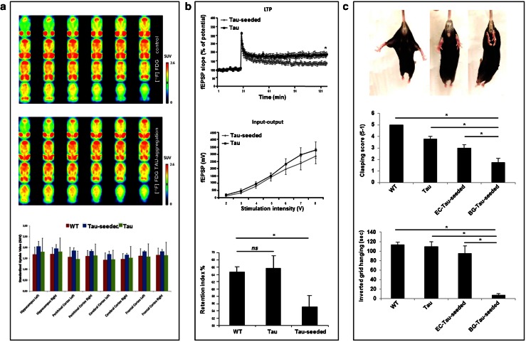
Fig. 6 Functional repercussions of Tau seed induced Tau-aggregation along unique neuronal networks, analyzed at 6 months post-injection. a FDG-PET analysis: mean horizontal small-animal FDG-PET images of the brain showing two representative samples of Tau-seeded and non-seeded Tau mice. Color bars indicate standardized uptake values for [18F]FDG. Quantitative analysis of the standardized uptake value revealed no significant difference between the different analyzed groups [non-transgenic (WT), non-seeded Tau transgenic (Tau), Tau-seeded Tau transgenic mice (Tau-seeded); n = 3 per group]. b Tau-seeded Tau-aggregation causes synaptic and cognitive dysfunction. Electrophysiological analysis of Tau transgenic mice seeded in entorhinal cortex or non-seeded analyzed 6 months post-injection reveals no significantly altered basic synaptic transmission in hippocampal CA1 region [Tau-seeded (n = 12); Tau (n = 12)]. Long term potentiation (LTP) induced by tetanic stimulation of Schaffer collaterals was significantly impaired following seeding in EC, 6 months post-injection [Tau-seeded (n = 5); Tau (n = 6); mean ± SEM are presented; p value <0.05; Student’s t test]. Cognitive function measured by the object recognition task (b, lower panel). Tau-transgenic mice with bilateral injection of Tau-seeds displayed significantly impaired object recognition memory compared to non-seeded Tau-transgenic mice or non-transgenic WT mice, while Tau transgenic mice were not significantly different compared to non-transgenic WT mice [n = 10 (control), n = 8 (non-seeded Tau transgenic mice), n = 8 (Tau-seeded Tau transgenic mice); mean ± SEM are presented; *p value <0.05; single ANOVA, post-Dunnett’s test]. c Motor impairment following initiation of Tau-seeding in basal ganglia, but not in entorhinal cortex. Representative images of clasping scores are presented (upper panel) from left to right: score 5 (no clasping); score 3 (intermediate clasping); score 1 (severe clasping). Clasping and inverted grid hanging was assessed for wild type mice (WT; n = 12), non-injected Tau mice (Tau; n = 6), Tau mice following Tau-seeding in entorhinal cortex (EC-Tau-seeded; n = 5), and Tau-mice following seeding in basal ganglia (BG-Tau-seeded; n = 7) 6 months post-seeding. Quantitative analysis revealed significantly increased clasping and impaired inverted grid hanging in Tau mice following seeding in basal ganglia, compared to Tau mice seeded in EC, Tau mice and non-transgenic mice (mean ± SEM are presented; *p value <0.05; one-way ANOVA, post-Dunnett’s test). In the graph in panel c, non-injected Tau-mice are presented for comparison with Tau-seeds injections in EC and basal ganglia. Sham injections in Tau mice in basal ganglia [clasping score: 3.9 ± 0.4, inverted grid hanging: 98.9 ± 12.7 (mean ± SEM, n = 9)] did not alter clasping or inverted grid hanging scores compared to non-injected Tau mice [clasping score: 3.8 ± 0.2, inverted grid hanging: 109.4 ± 10.0 (mean ± SEM, n = 6)] (not presented in the graph)
Fig. 6 Functional repercussions of Tau seed induced Tau-aggregation along unique neuronal networks, analyzed at 6 months post-injection. a FDG-PET analysis: mean horizontal small-animal FDG-PET images of the brain showing two representative samples of Tau-seeded and non-seeded Tau mice. Color bars indicate standardized uptake values for [18F]FDG. Quantitative analysis of the standardized uptake value revealed no significant difference between the different analyzed groups [non-transgenic (WT), non-seeded Tau transgenic (Tau), Tau-seeded Tau transgenic mice (Tau-seeded); n = 3 per group]. b Tau-seeded Tau-aggregation causes synaptic and cognitive dysfunction. Electrophysiological analysis of Tau transgenic mice seeded in entorhinal cortex or non-seeded analyzed 6 months post-injection reveals no significantly altered basic synaptic transmission in hippocampal CA1 region [Tau-seeded (n = 12); Tau (n = 12)]. Long term potentiation (LTP) induced by tetanic stimulation of Schaffer collaterals was significantly impaired following seeding in EC, 6 months post-injection [Tau-seeded (n = 5); Tau (n = 6); mean ± SEM are presented; p value <0.05; Student’s t test]. Cognitive function measured by the object recognition task (b, lower panel). Tau-transgenic mice with bilateral injection of Tau-seeds displayed significantly impaired object recognition memory compared to non-seeded Tau-transgenic mice or non-transgenic WT mice, while Tau transgenic mice were not significantly different compared to non-transgenic WT mice [n = 10 (control), n = 8 (non-seeded Tau transgenic mice), n = 8 (Tau-seeded Tau transgenic mice); mean ± SEM are presented; *p value <0.05; single ANOVA, post-Dunnett’s test]. c Motor impairment following initiation of Tau-seeding in basal ganglia, but not in entorhinal cortex. Representative images of clasping scores are presented (upper panel) from left to right: score 5 (no clasping); score 3 (intermediate clasping); score 1 (severe clasping). Clasping and inverted grid hanging was assessed for wild type mice (WT; n = 12), non-injected Tau mice (Tau; n = 6), Tau mice following Tau-seeding in entorhinal cortex (EC-Tau-seeded; n = 5), and Tau-mice following seeding in basal ganglia (BG-Tau-seeded; n = 7) 6 months post-seeding. Quantitative analysis revealed significantly increased clasping and impaired inverted grid hanging in Tau mice following seeding in basal ganglia, compared to Tau mice seeded in EC, Tau mice and non-transgenic mice (mean ± SEM are presented; *p value <0.05; one-way ANOVA, post-Dunnett’s test). In the graph in panel c, non-injected Tau-mice are presented for comparison with Tau-seeds injections in EC and basal ganglia. Sham injections in Tau mice in basal ganglia [clasping score: 3.9 ± 0.4, inverted grid hanging: 98.9 ± 12.7 (mean ± SEM, n = 9)] did not alter clasping or inverted grid hanging scores compared to non-injected Tau mice [clasping score: 3.8 ± 0.2, inverted grid hanging: 109.4 ± 10.0 (mean ± SEM, n = 6)] (not presented in the graph)CC BY · PubMed Central
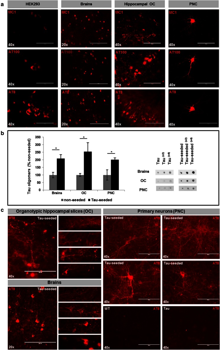
Fig. 7 Different pathological forms of Tau are generated by Tau-seed induced Tau-aggregation. a Immunostaining of pathological forms of Tau reveals the presence of hyperphosphorylated Tau forms phosphorylated at Ser202/Thr205 (AT8) and T212/S214 (AT100), as well as misfolded Tau (MC1) antibody in all models, i.e. HEK293, brains, organotypic cultures and primary neurons, following Tau-seeding (scale bar ×20—200 µm, ×40—100 µm). b Tau-seeding increased Tau-oligomer concentrations assessed by dot blotting analysis with the Tau-oligomer specific T22 antibody on total homogenates. Quantitative analysis demonstrated accumulation of oligomeric Tau forms following Tau-seeding in brains [(n = 4; n = 4), (non-seeded; Tau-seeded)], organotypic cultures (OC) (n = 5; n = 5) and primary neurons (PNC) (n = 6; n = 5) (mean ± SEM are presented; *p value < 0.05). Dilution curves of samples of the different models are presented. c Immunohisto/cytological analysis with AT8 reveals different forms of AT8 stained Tau. In addition to mature and immature NFTs, diffuse and punctated staining are prominently observed by AT8-staining in organotypic hippocampal cultures and brains following Tau-seeding (left panels provide an overview picture and details of different types of AT8 staining observed in Tau-seeded OC and brains). In PNC, AT8 staining following standard fixation in 4 % PAF, demonstrates strong diffuse and punctated AT8 staining present throughout the culture dish in PNC, and robustly induced following Tau-seeding in TauP301S expressing PNC. Lower panels show AT8 staining (standard fixation) in non-seeded primary neurons derived from TauP301S mice (Tau) or non-transgenic mice (WT) (scale bar ×20–200 µm, ×40–100 µm)
Fig. 7 Different pathological forms of Tau are generated by Tau-seed induced Tau-aggregation. a Immunostaining of pathological forms of Tau reveals the presence of hyperphosphorylated Tau forms phosphorylated at Ser202/Thr205 (AT8) and T212/S214 (AT100), as well as misfolded Tau (MC1) antibody in all models, i.e. HEK293, brains, organotypic cultures and primary neurons, following Tau-seeding (scale bar ×20—200 µm, ×40—100 µm). b Tau-seeding increased Tau-oligomer concentrations assessed by dot blotting analysis with the Tau-oligomer specific T22 antibody on total homogenates. Quantitative analysis demonstrated accumulation of oligomeric Tau forms following Tau-seeding in brains [(n = 4; n = 4), (non-seeded; Tau-seeded)], organotypic cultures (OC) (n = 5; n = 5) and primary neurons (PNC) (n = 6; n = 5) (mean ± SEM are presented; *p value < 0.05). Dilution curves of samples of the different models are presented. c Immunohisto/cytological analysis with AT8 reveals different forms of AT8 stained Tau. In addition to mature and immature NFTs, diffuse and punctated staining are prominently observed by AT8-staining in organotypic hippocampal cultures and brains following Tau-seeding (left panels provide an overview picture and details of different types of AT8 staining observed in Tau-seeded OC and brains). In PNC, AT8 staining following standard fixation in 4 % PAF, demonstrates strong diffuse and punctated AT8 staining present throughout the culture dish in PNC, and robustly induced following Tau-seeding in TauP301S expressing PNC. Lower panels show AT8 staining (standard fixation) in non-seeded primary neurons derived from TauP301S mice (Tau) or non-transgenic mice (WT) (scale bar ×20–200 µm, ×40–100 µm)CC BY · PubMed Central

Verification

Claims the paper makes

The claims and statistics of this paper haven’t been checked yet.

Run the check — free, no account needed. It reads the open-access text and checks the sources it cites.

A few free checks a day · papers already verified open instantly · results are public

Claims other papers make about this one

Each is checked against this paper’s own text: does it say what the citing paper says it does?

Self-check

Percentages, sums and reported differences re-computed from the paper’s own numbers.

The self-check needs the paper’s full text.

Citations

Cites

Show all 76 references

Cited by

Sorted by citations

Items 101–143 of 143

Metadata

Authors
Stancu, I.-C., Vasconcelos, B., Ris, L., Wang, P., Villers, A., Peeraer, E., Buist, A., Terwel, D., Baatsen, P., Oyelami, T., Pierrot, N., Casteels, C., Bormans, G., Kienlen-Campard, P., Octave, J.-N., Moechars, D., & Dewachter, I.
Affiliations
  • UCLouvain (BE)
  • University of Mons (BE)
  • Janssen (Belgium) (BE)
  • KU Leuven (BE)
Corresponding author
Dewachter, Ilse
Venue
Acta Neuropathologica
Published
2015-04-11
Volume
129
Issue
6
Pages
875-894
ISSN
  • 0001-6322 (ISSN-L)
  • 1432-0533
OpenAlex
W2109355761
Cited by
143
References
76
Citations by year
2026: 4 · 2025: 6 · 2024: 12 · 2023: 10 · 2022: 12 · 2021: 15 · 2020: 16 · 2019: 15 · 2018: 14 · 2017: 18 · 2016: 18 · 2015: 3
FWCI
13.01
Type
journal article
Language
en
Open access
Locations
  • Acta Neuropathologica (publishedVersion, cc-by)
  • PubMed Central (publishedVersion, cc-by)
  • PubMed (publishedVersion)
  • Lirias (submittedVersion, other-oa)
  • Digital Access to Libraries (submittedVersion)
  • Document Server@UHasselt (UHasselt) (submittedVersion, cc-by)
  • ORBi UMONS (submittedVersion)
Funders
  • Fonds De La Recherche Scientifique - FNRS
  • Vlaamse regering
Topics
  • Life Sciences › Neuroscience › Cellular and Molecular Neuroscience › Alzheimer's disease research and treatments
  • Life Sciences › Biochemistry, Genetics and Molecular Biology › Molecular Biology › Prion Diseases and Protein Misfolding
  • Life Sciences › Neuroscience › Neurology › Neuroinflammation and Neurodegeneration Mechanisms
Keywords
tau protein, prion-like seeding, tauopathy, neuronal network dysfunction, synaptic transmission, long-term potentiation, synaptic plasticity, hippocampus, entorhinal cortex, basal ganglia, motor impairment, cognitive impairment, object recognition, tau oligomers, neurofibrillary tangles, GABA-A receptor antagonists, organotypic hippocampal slices
MeSH terms
Animals, Animals, Newborn, Calcium, Cognition Disorders, Disease Models, Animal, Exploratory Behavior, Hippocampus, Membrane Potentials, Mice, Inbred C57BL, Mice, Transgenic, Mutation, Nerve Net, Prions, Fura-2, Neurofibrillary Tangles, tau Proteins, Tauopathies, Mice, Proteostasis Deficiencies, In Vitro Techniques

About these checks

Registry, retraction, Cites and statistics results are computed, not judged, with one exception: a printed reference that no index lists is read once by a model before it counts as a citation error. Claims are extracted by a model and kept only when their quote appears verbatim in the text; each cited source is then checked, with a model’s help, for whether it says what the claim relies on. Those verdicts can be wrong — read the claim’s quote and the cited source before relying on one.

Statistics and arithmetic are recomputed, not judged.

Confidence bands are shown only for fields calibrated on our annotated test set. None is calibrated yet, so no band is shown.

What counts as a claim ›How we measure confidence ›How we check this ›

Are you an author, or do you see a problem with this page? Request a review