

The DOI matches its Crossref record; 76 of the 76 references it cites were found in a registry or an index.
Templated misfolding of Tau by prion-like seeding along neuronal connections impairs neuronal network function and associated behavioral outcomes in Tau transgenic mice
Open accessAbstract
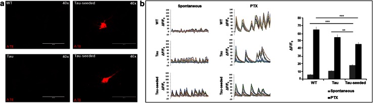
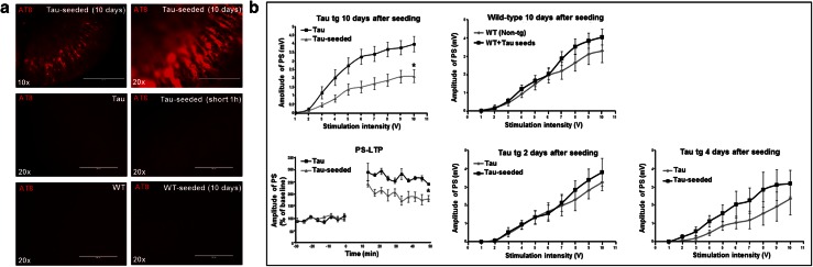
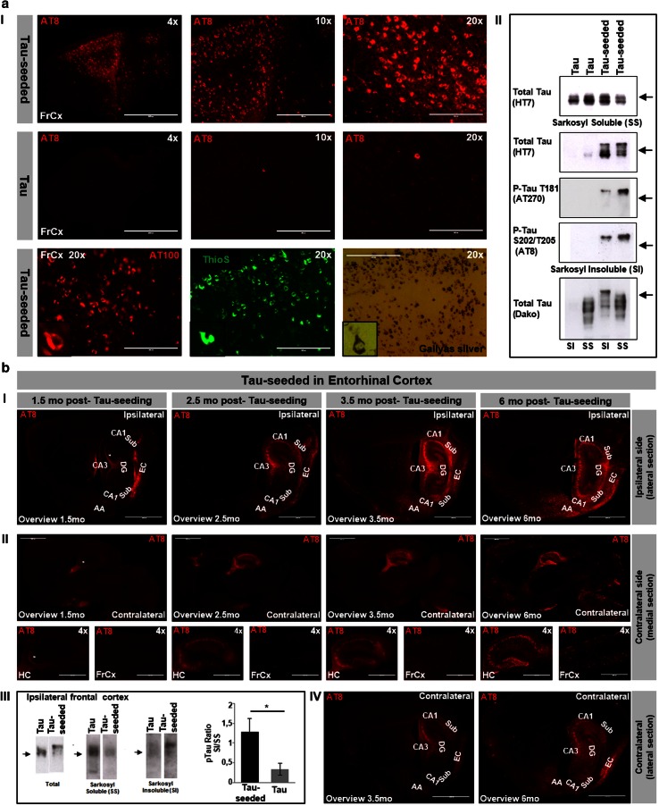
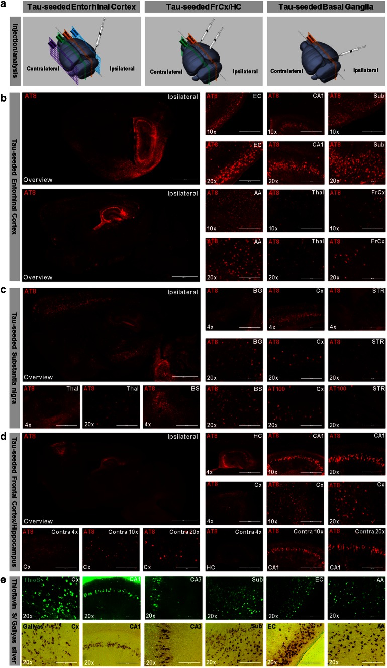
Prion-like seeding and propagation of Tau-pathology have been demonstrated experimentally and may underlie the stereotyped progression of neurodegenerative Tauopathies. However, the involvement of templated misfolding of Tau in neuronal network dysfunction and behavioral outcomes remains to be explored in detail. Here we analyzed the repercussions of prion-like spreading of Tau-pathology via neuronal connections on neuronal network function in TauP301S transgenic mice. Spontaneous and GABA(A)R-antagonist-induced neuronal network activity were affected following templated Tau-misfolding using synthetic preformed Tau fibrils in cultured primary neurons. Electrophysiological analysis in organotypic hippocampal slices of Tau transgenic mice demonstrated impaired synaptic transmission and impaired long-term potentiation following Tau-seed induced Tau-aggregation. Intracerebral injection of Tau-seeds in TauP301S mice, caused prion-like spreading of Tau-pathology through functionally connected neuroanatomical pathways. Electrophysiological analysis revealed impaired synaptic plasticity in hippocampal CA1 region 6 months after Tau-seeding in entorhinal cortex (EC). Furthermore, templated Tau aggregation impaired cognitive function, measured in the object recognition test 6 months post-seeding. In contrast, Tau-seeding in basal ganglia and subsequent spreading through functionally connected neuronal networks involved in motor control, resulted in motoric deficits reflected in clasping and impaired inverted grid hanging, not significantly affected following Tau-seeding in EC. Immunostaining, biochemical and electron microscopic analysis in the different models suggested early pathological forms of Tau, including Tau-oligomers, rather than fully mature neurofibrillary tangles (NFTs) as culprits of neuronal dysfunction. We here demonstrate for the first time using in vitro, ex vivo and in vivo models, that prion-like spreading of Tau-misfolding by Tau seeds, along unique neuronal connections, causes neuronal network dysfunction and associated behavioral dysfunction. Our data highlight the potential relevance of this mechanism in the symptomatic progression in Tauopathies. We furthermore demonstrate that the initial site of Tau-seeding thereby determines the behavioral outcome, potentially underlying the observed heterogeneity in (familial) Tauopathies, including in TauP301 mutants.
Overview
The claims and statistics of this paper haven’t been checked yet.
Run the check — free, no account needed. It reads the open-access text and checks the sources it cites.
Integrity signals
Registry
Cites
Statistics
Figures
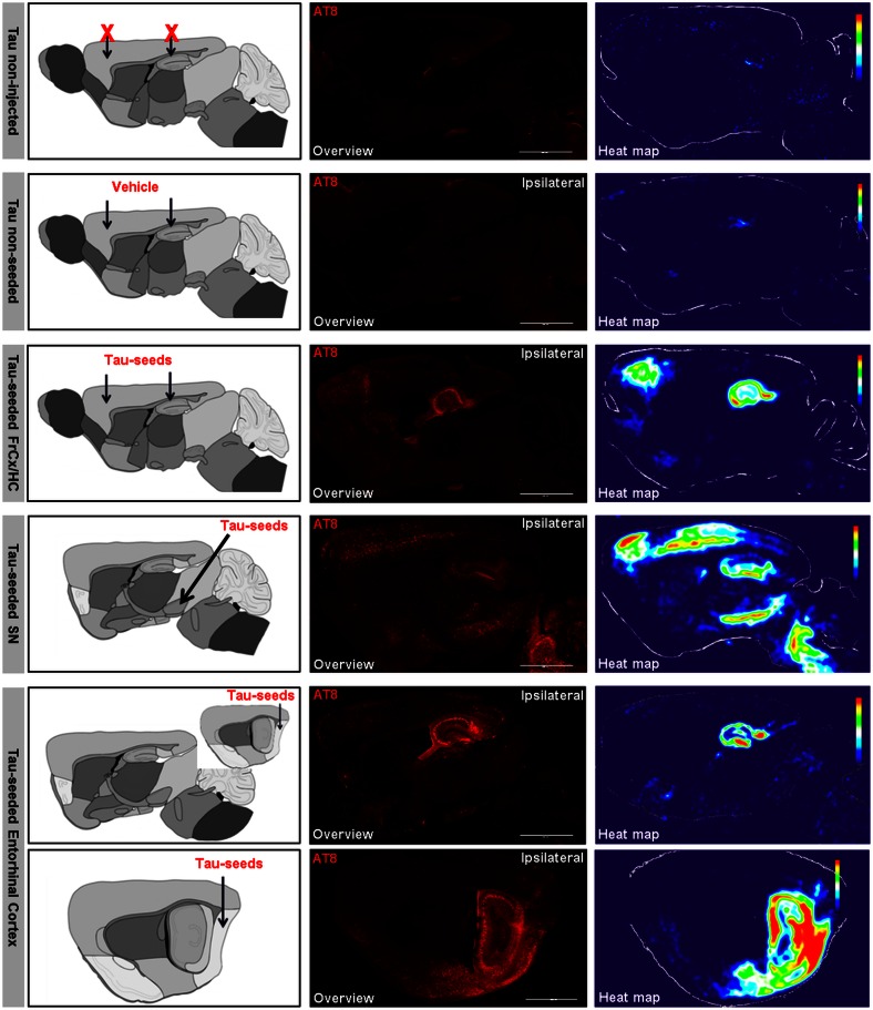
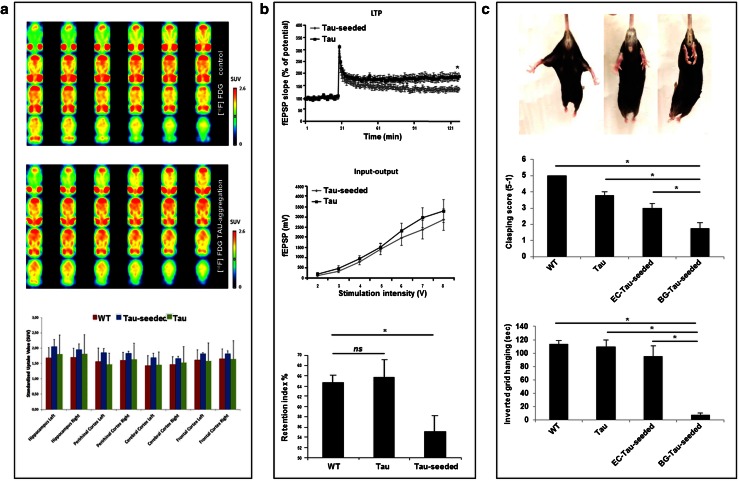
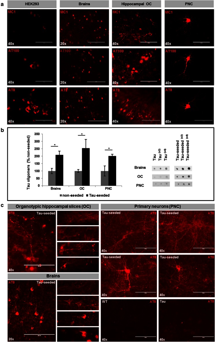
Verification
Claims the paper makes
The claims and statistics of this paper haven’t been checked yet.
Run the check — free, no account needed. It reads the open-access text and checks the sources it cites.
Claims other papers make about this one
Each is checked against this paper’s own text: does it say what the citing paper says it does?
The source backs part of the claim, not all of it — or backs it for a different population, condition or comparison.
In the citing paper
“These observations raise the prospect that degeneration could be initiated through synaptic or axonal mechanisms, independent of apoptosis, a mechanism which has been widely studied in other model systems, such as spinal cord injury and Multiple Sclerosis 59–61.”
From Amyloid-beta induced retrograde axonal degeneration in a mouse tauopathy model (2019) · cites this paper as [60]
Experimental — accuracy not yet measured
Self-check
Percentages, sums and reported differences re-computed from the paper’s own numbers.
The self-check needs the paper’s full text.
Citations
Cites
Neuropathological stageing of Alzheimer-related changes.
Braak, H., & Braak, E. (1991) · Acta Neuropathologica · doi:10.1007/bf00308809 · Cited by 16,123
The Mouse Brain in Stereotaxic Coordinates
Paxinos, G. (2001) · Cited by 12,859
Amyloid oligomers exacerbate tau pathology in a mouse model of tauopathy
Selenica, M.-L. B., Brownlow, M., Jimenez, J. P., Lee, D. C., Pena, G., Dickey, C. A., Gordon, M. N., & Morgan, D. (2013) · Neurodegenerative Diseases · doi:10.1159/000337230 · Cited by 10,359
Neuropathological alterations in Alzheimer disease
Serrano-Pozo, A., Frosch, M. P., Masliah, E., & Hyman, B. T. (2011) · Cold Spring Harbor Perspectives in Medicine · doi:10.1101/cshperspect.a006189 · Cited by 3,345
A simple method for organotypic cultures of nervous tissue
Stoppini, L., Buchs, P., & Muller, D. (1991) · Journal of Neuroscience Methods · doi:10.1016/0165-0270(91)90128-m · Cited by 3,096
Lee, V. M.-Y., Goedert, M., & Trojanowski, J. Q. (2001) · Annual Review of Neuroscience · doi:10.1146/annurev.neuro.24.1.1121 · Cited by 2,480
Neurodegenerative Diseases Target Large-Scale Human Brain Networks
Seeley, W. W., Crawford, R. K., Zhou, J., Miller, B. L., & Greicius, M. D. (2009) · Neuron · doi:10.1016/j.neuron.2009.03.024 · Cited by 2,350
Synapse Loss and Microglial Activation Precede Tangles in a P301S Tauopathy Mouse Model
Yoshiyama, Y., Higuchi, M., Zhang, B., Huang, S.-M., Iwata, N., Saido, T. C., Maeda, J., Suhara, T., Trojanowski, J. Q., & Lee, V. M.-Y. (2007) · Neuron · doi:10.1016/j.neuron.2007.01.010 · Cited by 2,208
Tau Suppression in a Neurodegenerative Mouse Model Improves Memory Function
SantaCruz, K., Lewis, J., Spires, T., Paulson, J., Kotilinek, L., Ingelsson, M., Guimaraes, A., DeTure, M., Ramsden, M., McGowan, E., et al. (2005) · Science · doi:10.1126/science.1113694 · Cited by 2,014
Learning induces long-term potentiation in the hippocampus
Whitlock, J. R., Heynen, A. J., Shuler, M. G., & Bear, M. F. (2006) · Science · doi:10.1126/science.1128134 · Cited by 1,913
Cited by
Sorted by citations
Stancu, I.-C., Cremers, N., Vanrusselt, H., Couturier, J., Vanoosthuyse, A., Kessels, S., Lodder, C., Brône, B., Huaux, F., Octave, J.-N., et al. (2019) · Acta Neuropathologica · doi:10.1007/s00401-018-01957-y · Cited by 447
Self-citationPropagation and spread of pathogenic protein assemblies in neurodegenerative diseases
Jucker, M., & Walker, L. C. (2018) · Nature Neuroscience · doi:10.1038/s41593-018-0238-6 · Cited by 450
Guo, J. L., Narasimhan, S., Changolkar, L., He, Z., Stieber, A., Zhang, B., Gathagan, R. J., Iba, M., McBride, J. D., Trojanowski, J. Q., et al. (2016) · Journal of Experimental Medicine · doi:10.1084/jem.20160833 · Cited by 440
MAPT mutations, tauopathy, and mechanisms of neurodegeneration
Strang, K. H., Golde, T. E., & Giasson, B. I. (2019) · Laboratory Investigation · doi:10.1038/s41374-019-0197-x · Cited by 424
Rodent models for Alzheimer disease
Götz, J., Bodea, L., & Goedert, M. (2018) · Nature reviews. Neuroscience · doi:10.1038/s41583-018-0054-8 · Cited by 422
What is the evidence that tau pathology spreads through prion-like propagation?
Mudher, A., Colin, M., Dujardin, S., Medina, M., Dewachter, I., Alavi Naini, S. M., Mandelkow, E.-M., Mandelkow, E., Buée, L., Goedert, M., et al. (2017) · Acta Neuropathologica Communications · doi:10.1186/s40478-017-0488-7 · Cited by 384
Self-citationThe complexity of tau in Alzheimer’s disease
Naseri, N. N., Wang, H., Guo, J. D., Sharma, M., & Luo, W. (2019) · Neuroscience Letters · doi:10.1016/j.neulet.2019.04.022 · Cited by 393
Lewczuk, P., Riederer, P. F., O’Bryant, S. E., Verbeek, M. M., Dubois, B., Visser, P. J., Jellinger, K. A., Engelborghs, S., Ramírez, A., Parnetti, L., et al. (2017) · The World Journal of Biological Psychiatry · doi:10.1080/15622975.2017.1375556 · Cited by 318
Widespread brain tau and its association with ageing, Braak stage and Alzheimer’s dementia
Lowe, V. J., Wiste, H. J., Senjem, M. L., Weigand, S. D., Therneau, T. M., Boeve, B. F., Josephs, K. A., Fang, P., Pandey, M. K., Murray, M. E., et al. (2017) · Brain · doi:10.1093/brain/awx320 · Cited by 311
A critical appraisal of the pathogenic protein spread hypothesis of neurodegeneration
Walsh, D. M., & Selkoe, D. J. (2016) · Nature reviews. Neuroscience · doi:10.1038/nrn.2016.13 · Cited by 300
Synaptic Tau Seeding Precedes Tau Pathology in Human Alzheimer’s Disease Brain
DeVos, S. L., Corjuc, B. T., Oakley, D. H., Nobuhara, C. K., Bannon, R. N., Chase, A., Commins, C., Gonzalez, J. A., Dooley, P. M., Frosch, M. P., et al. (2018) · Frontiers in Neuroscience · doi:10.3389/fnins.2018.00267 · Cited by 285
Dendritic Tau in Alzheimer’s Disease
Ittner, A., & Ittner, L. M. (2018) · Neuron · doi:10.1016/j.neuron.2018.06.003 · Cited by 278
The human connectome in Alzheimer disease — relationship to biomarkers and genetics
Yu, M., Sporns, O., & Saykin, A. J. (2021) · Nature Reviews Neurology · doi:10.1038/s41582-021-00529-1 · Cited by 265
Iba, M., McBride, J. D., Guo, J. L., Zhang, B., Trojanowski, J. Q., & Lee, V. M.-Y. (2015) · Acta Neuropathologica · doi:10.1007/s00401-015-1458-4 · Cited by 223
Lewis, J. M., & Dickson, D. W. (2015) · Acta Neuropathologica · doi:10.1007/s00401-015-1507-z · Cited by 229
Traumatic Brain Injury Induces Tau Aggregation and Spreading
Edwards, G., Zhao, J., Dash, P. K., Soto, C., & Moreno-Gonzalez, I. (2020) · Journal of Neurotrauma · doi:10.1089/neu.2018.6348 · Cited by 195
Tau Pathology in Parkinson's Disease
Zhang, X., Gao, F., Wang, D., Li, C., Fu, Y., He, W., & Zhang, J. (2018) · Frontiers in Neurology · doi:10.3389/fneur.2018.00809 · Cited by 210
Vasconcelos, B., Stancu, I.-C., Buist, A., Bird, M., Wang, P., Vanoosthuyse, A., Van Kolen, K., Verheyen, A., Kienlen-Campard, P., Octave, J.-N., et al. (2016) · Acta Neuropathologica · doi:10.1007/s00401-015-1525-x · Cited by 192
Self-citationThe basis of cellular and regional vulnerability in Alzheimer’s disease
Mrdjen, D., Fox, E. J., Bukhari, S. A., Montine, K. S., Bendall, S. C., & Montine, T. J. (2019) · Acta Neuropathologica · doi:10.1007/s00401-019-02054-4 · Cited by 162
Sustained synchronized neuronal network activity in a human astrocyte co-culture system
Kuijlaars, J., Oyelami, T., Diels, A., Rohrbacher, J., Versweyveld, S., Meneghello, G., Tuefferd, M., Verstraelen, P., Detrez, J. R., Verschuuren, M., et al. (2016) · Scientific Reports · doi:10.1038/srep36529 · Cited by 153
Self-citationDiscovery and characterization of stable and toxic Tau/phospholipid oligomeric complexes
Ait‐Bouziad, N., Lv, G., Mahul‐Mellier, A., Xiao, S., Zorludemir, G., Eliezer, D., Walz, T., & Lashuel, H. A. (2017) · Nature Communications · doi:10.1038/s41467-017-01575-4 · Cited by 149
Widespread tau seeding activity at early Braak stages
Furman, J. L., Vaquer‐Alicea, J., White, C. L., Cairns, N. J., Nelson, P. T., & Diamond, M. I. (2016) · Acta Neuropathologica · doi:10.1007/s00401-016-1644-z · Cited by 146
Strang, K. H., Croft, C. L., Sorrentino, Z. A., Chakrabarty, P., Golde, T. E., & Giasson, B. I. (2018) · Journal of Biological Chemistry · doi:10.1074/jbc.m117.815357 · Cited by 124
Tau pathology in neurodegenerative disease: disease mechanisms and therapeutic avenues
Samudra, N. P., Lane‐Donovan, C., VandeVrede, L. A., & Boxer, A. L. (2023) · Journal of Clinical Investigation · doi:10.1172/jci168553 · Cited by 142
Valera, E., Spencer, B. J., & Masliah, E. (2015) · Neurotherapeutics · doi:10.1007/s13311-015-0397-z · Cited by 135
Hyperphosphorylation determines both the spread and the morphology of tau pathology
Hu, W. H., Zhang, X., Tung, Y. C., Xie, S.-T., Liu, F., & Iqbal, K. (2016) · Alzheimer s & Dementia · doi:10.1016/j.jalz.2016.01.014 · Cited by 137
The role of exosomes in the pathogenesis of Alzheimer’ disease
Xiao, T., Zhang, W., Jiao, B., Pan, C., Liu, X., & Shen, L. H. (2017) · Translational Neurodegeneration · doi:10.1186/s40035-017-0072-x · Cited by 133
Wang, P., Joberty, G., Buist, A., Vanoosthuyse, A., Stancu, I., Vasconcelos, B., Pierrot, N., Faelth-Savitski, M., Kienlen‐Campard, P., Octave, J., et al. (2017) · Acta Neuropathologica · doi:10.1007/s00401-016-1663-9 · Cited by 120
Self-citationSpreading of α-Synuclein and Tau: A Systematic Comparison of the Mechanisms Involved
Vasili, E., Dominguez‐Meijide, A., & Outeiro, T. F. (2019) · Frontiers in Molecular Neuroscience · doi:10.3389/fnmol.2019.00107 · Cited by 121
Tau biology, tauopathy, traumatic brain injury, and diagnostic challenges
Castellani, R. J., & Perry, G. (2019) · Journal of Alzheimer’s Disease · doi:10.3233/jad-180721 · Cited by 110
Different tau species lead to heterogeneous tau pathology propagation and misfolding
Dujardin, S., Bégard, S., Caillierez, R., Lachaud, C., Carrier, S., Lieger, S., Gonzalez, J. A., Deramecourt, V., Déglon, N., Maurage, C.-A., et al. (2018) · Acta Neuropathologica Communications · doi:10.1186/s40478-018-0637-7 · Cited by 109
In vivo tau pathology is associated with synaptic loss and altered synaptic function
Coomans, E. M., Schoonhoven, D. N., Tuncel, H., Verfaillie, S. C. J., Wolters, E. E., Boellaard, R., Ossenkoppele, R., den Braber, A., Scheper, W., Schober, P. R., et al. (2021) · Alzheimer s Research & Therapy · doi:10.1186/s13195-021-00772-0 · Cited by 106
Shulman, M. B., Kong, J., O'Gorman, J. T., Ratti, E., Rajagovindan, R., Viollet, L., Huang, E., Sharma, S., Racine, A. M., Czerkowicz, J., et al. (2023) · Nature Aging · doi:10.1038/s43587-023-00523-w · Cited by 100
Filamentous recombinant human Tau activates primary astrocytes via an integrin receptor complex
Wang, P., & Ye, Y. (2021) · Nature Communications · doi:10.1038/s41467-020-20322-w · Cited by 93
Self-citationPrions and Protein Assemblies that Convey Biological Information in Health and Disease
Sanders, D. W., Kaufman, S. K., Holmes, B. B., & Diamond, M. I. (2016) · Neuron · doi:10.1016/j.neuron.2016.01.026 · Cited by 92
Dutta, D., Jana, M., Paidi, R. K., Majumder, M., Raha, S., Dasarathy, S., & Pahan, K. (2023) · Journal of Clinical Investigation · doi:10.1172/jci161987 · Cited by 87
Tau prion-like propagation: state of the art and current challenges
Dujardin, S., & Hyman, B. T. (2019) · Advances in Experimental Medicine and Biology · doi:10.1007/978-981-32-9358-8_23 · Cited by 84
Involvement of Oligodendrocytes in Tau Seeding and Spreading in Tauopathies
Ferrer, I., Aguiló, M., Carmona, M., Andrés‐Benito, P., Torrejón-Escribano, B., Garcia‐Esparcia, P., & del Rı́o, J. A. (2019) · Frontiers in Aging Neuroscience · doi:10.3389/fnagi.2019.00112 · Cited by 78
Wu, X., Piña-Crespo, J., Zhang, Y., Chen, X., & Xu, H. (2017) · Chinese Medical Journal · doi:10.4103/0366-6999.220313 · Cited by 81
Andersson, C. R., Falsig, J., Stavenhagen, J. B., Christensen, S., Kartberg, F., Rosenqvist, N., Finsen, B. R., & Pedersen, J. T. (2019) · Scientific Reports · doi:10.1038/s41598-019-41105-4 · Cited by 78
Neuronally derived extracellular vesicles: an emerging tool for understanding Alzheimer’s disease
Watson, L. S., Hamlett, E. D., Stone, T. D., & Sims‐Robinson, C. (2019) · Molecular Neurodegeneration · doi:10.1186/s13024-019-0317-5 · Cited by 75
The NLRP3 inflammasome modulates tau pathology and neurodegeneration in a tauopathy model
Stancu, I. C., Lodder, C., Botella Lucena, P., Vanherle, S., Gutiérrez de Ravé, M., Terwel, D., Bottelbergs, A., & Dewachter, I. (2022) · Glia · doi:10.1002/glia.24160 · Cited by 72
Self-citationLodder, C., Scheyltjens, I., Stancu, I., Lucena, P. B., de Ravé, M. G., Vanherle, S., Vanmierlo, T., Cremers, N., Vanrusselt, H., Brône, B., et al. (2021) · Acta Neuropathologica Communications · doi:10.1186/s40478-021-01204-8 · Cited by 71
Self-citationAudouard, E., Houben, S., Masaracchia, C., Yilmaz, Z., Suain, V., Authelet, M., Decker, R. D., Buée, L., Boom, A., Leroy, K., et al. (2016) · American Journal Of Pathology · doi:10.1016/j.ajpath.2016.06.008 · Cited by 73
Prion-like mechanisms in Alzheimer disease
Walker, L. C. (2018) · Handbook of clinical neurology · doi:10.1016/b978-0-444-63945-5.00016-7 · Cited by 67
In vivoimaging reveals reduced activity of neuronal circuits in a mouse tauopathy model
Marinković, P., Blumenstock, S., Goltstein, P. M., Korzhova, V., Peters, F., Knebl, A., & Herms, J. W. (2019) · Brain · doi:10.1093/brain/awz035 · Cited by 63
Ferrer, I., Andrés‐Benito, P., Zelaya, M. V., Aguirre, M. E. E., Carmona, M., Ausín, K., Lachén‐Montes, M., Fernández‐Irigoyen, J., Santamaría, E., & del Río, J. A. (2020) · Acta Neuropathologica · doi:10.1007/s00401-019-02122-9 · Cited by 58
Elevated matrix metalloproteinase‐9 levels in neuronal extracellular vesicles in Alzheimer’s disease
Gu, D., Liu, F., Meng, M., Zhang, L., Gordon, M. L., Wang, Y., Cai, L., & Zhang, N. (2020) · Annals of Clinical and Translational Neurology · doi:10.1002/acn3.51155 · Cited by 58
Tau-Cofactor Complexes as Building Blocks of Tau Fibrils
Fichou, Y., Oberholtzer, Z. R., Ngo, H., Cheng, C., Keller, T. J., Eschmann, N. A., & Han, S. (2019) · Frontiers in Neuroscience · doi:10.3389/fnins.2019.01339 · Cited by 57
The evolutionary scope and neurological disease linkage of yeast-prion-like proteins in humans
An, L., & Harrison, P. M. (2016) · Biology Direct · doi:10.1186/s13062-016-0134-5 · Cited by 53
Effects of pharmacological modulators of α-synuclein and tau aggregation and internalization
Dominguez‐Meijide, A., Vasili, E., König, A., Cima‐Omori, M., de Opakua, A. I., Leonov, A., Ryazanov, S., Zweckstetter, M., Griesinger, C., & Outeiro, T. F. (2020) · Scientific Reports · doi:10.1038/s41598-020-69744-y · Cited by 52
Chai, A. B., Hartz, A. M. S., Gao, X., Yang, A., Callaghan, R., & Gelissen, I. C. (2020) · International Journal of Molecular Sciences · doi:10.3390/ijms22010246 · Cited by 51
The evolving landscape of Alzheimer’s disease therapy: From Aβ to tau
Courade, J., Zetterberg, H., Höglinger, G. U., & Dewachter, I. (2025) · Cell · doi:10.1016/j.cell.2025.11.033 · Cited by 57
Self-citationMcAllister, B. B., Lacoursiere, S. G., Sutherland, R. J., & Mohajerani, M. H. (2020) · Neuroscience & Biobehavioral Reviews · doi:10.1016/j.neubiorev.2020.01.026 · Cited by 48
Neuropathology of the Alzheimer's continuum: an update
Jellinger, K. A. (2020) · Free Neuropathology · doi:10.17879/freeneuropathology-2020-3050 · Cited by 52
The Prion-Like Behavior of Assembled Tau in Transgenic MicePreprint
Clavaguera, F., Tolnay, M., & Goedert, M. (2016) · Cold Spring Harbor Perspectives in Medicine · doi:10.1101/cshperspect.a024372 · Cited by 45
Schultz, M. K., Gentzel, R. C., Usenovic, M., Gretzula, C. A., Ware, C., Parmentier‐Batteur, S., Schachter, J. B., & Zariwala, H. A. (2018) · Neurobiology of Disease · doi:10.1016/j.nbd.2018.07.003 · Cited by 45
CSF biomarkers in neurodegenerative and vascular dementias
Llorens, F., Schmitz, M., Ferrer, I., & Zerr, I. (2016) · Progress in Neurobiology · doi:10.1016/j.pneurobio.2016.03.003 · Cited by 44
Chronic Traumatic Encephalopathy: Is Latency in Symptom Onset Explained by Tau Propagation?Preprint
Kriegel, J., Papadopoulos, Z., & McKee, A. C. (2017) · Cold Spring Harbor Perspectives in Medicine · doi:10.1101/cshperspect.a024059 · Cited by 38
Relevance of host tau in tau seeding and spreading in tauopathies
Ferrer, I., Zelaya, M. V., Aguiló, M., Carmona, M., López‐González, I., Andrés‐Benito, P., Lidón, L., Gavı́n, R., Garcia‐Esparcia, P., & del Rı́o, J. A. (2019) · Brain Pathology · doi:10.1111/bpa.12778 · Cited by 37
Kim, Y. D., Jeong, E. I., Nah, J., Yoo, S., Lee, W. J., Kim, Y., Moon, S., Hong, S., & Jung, Y. (2017) · Journal of Neurochemistry · doi:10.1111/jnc.14109 · Cited by 35
Amyloid-beta induced retrograde axonal degeneration in a mouse tauopathy model
Nishioka, C., Liang, H.-F., Barsamian, B., & Sun, S.-W. (2019) · NeuroImage · doi:10.1016/j.neuroimage.2019.01.007 · Cited by 30
“Although tau aggregates are hallmarks of AD, several studies have suggested that oligomeric tau in soluble forms may be the most potent neurotoxic form (Wu et al., 2013; Iba et al., 2013, 2015; Stancu et al., 2015; Peeraer et al., 2015; Kopeikina et al., 2012).”
Hosokawa, M., Masuda‐Suzukake, M., Shitara, H., Shimozawa, A., Suzuki, G., Kondo, H., Nonaka, T., Campbell, W. D., Arai, T., & Hasegawa, M. (2021) · Brain · doi:10.1093/brain/awab289 · Cited by 30
Elevated medial temporal lobe and pervasive brain tau‐PET signal in normal participants
Lowe, V. J., Bruinsma, T. J., Min, H.-K. P., Lundt, E. S., Fang, P., Senjem, M. L., Boeve, B. F., Josephs, K. A., Pandey, M. K., Murray, M. E., et al. (2018) · Alzheimer s & Dementia Diagnosis Assessment & Disease Monitoring · doi:10.1016/j.dadm.2018.01.005 · Cited by 32
Multicenter 18F-PI-2620 PET for In Vivo Braak Staging of Tau Pathology in Alzheimer’s Disease
Rullmann, M., Brendel, M., Schroeter, M. L., Saur, D., Levin, J., Perneczky, R. G., Tiepolt, S., Patt, M., Mueller, A., Villemagne, V. L., et al. (2022) · Biomolecules · doi:10.3390/biom12030458 · Cited by 29
Jiang, Z., Wang, J., Qin, Y., Liu, S., Luo, B., Bai, F., Wei, H., Zhang, S., Wei, J., Ding, G., et al. (2024) · Alzheimer s Research & Therapy · doi:10.1186/s13195-024-01392-0 · Cited by 32
Visualization of prion-like transfer in Huntington's disease models
Jansen, A. H. P., Batenburg, K. L., Pecho‐Vrieseling, E., & Reits, E. A. (2016) · Biochimica et Biophysica Acta (BBA) - Molecular Basis of Disease · doi:10.1016/j.bbadis.2016.12.015 · Cited by 26
Amyloid-β oligomers increase the binding and internalization of tau oligomers in human synapses
Kadamangudi, S., Marcatti, M., Zhang, W.-R., Fracassi, A., Kayed, R., Limon, A., & Taglialatela, G. (2024) · Acta Neuropathologica · doi:10.1007/s00401-024-02839-2 · Cited by 25
Interactions between Microtubule-Associated Protein Tau (MAPT) and Small MoleculesPreprint
Rauch, J. N., Olson, S. H., & Gestwicki, J. E. (2016) · Cold Spring Harbor Perspectives in Medicine · doi:10.1101/cshperspect.a024034 · Cited by 22
Quantitative proteomics of tau and Aβ in detergent fractions from Alzheimer's disease brains
Mukherjee, S., Dubois, C., Perez, K. A., Varghese, S., Birchall, I. E., Leckey, M., Davydova, N., McLean, C., Nisbet, R. M., Roberts, B. R., et al. (2022) · Journal of Neurochemistry · doi:10.1111/jnc.15713 · Cited by 22
Eskandari-Sedighi, G., Daude, N., Gapeshina, H., Sanders, D. W., Kamali-Jamil, R., Yang, J., Shi, B., Wille, H., Ghetti, B., Diamond, M. I., et al. (2017) · Molecular Neurodegeneration · doi:10.1186/s13024-017-0215-7 · Cited by 22
Intraneuronal tau aggregation induces the integrated stress response in astrocytes
Batenburg, K. L., Kasri, N. N., Heine, V. M., & Scheper, W. (2022) · Journal of Molecular Cell Biology · doi:10.1093/jmcb/mjac071 · Cited by 21
Koch, Y., Helferich, A. M., Steinacker, P., Oeckl, P., Walther, P., Weishaupt, J. H., Danzer, K. M., & Otto, M. (2016) · American Journal Of Pathology · doi:10.1016/j.ajpath.2016.04.008 · Cited by 21
Zhang, G., Shen, Y., Phipps, J., Sun, L., & Ma, S. (2024) · Coordination Chemistry Reviews · doi:10.1016/j.ccr.2024.216059 · Cited by 21
Candelise, N., Schmitz, M., Correia, S., Arora, A. S., Villar‐Piqué, A., Zafar, S., Llorens, F., Cramm, M., & Zerr, I. (2017) · Expert Review of Molecular Diagnostics · doi:10.1080/14737159.2017.1368389 · Cited by 20
Astrocytes in Neurodegenerative Diseases: A Perspective from Tauopathy and α-Synucleinopathy
Wang, P., & Ye, Y. (2021) · Life · doi:10.3390/life11090938 · Cited by 21
Self-citationLucena, P. B., Vanherle, S., Lodder, C., de Ravé, M. G., Stancu, I., Lambrichts, I., Vangheluwe, R., Bruffaerts, R., & Dewachter, I. (2022) · Acta Neuropathologica · doi:10.1007/s00401-022-02458-9 · Cited by 22
Self-citationHromádková, L., Siddiqi, M. K., Liu, H., & Safar, J. (2022) · Cells · doi:10.3390/cells11192997 · Cited by 19
Zhang, X., Zou, L., Tang, L., Xiong, M., Yan, X., Meng, L., Chen, G., Xiong, J., Nie, S., Zhang, Z., et al. (2024) · PLoS Biology · doi:10.1371/journal.pbio.3002470 · Cited by 20
Indication of retrograde tau spreading along Braak stages and functional connectivity pathways
Seemiller, J., Bischof, G. N., Hoenig, M. C., Tahmasian, M., van Eimeren, T., Drzezga, A. E., & Initiative, A. T. A. D. N. (2021) · European Journal of Nuclear Medicine and Molecular Imaging · doi:10.1007/s00259-020-05183-1 · Cited by 20
Inflammasome links traumatic brain injury, chronic traumatic encephalopathy, and Alzheimer’s disease
Seplovich, G., Bouchi, Y., Vaccari, J. P. D. R., Pareja, J. C. M., Reisner, A., Blackwell, L. S., Mechref, Y. S., Wang, K., Tyndall, J. A., Tharakan, B., et al. (2024) · Neural Regeneration Research · doi:10.4103/nrr.nrr-d-24-00107 · Cited by 21
Umeda, T., Uekado, R., Shigemori, K., Eguchi, H., & Tomiyama, T. (2022) · Biomedicines · doi:10.3390/biomedicines10020297 · Cited by 18
Tau: More than a microtubule‐binding protein in neurons
Alonso, A. D. C., Idrissi, A. E., Candia, R. F., Morozova, V., & Kleiman, F. E. (2023) · Cytoskeleton · doi:10.1002/cm.21795 · Cited by 17
Masuda‐Suzukake, M., Suzuki, G., Hosokawa, M., Nonaka, T., Goedert, M., & Hasegawa, M. (2020) · Brain Communications · doi:10.1093/braincomms/fcaa091 · Cited by 16
The use of mouse models to study cell-to-cell transmission of pathological tau
Narasimhan, S., & Lee, V. M. ‐. (2017) · Methods in cell biology · doi:10.1016/bs.mcb.2017.06.009 · Cited by 16
Spreading of Pathology in Alzheimer’s Disease
Lv, Z., Tan, C., Yu, J., & Tan, L. (2017) · Neurotoxicity Research · doi:10.1007/s12640-017-9765-2 · Cited by 15
Pineau, H., & Sim, V. L. (2020) · Biomolecules · doi:10.3390/biom10071079 · Cited by 15
Veys, L., houcke, J. V., Aerts, J., Pottelberge, S. V., Mahieu, M., Coens, A., Melki, R., Moechars, D., Muynck, L. D., & Groef, L. D. (2021) · Frontiers in Aging Neuroscience · doi:10.3389/fnagi.2020.614587 · Cited by 16
Self-citationA seeding-based neuronal model of tau aggregation for use in drug discovery
Amorim, I. S., Challal, S., Cistarelli, L., Dorval, T., Abjean, L., Touzard, M., Arbez, N., François, A., Panayi, F., Jeggo, R., et al. (2023) · PLoS ONE · doi:10.1371/journal.pone.0283941 · Cited by 14
The role of wild-type tau in Alzheimer’s disease and related tauopathies
Lo, C. H., & Sachs, J. N. (2020) · JoLS Journal of Life Sciences · doi:10.36069/jols/20201201 · Cited by 15
Yang, X., Yao, K., Zhang, M., Zhang, W., & Zu, H. (2025) · Brain Research Bulletin · doi:10.1016/j.brainresbull.2025.111321 · Cited by 12
Martins, D. R., Sha, F., der Elst, W. V., Shih, P., Devoght, J., Kolen, K. V., Mercken, M. H., Broeck, B. V., Declerck, P. J., & Theunis, C. (2023) · Molecular Therapy — Methods & Clinical Development · doi:10.1016/j.omtm.2023.101158 · Cited by 11
Unknown roles of tau pathology in neurological disorders. Challenges and new perspectives
Olesen, M. A., Villavicencio-Tejo, F., Cuevas-Espinoza, V., & Quintanilla, R. A. (2024) · Ageing Research Reviews · doi:10.1016/j.arr.2024.102594 · Cited by 12
Cho, H., Baek, M. S., Lee, H. S., Lee, J., Ryu, Y. H., & Lyoo, C. H. (2020) · Alzheimer s Research & Therapy · doi:10.1186/s13195-020-00685-4 · Cited by 13
Apolipoprotein E isoform does not influence trans-synaptic spread of tau pathology in a mouse model
Davies, C., Tulloch, J., Yip, E., Currie, L., Colom‐Cadena, M., Wegmann, S., Hyman, B. T., Wilkins, L., Hooley, M., Tzioras, M., et al. (2023) · Brain and Neuroscience Advances · doi:10.1177/23982128231191046 · Cited by 11
Nam, W., & Choi, Y. P. (2018) · Prion · doi:10.1080/19336896.2018.1545524 · Cited by 13
Basheer, N., Buée, L., Brion, J., Smolek, T., Muhammadi, M. K., Hritz, J., Hromádka, T., Dewachter, I., Wegmann, S., Landrieu, I., et al. (2024) · Acta Neuropathologica Communications · doi:10.1186/s40478-024-01748-5 · Cited by 12
Self-citationDetrez, J. R., Ben-Nejma, I. R. H., Kolen, K. V., Dam, D. V., Deyn, P. P. D., Fransén, E., Verhoye, M., Timmermans, J., Nuydens, R., der Linden, A. V., et al. (2020) · Neurobiology of Disease · doi:10.1016/j.nbd.2020.105011 · Cited by 10
Dynamics of microtubules and their associated proteins
Benarroch, E. E. (2016) · Neurology · doi:10.1212/wnl.0000000000002686 · Cited by 9
Cerebrospinal fluid in Creutzfeldt–Jakob disease
Zerr, I., Zafar, S., Schmitz, M., & Llorens, F. (2017) · Handbook of clinical neurology · doi:10.1016/b978-0-12-804279-3.00008-3 · Cited by 9
Metadata
- Authors
- Stancu, I.-C., Vasconcelos, B., Ris, L., Wang, P., Villers, A., Peeraer, E., Buist, A., Terwel, D., Baatsen, P., Oyelami, T., Pierrot, N., Casteels, C., Bormans, G., Kienlen-Campard, P., Octave, J.-N., Moechars, D., & Dewachter, I.
- Affiliations
- UCLouvain (BE)
- University of Mons (BE)
- Janssen (Belgium) (BE)
- KU Leuven (BE)
- Corresponding author
- Dewachter, Ilse
- ORCID
- Venue
- Acta Neuropathologica
- Published
- 2015-04-11
- Volume
- 129
- Issue
- 6
- Pages
- 875-894
- ISSN
- 0001-6322 (ISSN-L)
- 1432-0533
- PMC
- PMC4436846
- OpenAlex
- W2109355761
- Cited by
- 143
- References
- 76
- Citations by year
- 2026: 4 · 2025: 6 · 2024: 12 · 2023: 10 · 2022: 12 · 2021: 15 · 2020: 16 · 2019: 15 · 2018: 14 · 2017: 18 · 2016: 18 · 2015: 3
- FWCI
- 13.01
- Type
- journal article
- Language
- en
- Open access
- hybrid
- doi.org
- Locations
- Acta Neuropathologica (publishedVersion, cc-by)
- PubMed Central (publishedVersion, cc-by)
- PubMed (publishedVersion)
- Lirias (submittedVersion, other-oa)
- Digital Access to Libraries (submittedVersion)
- Document Server@UHasselt (UHasselt) (submittedVersion, cc-by)
- ORBi UMONS (submittedVersion)
- Funders
- Fonds De La Recherche Scientifique - FNRS
- Vlaamse regering
- Topics
- Life Sciences › Neuroscience › Cellular and Molecular Neuroscience › Alzheimer's disease research and treatments
- Life Sciences › Biochemistry, Genetics and Molecular Biology › Molecular Biology › Prion Diseases and Protein Misfolding
- Life Sciences › Neuroscience › Neurology › Neuroinflammation and Neurodegeneration Mechanisms
- Keywords
- tau protein, prion-like seeding, tauopathy, neuronal network dysfunction, synaptic transmission, long-term potentiation, synaptic plasticity, hippocampus, entorhinal cortex, basal ganglia, motor impairment, cognitive impairment, object recognition, tau oligomers, neurofibrillary tangles, GABA-A receptor antagonists, organotypic hippocampal slices
- MeSH terms
- Animals, Animals, Newborn, Calcium, Cognition Disorders, Disease Models, Animal, Exploratory Behavior, Hippocampus, Membrane Potentials, Mice, Inbred C57BL, Mice, Transgenic, Mutation, Nerve Net, Prions, Fura-2, Neurofibrillary Tangles, tau Proteins, Tauopathies, Mice, Proteostasis Deficiencies, In Vitro Techniques
About these checks
Registry, retraction, Cites and statistics results are computed, not judged, with one exception: a printed reference that no index lists is read once by a model before it counts as a citation error. Claims are extracted by a model and kept only when their quote appears verbatim in the text; each cited source is then checked, with a model’s help, for whether it says what the claim relies on. Those verdicts can be wrong — read the claim’s quote and the cited source before relying on one.
Statistics and arithmetic are recomputed, not judged.
Confidence bands are shown only for fields calibrated on our annotated test set. None is calibrated yet, so no band is shown.
What counts as a claim ›How we measure confidence ›How we check this ›
Are you an author, or do you see a problem with this page? Request a review
