

The DOI matches its Crossref record; 276 of the 277 references it cites were found in a registry or an index.
Leukodystrophies: a proposed classification system based on pathological changes and pathogenetic mechanisms
Open accessAbstract
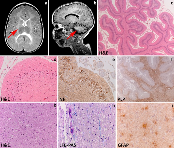
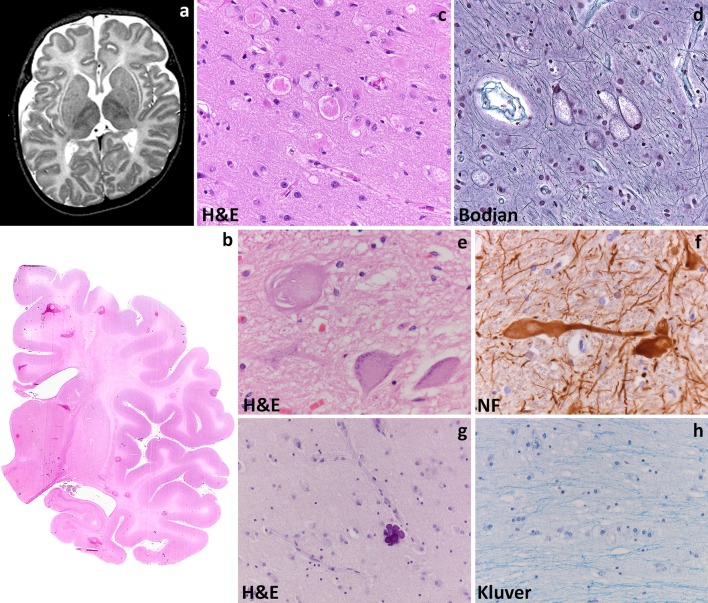
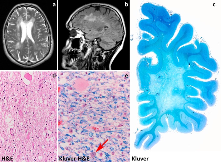
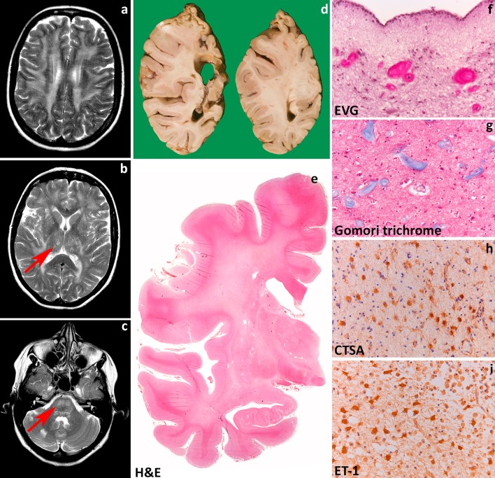
Leukodystrophies are genetically determined disorders characterized by the selective involvement of the central nervous system white matter. Onset may be at any age, from prenatal life to senescence. Many leukodystrophies are degenerative in nature, but some only impair white matter function. The clinical course is mostly progressive, but may also be static or even improving with time. Progressive leukodystrophies are often fatal, and no curative treatment is known. The last decade has witnessed a tremendous increase in the number of defined leukodystrophies also owing to a diagnostic approach combining magnetic resonance imaging pattern recognition and next generation sequencing. Knowledge on white matter physiology and pathology has also dramatically built up. This led to the recognition that only few leukodystrophies are due to mutations in myelin- or oligodendrocyte-specific genes, and many are rather caused by defects in other white matter structural components, including astrocytes, microglia, axons and blood vessels. We here propose a novel classification of leukodystrophies that takes into account the primary involvement of any white matter component. Categories in this classification are the myelin disorders due to a primary defect in oligodendrocytes or myelin (hypomyelinating and demyelinating leukodystrophies, leukodystrophies with myelin vacuolization); astrocytopathies; leuko-axonopathies; microgliopathies; and leuko-vasculopathies. Following this classification, we illustrate the neuropathology and disease mechanisms of some leukodystrophies taken as example for each category. Some leukodystrophies fall into more than one category. Given the complex molecular and cellular interplay underlying white matter pathology, recognition of the cellular pathology behind a disease becomes crucial in addressing possible treatment strategies.
Overview
The claims and statistics of this paper haven’t been checked yet.
Run the check — free, no account needed. It reads the open-access text and checks the sources it cites.
Integrity signals
Registry
Cites
Statistics
Figures
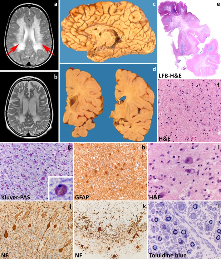
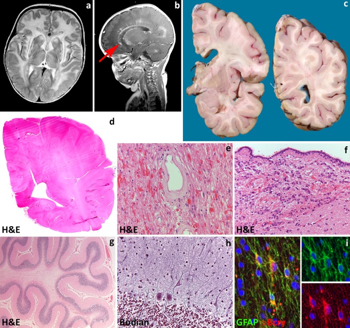
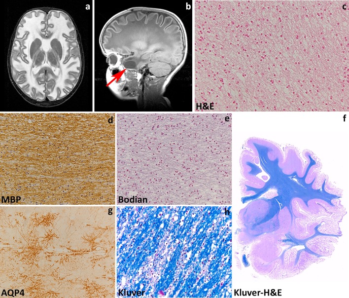
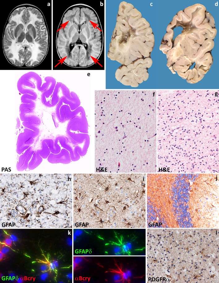
Verification
Claims the paper makes
The claims and statistics of this paper haven’t been checked yet.
Run the check — free, no account needed. It reads the open-access text and checks the sources it cites.
Claims other papers make about this one
Each is checked against this paper’s own text: does it say what the citing paper says it does?
The source backs part of the claim, not all of it — or backs it for a different population, condition or comparison.
In the citing paper
“The astrocytic contribution of the TSPO signal could also complicate differentiation of multiple sclerosis from leukodystrophies that have significant astrocytic but not microglial involvement (van der Knaap and Bugiani, 2017).”
From A quantitative neuropathological assessment of translocator protein expression in multiple sclerosis (2019) · cites this paper as van der Knaap and Bugiani, 2017
Experimental — accuracy not yet measured
Self-check
Percentages, sums and reported differences re-computed from the paper’s own numbers.
The self-check needs the paper’s full text.
Citations
Cites
The Metabolic and Molecular Bases of Inherited Disease
Scriver, R. C. (1995) · Medical Entomology and Zoology · Cited by 12,101
Polazzi, E., Mengoni, I., Caprini, M., Peña‐Altamira, E., Kurtys, E., & Monti, B. (2012) · Neurosignals · doi:10.1159/000337115 · Cited by 9,023
Astrocytes: Biology and pathology
Sofroniew, M. V., & Vinters, H. V. (2010) · Acta Neuropathologica · doi:10.1007/s00401-009-0619-8 · Cited by 5,196
Microglia: Active sensor and versatile effector cells in the normal and pathologic brain
Hanisch, U.-K., & Kettenmann, H. (2007) · Nature Neuroscience · doi:10.1038/nn1997 · Cited by 3,676
Pantoni, L. (2010) · The Lancet Neurology · doi:10.1016/s1474-4422(10)70104-6 · Cited by 3,405
Dynamic instability of microtubule growth
Mitchison, T., & Kirschner, M. (1984) · Nature · doi:10.1038/312237a0 · Cited by 3,337
M2 microglia and macrophages drive oligodendrocyte differentiation during CNS remyelination
Miron, V. E., Boyd, A., Zhao, J.-W., Yuen, T. J., Ruckh, J. M., Shadrach, J. L., van Wijngaarden, P., Wagers, A. J., Williams, A., Franklin, R. J. M., et al. (2013) · Nature Neuroscience · doi:10.1038/nn.3469 · Cited by 1,777
Mechanisms of sporadic cerebral small vessel disease: insights from neuroimaging
Wardlaw, J. M., Smith, C., & Dichgans, M. (2013) · The Lancet Neurology · doi:10.1016/s1474-4422(13)70060-7 · Cited by 1,657
Glycolytic oligodendrocytes maintain myelin and long-term axonal integrity
Fünfschilling, U., Supplie, L. M., Mahad, D., Boretius, S., Saab, A. S., Edgar, J., Brinkmann, B. G., Kassmann, C. M., Tzvetanova, I. D., Möbius, W., et al. (2012) · Nature · doi:10.1038/nature11007 · Cited by 1,523
Neuronal activity promotes oligodendrogenesis and adaptive myelination in the mammalian brain
Gibson, E. M., Purger, D., Mount, C. W., Goldstein, A. K., Lin, G. L., Wood, L. S., Inema, I., Miller, S. E., Bieri, G., Zuchero, J. B., et al. (2014) · Science · doi:10.1126/science.1252304 · Cited by 1,472
Research works printed but not linked: 6
- Agamanolis DP (2004) Pathology & genetics. Developmental neuropathology. ISN Neuropath, City
- Bielschowsky MH, Henneberg R (1928) Über familiäre diffuse Sklerose (Leukodystrophia cerebri progressiva hereditaria). J Psychol Neurol 36:131–181
- Pelizaeus F (1899) Überr eine eigenartige faimiliäre Entwicklungshemmung cornehmlich auf motorischem Gebiete. Archiv Psych 31:100
- Powers JM (2004) Peroxisomal disorders. In: Pathology & genetics. Developmental neuropathology. JA Golden, BN Harding (eds). ISN Neuropath Press, Basel. pp.287–295
- Suzuki K, Suzuki K (1996) The gangliosidoses. In: handbook of clinical neurology, RJ Vinken and GW Bruyn, Revised series Vol 22 (66) Neurodystrophies and neurolipidoses, HW Moser (ed) Elsevier Science B.V., Amsterdam, pp.247-280.
- Suzuki KV, Vanier M.T. (2004) Sphingolipidoses. In: Pathology & genetics. Developmental neuropathology. JA Golden, BN Arding (eds). ISN Neuropath, Basel. pp.235-236.
Cited by
Sorted by citations
Items 301–400 of 414
Ataxie, Leukenzephalopathie und Medulla oblongata-Atrophie bei einem 47-jährigen Mann
Schmidt, S. (2019) · DGNeurologie · doi:10.1007/s42451-019-0063-5
Neurodegenerative Erkrankungen der weißen Hirnsubstanz
Henneke, M., & Gärtner, J. (2020) · Springer Reference Medizin · doi:10.1007/978-3-662-60300-0_255
de Paiva, A. R. B. (2019) · Digital Library of Theses and Dissertations (Universidade de São Paulo) · doi:10.11606/t.5.2019.tde-16102019-142301
Neurodegeneration: General Aspects
Weis, S., Sonnberger, M., Dunzinger, A., Voglmayr, E., Aichholzer, M., Kleiser, R., & Strasser, P. (2019) · Springer eBooks · doi:10.1007/978-3-7091-1544-2_30
Hypomyelinating leukodystrophies: Navigating the diagnostic maze
Dixon, L. V. M. (2020) · European Journal of Paediatric Neurology · doi:10.1016/j.ejpn.2020.06.015
Type 6 hypomyelinating leukodystrophy caused by a de novo mutation in the tubulin beta-4A gene
Ozhegova, I. Y., Asanov, A. Y., Voskresenskaya, O. N., Razheva, D. S., Kondakova, O. B., & Nikolaeva, E. A. (2020) · Rossiyskiy Vestnik Perinatologii i Pediatrii (Russian Bulletin of Perinatology and Pediatrics) · doi:10.21508/1027-4065-2020-65-4-128-133
Progressive cavitational leukoencephalopathy: An enigma
Divahia, G., Natalia, M., Sara, L., & Espinosa, E. (2021) · Clinical Case Reports and Reviews · doi:10.15761/ccrr.1000497
Wolf, N. I. (2021) · Elsevier eBooks · doi:10.1016/b978-0-12-821635-4.00124-7
Trinkle, S., Wildenberg, G. A., Kasthuri, N., Rivière, P. J. L., & Foxley, S. (2022) · bioRxiv (Cold Spring Harbor Laboratory) · doi:10.1101/2022.02.24.481824
Rowitch, D. H. (2019) · Faculty Opinions – Post-Publication Peer Review of the Biomedical Literature · doi:10.3410/f.735528266.793559376
Metabolic, Toxic and Nutritional Diseases
Hilton, D. A., & Shivane, A. G. (2021) · Springer eBooks · doi:10.1007/978-3-030-66830-3_11
Neurodegenerative Erkrankungen der weißen Hirnsubstanz
Henneke, M., & Gärtner, J. (2019) · Springer Reference Medizin · doi:10.1007/978-3-642-54671-6_255-2
Progressive Cavitational Leukoencephalopathy: An Enigma
(2021) · Journal of Pediatrics & Child Care · doi:10.13188/2380-0534.1000029
Gaillard, F., Sharma, R., & Walizai, T. (2008) · Radiopaedia.org · doi:10.53347/rid-5266
[Research advances in the clinical genetics of leukodystrophy in children].
Huang, Z.-L., & Zhou, W. (2022) · PubMed Central · doi:10.7499/j.issn.1008-8830.2202020
Ventura, N. (2022) · Radiologia Brasileira · doi:10.1590/0100-3984.2022.55.3e2
MRI and In Vivo Spectroscopy of the Brain
Whitehead, M. T., & Gropman, A. L. (2022) · Springer eBooks · doi:10.1007/978-3-030-67727-5_8
Popovich, S. G., Kuzenkova, L. M., Kondakova, O. B., Firumyants, A. I., Владимировна, П. Т., & Uvakina, E. V. (2022) · L O Badalyan Neurological Journal · doi:10.46563/2686-8997-2022-3-3-122-126
Marashly, A. (2022) · Elsevier eBooks · doi:10.1016/b978-0-323-76174-1.00038-9
Leucoencefalopatie ereditarie e leucodistrofie dell’adulto
Ayrignac, X., Dalliere, C. C., & Labauge, P. M. (2022) · EMC - Neurologia · doi:10.1016/s1634-7072(22)47096-6
A Case of Unilateral Closed-lip Schizencephaly Combined with Hypomyelination In NeonatePreprint
Zhou, Y., Liu, L., Shen, X., Lin, G., Wen, X., Zhou, Y., & Wang, S. (2023) · Research Square · doi:10.21203/rs.3.rs-2711185/v1
Positron emission tomography imaging of neuroinflammation
(2023) · Advances in magnetic resonance technology and applications · doi:10.1016/b978-0-323-91771-1.00007-1
Neurometabolische Erkrankungen inklusive Neurotransmitterstörungen
Puchwein-Schwepcke, A. (2023) · Springer eBooks · doi:10.1007/978-3-662-65724-9_16
Verkhratsky, A., & Butt, A. M. (2023) · Elsevier eBooks · doi:10.1016/b978-0-12-821565-4.00018-3
Verkhratsky, A., & Butt, A. M. (2023) · Elsevier eBooks · doi:10.1016/b978-0-12-821565-4.00005-5
The Role of eIF2B Localisation in Cell-specific Stress Responses
Hanson, F. M. (2023) · Sheffield Hallam University · doi:10.7190/shu-thesis-00555
Understanding Van der Knaap’s Disease: A Case Report on Megalencephalic LeukoencephalopathyPreprint
Gami, V. M., Teraiya, T., Shah, S., & Desai, D. H. (2023) · Preprints.org · doi:10.20944/preprints202312.1273.v1
Démarche diagnostique devant une leucodystrophie métabolique de l’adulte
Mochel, F. (2024) · Pratique Neurologique - FMC · doi:10.1016/j.praneu.2024.01.003
Gowda, V., Suryanarayana, S. G., Srinivasan, V. M., Shivappa, S. K., Benakappa, N., Bhat, M. D., & Muthane, Y. T. C. (2021) · Journal of Pediatric Neurosciences · doi:10.4103/jpn.jpn_331_20
Ramos‐Fernández, J. M., Cano-Martínez, M., & Martínez-León, M. I. (2024) · Revista de Neurología · doi:10.33588/rn.7803.2023349
Therapie der X-chromosomalen Adrenoleukodystrophie
Köhler, W. (2024) · DGNeurologie · doi:10.1007/s42451-024-00645-4
Brouwers, E., Breur, M., Jorge, M. S., Mansvelder, H. D., van der Knaap, M. S., Bugiani, M., & Min, R. (2024) · bioRxiv (Cold Spring Harbor Laboratory) · doi:10.1101/2024.04.23.590685
Self-citationChildhood-onset leukodystrophy
Federico, A. (2024) · Elsevier eBooks · doi:10.1016/b978-0-323-99417-0.00001-x
Green, L., Hamilton, N., Elpidorou, M., Maroofian, R., Douglas, A. G. L., Õunap, K., Rose, A. M. S., Harris, E. L., Elworthy, S., Renshaw, S. A., et al. (2024) · Research Square · doi:10.21203/rs.3.rs-4356120/v1
Metachromatic leukodystrophy in infant presenting as acute febrile illness: a case report
Acharya, S., Regmi, B. U., Adhikari, Y., Thapa, P. B., Basnet, D., & Paudel, B. (2024) · Annals of Medicine and Surgery · doi:10.1097/ms9.0000000000002144
Mutações no Gene IBA57 como Causa de Leucodistrofia na Idade Pediátrica: Um Caso Clínico
Rodrigues, L. L., Janeiro, P. C. R. B., Santos, T., & Coelho, J. M. C. L. (2024) · Sinapse · doi:10.46531/sinapse/cc/230035/2024
CASE HISTORY OF METACHROMATIC LEUKODYSTROPHY OF 5 YEAR OLD CHILD
Boiarskа, L. M., & Redko, I. I. (2024) · Neonatology surgery and perinatal medicine · doi:10.24061/2413-4260.xiv.2.52.2024.25
Rare forms of hypomyelination and delayed myelination
Mura, E., Parazzini, C., & Tonduti, D. (2024) · Handbook of clinical neurology · doi:10.1016/b978-0-323-99209-1.00002-8
Engelen, M., & Wolf, N. I. (2024) · Elsevier eBooks · doi:10.1016/b978-0-443-19041-4.00074-1
Maladies neurologiques dues à une erreur innée du métabolisme
Cambier, J., Masson, M., Masson-Boivin, C., & Dehen, H. (2024) · Elsevier eBooks · doi:10.1016/b978-2-294-78318-0.00026-4
Review of investigations and possible therapies to address underlying disease manifestations
Smith, M. (2024) · Elsevier eBooks · doi:10.1016/b978-0-443-23648-8.00005-x
Al‐Saady, M. L. (2024) · doi:10.5463/thesis.775
Impact of Gut Microbiota on Neurogenesis and Neurological Diseases During Early Life
(2025) · Frontiers research topics · doi:10.3389/978-2-8325-5953-6
Deng, L., Feng, L., Li, J., Huang, Y., Ou, P., Shi, L., Chen, H., Zhang, Y., Dai, L., He, Y., et al. (2025) · Neurobiology of Disease · doi:10.1016/j.nbd.2025.106816
Giant Axonal Neuropathy in a Child with Stop-Gain Variant in GAN Gene
Srinivasan, V. M., Manisha, R., Nilay, M., Bhakhri, B. K., & Singh, D. K. (2025) · Indian Pediatrics Case Reports · doi:10.4103/ipcares.ipcares_241_24
Jauregui, R., Garcia, M. R., Mehuron, T., Galetta, S. L., & Segal, D. (2025) · Neurology International · doi:10.3390/neurolint17020028
Benzoni, C., Moscatelli, M. E. M., Bersano, A., Sole, A. D., & Salsano, E. (2025) · European Journal of Neurology · doi:10.1111/ene.70141
Genetic and Metabolic Conditions Presenting as Pediatric Leukodystrophies
Radhakrishnan, R., Kralik, S. F., Class, J. A., Sivam, S., Sivam, I., & Patel, R. (2025) · Seminars in Ultrasound CT and MRI · doi:10.1053/j.sult.2025.04.001
Inherited white matter disorders in children
Kim, N. N., Biswas, A., & Hemingway, C. A. (2025) · Paediatrics and Child Health · doi:10.1016/j.paed.2025.02.008
Genetic and Metabolic Disorders of the White Matter
Adang, L. A., Vanderver, A. L., & Wolf, N. I. (2025) · Elsevier eBooks · doi:10.1016/b978-0-443-10944-7.00119-0
Wang, J., Xu, Z., Chen, D., & Xiang, H. (2025) · Taiwanese Journal of Obstetrics and Gynecology · doi:10.1016/j.tjog.2025.01.007
Köhler, W. (2022) · Elsevier eBooks · doi:10.1016/b978-3-437-21884-2.00010-6
Ahmad, S. R., Zeyaullah, M., Zahrani, Y. I., Altijani, A. A. G., Dawria, A., & Salih, A. A. (2025) · Journal of Human Genetics · doi:10.1038/s10038-025-01386-2
Nichol, A. A., Ngo, A. B., Alharthi, M., Hird, K., Owen, M. J., Raefsky, S. M., & Yang, J. H. (2025) · The Neurohospitalist · doi:10.1177/19418744251377118
Kokaly, N., Guerreiro, H., Bredow, J., Dreha‐Kulaczewski, S. F., Ohlenbusch, A., Köhler, W., Reinhardt, T., Schön, G., Volk, A. E., Sigel, H., et al. (2025) · Journal of Clinical Medicine · doi:10.3390/jcm14196918
Bandi, T. (2025) · Springer eBooks · doi:10.1007/978-981-96-8614-8_7
Risk of Seizures and Epilepsy in the Leukodystrophies
Hackett, A., Yan, H., Liu, K., Corre, C. S., Gao, K., Grzyb, C., Goodman, J., Kelly, D. C., Ingemansson, C. R., Ghabash, G., et al. (2025) · Pediatric Neurology · doi:10.1016/j.pediatrneurol.2025.09.021
TFEB-depletion rescues TMEM106B-related myelin defects in zebrafishPreprint
Berdowski, W. M., van der Linde, H. C., Wolf, N. I., & van Ham, T. J. (2025) · bioRxiv (Cold Spring Harbor Laboratory) · doi:10.1101/2025.10.10.681547
Endogenous Repair in Vanishing White Matter
Plug, B. C., Man, J. H. K., Breur, M., Nutma, E., Stellingwerff, M. D., Abbink, T. E. M., Bugiani, M., & van der Knaap, M. S. (2025) · Annals of Neurology · doi:10.1002/ana.78083
Self-citationSavy, S., Montes, F. A., Grosso, C. L., Laróvere, L. E., Silvera-Ruiz, S. M., Carro, G. H., Guelbert, G., Becerra, A., Morales, D., & Nicola, J. P. (2025) · Frontiers in Genetics · doi:10.3389/fgene.2025.1688885
Imagerie des affections métaboliques congénitales cérébrales
Ntorkou, A. A., Boespflug‐Tanguy, O., Adamsbaum, C., Alison, M., & Elmaleh, M. (2025) · EMC - Radiologie et imagerie médicale - Musculosquelettique - Neurologique - Maxillofaciale · doi:10.1016/s1879-8551(25)44948-3
Additional file 4 of Regional vulnerability of brain white matter in vanishing white matter
Man, J. H. K., van Gelder, C. A. G. H., Breur, M., Molenaar, D., Abbink, T. E. M., Altelaar, M. A. F., Bugiani, M., & van der Knaap, M. S. (2024) · Figshare · doi:10.6084/m9.figshare.26599010
Self-citationPlug, B. C., Revers, I. M., Breur, M., González, G. M., Timmerman, J. A., Meijns, N., Hamberg, D., Wagendorp, J., Nutma, E., Wolf, N. I., et al. (2024) · Figshare · doi:10.6084/m9.figshare.26728201
Self-citationAdditional file 6 of Regional vulnerability of brain white matter in vanishing white matter
Man, J. H. K., van Gelder, C. A. G. H., Breur, M., Molenaar, D., Abbink, T. E. M., Altelaar, M. A. F., Bugiani, M., & van der Knaap, M. S. (2024) · Figshare · doi:10.6084/m9.figshare.26599016
Self-citationYska, H. A. F., Engelen, M., & Bugiani, M. (2024) · Figshare · doi:10.6084/m9.figshare.26699159
Self-citationPan, J., Forés-Martos, J., Nouh, C. D., Jensen, T. D., Vallejo, K., Cayrol, R., Ahmadian, S. S., Ashley, E. A., Greicius, M. D., & Cobos, I. (2024) · Figshare · doi:10.6084/m9.figshare.26944581
Plug, B. C., Revers, I. M., Breur, M., González, G. M., Timmerman, J. A., Meijns, N., Hamberg, D., Wagendorp, J., Nutma, E., Wolf, N. I., et al. (2024) · Figshare · doi:10.6084/m9.figshare.26728192
Self-citationAdditional file 5 of Regional vulnerability of brain white matter in vanishing white matter
Man, J. H. K., van Gelder, C. A. G. H., Breur, M., Molenaar, D., Abbink, T. E. M., Altelaar, M. A. F., Bugiani, M., & van der Knaap, M. S. (2024) · Figshare · doi:10.6084/m9.figshare.26599013
Self-citationYska, H. A. F., Engelen, M., & Bugiani, M. (2024) · Figshare · doi:10.6084/m9.figshare.26699156
Self-citationPlug, B. C., Revers, I. M., Breur, M., González, G. M., Timmerman, J. A., Meijns, N., Hamberg, D., Wagendorp, J., Nutma, E., Wolf, N. I., et al. (2024) · Figshare · doi:10.6084/m9.figshare.26728189
Self-citationAdditional file 8 of Regional vulnerability of brain white matter in vanishing white matter
Man, J. H. K., van Gelder, C. A. G. H., Breur, M., Molenaar, D., Abbink, T. E. M., Altelaar, M. A. F., Bugiani, M., & van der Knaap, M. S. (2024) · Figshare · doi:10.6084/m9.figshare.26599022
Self-citationPlug, B. C., Revers, I. M., Breur, M., González, G. M., Timmerman, J. A., Meijns, N., Hamberg, D., Wagendorp, J., Nutma, E., Wolf, N. I., et al. (2024) · Figshare · doi:10.6084/m9.figshare.26728198
Self-citationAdditional file 7 of Regional vulnerability of brain white matter in vanishing white matter
Man, J. H. K., van Gelder, C. A. G. H., Breur, M., Molenaar, D., Abbink, T. E. M., Altelaar, M. A. F., Bugiani, M., & van der Knaap, M. S. (2024) · Figshare · doi:10.6084/m9.figshare.26599019
Self-citationAdditional file 2 of Regional vulnerability of brain white matter in vanishing white matter
Man, J. H. K., van Gelder, C. A. G. H., Breur, M., Molenaar, D., Abbink, T. E. M., Altelaar, M. A. F., Bugiani, M., & van der Knaap, M. S. (2024) · Figshare · doi:10.6084/m9.figshare.26599004
Self-citationPlug, B. C., Revers, I. M., Breur, M., González, G. M., Timmerman, J. A., Meijns, N., Hamberg, D., Wagendorp, J., Nutma, E., Wolf, N. I., et al. (2024) · Figshare · doi:10.6084/m9.figshare.26728204
Self-citationPlug, B. C., Revers, I. M., Breur, M., González, G. M., Timmerman, J. A., Meijns, N., Hamberg, D., Wagendorp, J., Nutma, E., Wolf, N. I., et al. (2024) · Figshare · doi:10.6084/m9.figshare.26728186
Self-citationAdditional file 3 of Regional vulnerability of brain white matter in vanishing white matter
Man, J. H. K., van Gelder, C. A. G. H., Breur, M., Molenaar, D., Abbink, T. E. M., Altelaar, M. A. F., Bugiani, M., & van der Knaap, M. S. (2024) · Figshare · doi:10.6084/m9.figshare.26599007
Self-citationPlug, B. C., Revers, I. M., Breur, M., González, G. M., Timmerman, J. A., Meijns, N., Hamberg, D., Wagendorp, J., Nutma, E., Wolf, N. I., et al. (2024) · Figshare · doi:10.6084/m9.figshare.26728183
Self-citationPan, J., Forés-Martos, J., Nouh, C. D., Jensen, T. D., Vallejo, K., Cayrol, R., Ahmadian, S. S., Ashley, E. A., Greicius, M. D., & Cobos, I. (2024) · Figshare · doi:10.6084/m9.figshare.26944587
Additional file 1 of Regional vulnerability of brain white matter in vanishing white matter
Man, J. H. K., van Gelder, C. A. G. H., Breur, M., Molenaar, D., Abbink, T. E. M., Altelaar, M. A. F., Bugiani, M., & van der Knaap, M. S. (2024) · Figshare · doi:10.6084/m9.figshare.26599001
Self-citationPan, J., Forés-Martos, J., Nouh, C. D., Jensen, T. D., Vallejo, K., Cayrol, R., Ahmadian, S. S., Ashley, E. A., Greicius, M. D., & Cobos, I. (2024) · Figshare · doi:10.6084/m9.figshare.26944590
Yska, H. A. F., Engelen, M., & Bugiani, M. (2024) · Figshare · doi:10.6084/m9.figshare.25506745
Self-citationPan, J., Forés-Martos, J., Nouh, C. D., Jensen, T. D., Vallejo, K., Cayrol, R., Ahmadian, S. S., Ashley, E. A., Greicius, M. D., & Cobos, I. (2024) · Figshare · doi:10.6084/m9.figshare.26944593
Yska, H. A. F., Engelen, M., & Bugiani, M. (2024) · Figshare · doi:10.6084/m9.figshare.25506610
Self-citationPan, J., Forés-Martos, J., Nouh, C. D., Jensen, T. D., Vallejo, K., Cayrol, R., Ahmadian, S. S., Ashley, E. A., Greicius, M. D., & Cobos, I. (2024) · Figshare · doi:10.6084/m9.figshare.26944584
A Child Who Drops Very Quickly in His School Performance
Arroyo, H. A., Torres, A. R., Vidaurre, J. A., Rugilo, C., & Ho, M.-L. (2026) · Elsevier eBooks · doi:10.1016/b978-0-443-28722-0.00037-x
Campistol, J., & Lopez, J. M. (2026) · Elsevier eBooks · doi:10.1016/b978-0-443-28722-0.00742-5
The Infant Who Loses Ambulation
Arroyo, H. A., Torres, A. R., Vidaurre, J. A., Rugilo, C., & Ho, M.-L. (2026) · Elsevier eBooks · doi:10.1016/b978-0-443-28722-0.00039-3
(2005) · Radiopaedia.org · doi:10.53347/rid-228852
Siala, S., Alves, C., & Guimaraes, C. V. A. (2026) · Neuroimaging Clinics of North America · doi:10.1016/j.nic.2026.02.004
Cucu, L.-E., Săcărescu, A., Grosu, C., Constantinescu, V. A., Baciu, L.-C., Asaftei-Titianu, G.-S., Gațcan, C., Chirica, C., Frăsinariu, O. E., & Ignat, B. E. (2026) · International Journal of Molecular Sciences · doi:10.3390/ijms27062810
Metabolic White Matter Disease
Gluščević, S. V. (2026) · doi:10.59537/saupress.2527
Tunç, A., Seferoğlu, M., & Samadzadeh, S. (2026) · doi:10.59537/saupress.2514
Multi-contrast dual-resolution UTE for myelin fraction mapping and structural imaging
Yin, N., Rau, A., Reisert, M., İlbey, S., Mehr, K. T., Liu, S., Helmstädter, M., Schwabenland, M., Emir, U. E., Demerath, T., et al. (2026) · Magnetic Resonance Materials in Physics Biology and Medicine · doi:10.1007/s10334-026-01362-7
Mohammadi, P., Fateh, S. T., Badv, R. S., Pak, N., Ashrafi, M. R., Heidari, M., Mohammadi, M., Zamani, G., Rezaei, Z., Nikbakht, S., et al. (2026) · Epilepsy Research · doi:10.1016/j.eplepsyres.2026.107868
Green, L., Hamilton, N., Elpidorou, M., Maroofian, R., Zaki, M. S., Douglas, A. G. L., Õunap, K., Rose, A. M. S., Harris, E., Candeias, D., et al. (2026) · Research Square · doi:10.21203/rs.3.rs-4356120/v2
Advances in Research on AARS1/AARS2-Related Disorders: A Focus on Leukodystrophies
Yang, Z., Zhang, W., Yu, F., Duan, X., Zhou, M., & Bs, T. (2026) · Genes · doi:10.3390/genes17080872
Oligodendroglial TANGO2 Regulates Lipid Metabolism to Control Motor Coordination
Chen, J., Liu, Z., Chen, F., Cui, Y., Qin, L., Wang, M., Zhu, X., Li, N., Huang, C., Zhang, B., et al. (2026) · The FASEB Journal · doi:10.1096/fj.202601407rr
White matter disorders at the intersection of transcription, RNA processing and translation
Chapleau, A., Tobón, F. V., & Bernard, G. (2026) · Nature Reviews Neurology · doi:10.1038/s41582-026-01248-1
Metadata
- Authors
- van der Knaap, M. S., & Bugiani, M.
- Affiliations
- Amsterdam Neuroscience (NL)
- Corresponding author
- Bugiani, Marianna
- Venue
- Acta Neuropathologica
- Published
- 2017-06-21
- Volume
- 134
- Issue
- 3
- Pages
- 351-382
- ISSN
- 0001-6322 (ISSN-L)
- 1432-0533
- PMC
- PMC5563342
- OpenAlex
- W2653495475
- Cited by
- 403
- References
- 276
- Citations by year
- 2026: 32 · 2025: 49 · 2024: 79 · 2023: 52 · 2022: 36 · 2021: 51 · 2020: 39 · 2019: 45 · 2018: 13 · 2017: 5
- FWCI
- 3.44
- Type
- journal article
- Language
- en
- Open access
- hybrid
- doi.org
- Locations
- Acta Neuropathologica (publishedVersion, cc-by)
- PubMed Central (publishedVersion, cc-by)
- PubMed (publishedVersion)
- Pure Amsterdam UMC (publishedVersion, other-oa)
- Amsterdam UMC (VU Amsterdam) - Institutional Repository (publishedVersion)
- VU Research Portal (publishedVersion, other-oa)
- Data Archiving and Networked Services (DANS) (submittedVersion, other-oa)
- Data Archiving and Networked Services (DANS) (submittedVersion)
- Funders
- Vrije Universiteit Amsterdam
- Fonds NutsOhra
- Topics
- Life Sciences › Neuroscience › Cellular and Molecular Neuroscience › Hereditary Neurological Disorders
- Life Sciences › Biochemistry, Genetics and Molecular Biology › Clinical Biochemistry › Metabolism and Genetic Disorders
- Health Sciences › Medicine › Clinical Neurology › Cerebrovascular and genetic disorders
- Keywords
- leukodystrophy, white matter pathology, hypomyelinating leukodystrophy, astrocytopathy, oligodendrocytes, myelin disorders, neuropathology, disease mechanisms
- MeSH terms
- Axons, Demyelinating Diseases, Humans, Myelin Sheath, Neuroglia, White Matter
About these checks
Registry, retraction, Cites and statistics results are computed, not judged, with one exception: a printed reference that no index lists is read once by a model before it counts as a citation error. Claims are extracted by a model and kept only when their quote appears verbatim in the text; each cited source is then checked, with a model’s help, for whether it says what the claim relies on. Those verdicts can be wrong — read the claim’s quote and the cited source before relying on one.
Statistics and arithmetic are recomputed, not judged.
Confidence bands are shown only for fields calibrated on our annotated test set. None is calibrated yet, so no band is shown.
What counts as a claim ›How we measure confidence ›How we check this ›
Are you an author, or do you see a problem with this page? Request a review
