

The DOI matches its Crossref record; 51 of the 51 references it cites were found in a registry or an index.
Chronic traumatic encephalopathy is a common co-morbidity, but less frequent primary dementia in former soccer and rugby players
Open accessAbstract
Chronic traumatic encephalopathy (CTE) is reported at high prevalence in selected autopsy case series of former contact sports athletes. Nevertheless, the contribution of CTE pathology to clinical presentation and its interaction with co-morbid neurodegenerative pathologies remain unclear. To address these issues, we performed comprehensive neuropathology assessments on the brains of former athletes with dementia and considered these findings together with detailed clinical histories to derive an integrated clinicopathological diagnosis for each case. Consecutive, autopsy-acquired brains from former soccer and rugby players with dementia were assessed for neurodegenerative pathologies using established and preliminary consensus protocols. Thereafter, next of kin interviews were conducted to obtain detailed accounts of the patient's clinical presentation and course of disease to inform a final, integrated clinicopathological diagnosis. Neuropathologic change consistent with CTE (CTE-NC) was confirmed in five of seven former soccer and three of four former rugby players' brains, invariably in combination with mixed, often multiple neurodegenerative pathologies. However, in just three cases was the integrated dementia diagnosis consistent with CTE, the remainder having alternate diagnoses, with the most frequent integrated diagnosis Alzheimer's disease (AD) (four cases; one as mixed AD and vascular dementia). This consecutive autopsy series identifies neuropathologic change consistent with preliminary diagnostic criteria for CTE (CTE-NC) in a high proportion of former soccer and rugby players dying with dementia. However, in the majority, CTE-NC appears as a co-morbidity rather than the primary, dementia causing pathology. As such, we suggest that while CTE-NC might be common in former athletes with dementia, in many cases its clinical significance remains uncertain.
Overview
The claims and statistics of this paper haven’t been checked yet.
Run the check — free, no account needed. It reads the open-access text and checks the sources it cites.
Integrity signals
Registry
Cites
Statistics
Figures
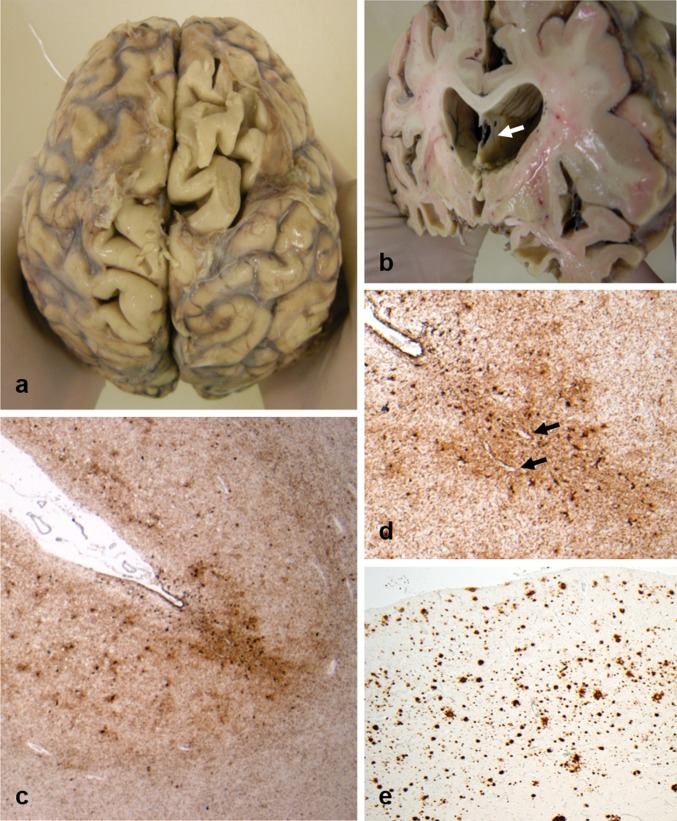
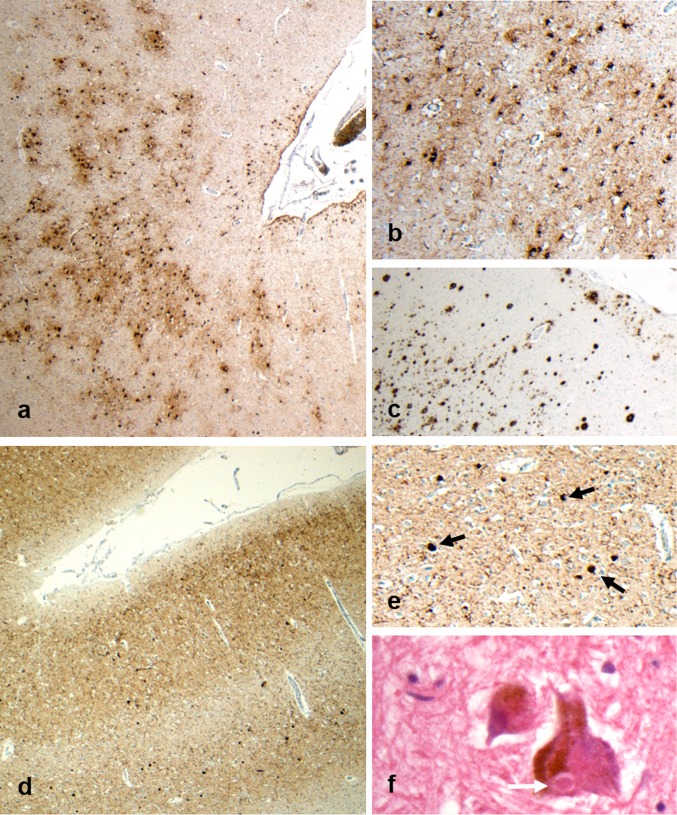
Verification
Claims the paper makes
The claims and statistics of this paper haven’t been checked yet.
Run the check — free, no account needed. It reads the open-access text and checks the sources it cites.
Claims other papers make about this one
Each is checked against this paper’s own text: does it say what the citing paper says it does?
In the citing paper
“Furthermore, the authors concluded that the clinical significance of CTE-pathology remains uncertain, and that it is likely a co-morbidity rather than the cause of dementia in former athletes [83].”
From DNA repair deficiency and senescence in concussed professional athletes involved in contact sports (2019) · cites this paper as [83]
In the citing paper
“Although CTE-consistent pathology was found in 73% of cases, all of these presented with multiple mixed neurodegenerative pathologies, most notably AD-consistent pathology.”
From DNA repair deficiency and senescence in concussed professional athletes involved in contact sports (2019) · cites this paper as [83]
Self-check
Percentages, sums and reported differences re-computed from the paper’s own numbers.
The self-check needs the paper’s full text.
Citations
Cites
Mirra, S. S., Heyman, A., McKeel, D., Sumi, S. M., Crain, B. J., Brownlee, L. M., Vogel, F. S., Hughes, J. P., Belle, G. V., Berg, L., et al. (1991) · Neurology · doi:10.1212/wnl.41.4.479 · Cited by 4,961
Diagnosis and management of dementia with Lewy bodies: Fourth consensus report of the DLB Consortium
McKeith, I. G., Boeve, B. F., Dickson, D. W., Halliday, G., Taylor, J.-P., Weintraub, D., Aarsland, D., Galvin, J., Attems, J., Ballard, C. G., et al. (2017) · Neurology · doi:10.1212/wnl.0000000000004058 · Cited by 4,476
Self-citationPhases of Aβ-deposition in the human brain and its relevance for the development of AD
Thal, D. R., Rüb, U., Orantes, M., & Braak, H. (2002) · Neurology · doi:10.1212/wnl.58.12.1791 · Cited by 3,490
Hyman, B. T., Phelps, C. H., Beach, T. G., Bigio, E. H., Cairns, N. J., Carrillo, M. C., Dickson, D. W., Duyckaerts, C., Frosch, M. P., Masliah, E., et al. (2012) · Alzheimer's & Dementia · doi:10.1016/j.jalz.2011.10.007 · Cited by 3,092
Self-citationMontine, T. J., Phelps, C. H., Beach, T. G., Bigio, E. H., Cairns, N. J., Dickson, D. W., Duyckaerts, C., Frosch, M. P., Masliah, E., Mirra, S. S., et al. (2012) · Acta Neuropathologica · doi:10.1007/s00401-011-0910-3 · Cited by 2,894
Self-citationStaging of alzheimer's disease-related neurofibrillary changes
Braak, H., & Braak, E. (1995) · Neurobiology of Aging · doi:10.1016/0197-4580(95)00021-6 · Cited by 2,357
Chronic traumatic encephalopathy in athletes: Progressive tauopathy after repetitive head injury
McKee, A. C., Cantu, R. C., Nowinski, C. J., Hedley-Whyte, E. T., Gavett, B. E., Budson, A. E., Santini, V. E., Lee, H.-S., Kubilus, C. A., & Stern, R. A. (2009) · Journal of Neuropathology & Experimental Neurology · doi:10.1097/nen.0b013e3181a9d503 · Cited by 2,295
The spectrum of disease in chronic traumatic encephalopathy
McKee, A. C., Stein, T. D., Nowinski, C. J., Stern, R. A., Daneshvar, D. H., Alvarez, V. E., Lee, H.-S., Hall, G., Wojtowicz, S. M., Baugh, C. M., et al. (2013) · Brain · doi:10.1093/brain/aws307 · Cited by 2,098
Jack, C. R., Albert, M. S., Knopman, D. S., McKhann, G. M., Sperling, R. A., Carrillo, M. C., Thies, B., & Phelps, C. H. (2011) · Alzheimer's & Dementia · doi:10.1016/j.jalz.2011.03.004 · Cited by 2,042
Clinicopathological evaluation of chronic traumatic encephalopathy in players of American football
Mez, J., Daneshvar, D. H., Kiernan, P. T., Abdolmohammadi, B., Alvarez, V. E., Huber, B. R., Alosco, M. L., Solomon, T. M., Nowinski, C. J., McHale, L., et al. (2017) · JAMA · doi:10.1001/jama.2017.8334 · Cited by 1,098
Research works printed but not linked: 1
- McKee AC, Stein TD, Nowinski CJ, Stern RA, Deneshvar DH, Alvarez VE et al (2012) The spectrum of disease in chronic traumatic encephalopathy. Brain 136:43–64 · DOI · Paper page
Cited by
Sorted by citations
Traumatic brain injury: progress and challenges in prevention, clinical care, and research
Maas, A. I. R., Menon, D. K., Manley, G. T., Abrams, M. B., Åkerlund, C. A. I., Anđelić, N., Aries, M. J. H., Bashford, T., Bell, M. J., Bodien, Y. G., et al. (2022) · The Lancet Neurology · doi:10.1016/s1474-4422(22)00309-x · Cited by 1,737
Neurodegenerative disease mortality among former professional soccer players
Mackay, D. F., Russell, E. R., Stewart, K., MacLean, J. A., Pell, J. P., & Stewart, W. (2019) · New England Journal of Medicine · doi:10.1056/nejmoa1908483 · Cited by 483
Traumatic Brain Injury and Risk of Neurodegenerative Disorder
Brett, B. L., Gardner, R. C., Godbout, J. P., Dams-O’Connor, K., & Keene, C. D. (2021) · Biological Psychiatry · doi:10.1016/j.biopsych.2021.05.025 · Cited by 454
Bieniek, K. F., Cairns, N. J., Crary, J. F., Dickson, D. W., Folkerth, R. D., Keene, C. D., Litvan, I., Perl, D. P., Stein, T. D., Vonsattel, J., et al. (2021) · Journal of Neuropathology & Experimental Neurology · doi:10.1093/jnen/nlab001 · Cited by 345
Tauopathies: new perspectives and challenges
Zhang, Y., Wu, K.-M., Yang, L., Dong, Q., & Yu, J.-T. (2022) · Molecular Neurodegeneration · doi:10.1186/s13024-022-00533-z · Cited by 323
Metadata
- Authors
- Lee, E. B., Kinch, K., Johnson, V. E., Trojanowski, J. Q., Smith, D. H., & Stewart, W.
- Affiliations
- University of Pennsylvania (US)
- Queen Elizabeth University Hospital (GB)
- ORCID
- Venue
- Acta Neuropathologica
- Published
- 2019-06-01
- Volume
- 138
- Issue
- 3
- Pages
- 389-399
- ISSN
- 0001-6322 (ISSN-L)
- 1432-0533
- PMC
- PMC6689293
- OpenAlex
- W2947253577
- Cited by
- 157
- References
- 51
- Citations by year
- 2026: 7 · 2025: 23 · 2024: 12 · 2023: 29 · 2022: 19 · 2021: 26 · 2020: 34 · 2019: 4
- FWCI
- 15.80
- Type
- journal article
- Language
- en
- Open access
- hybrid
- doi.org
- Locations
- Acta Neuropathologica (publishedVersion, cc-by)
- Europe PMC (PubMed Central) (publishedVersion, cc-by)
- PubMed Central (publishedVersion, cc-by)
- PubMed (publishedVersion)
- ENLIGHTEN (Jurnal Bimbingan dan Konseling Islam) (acceptedVersion, cc-by-sa)
- Funders
- National Institute of Neurological Disorders and Stroke (R01NS092398, R01NS094003, R01NS038104)
- The Football Association
- Topics
- Health Sciences › Medicine › Clinical Neurology › Traumatic Brain Injury Research
- Health Sciences › Medicine › Psychiatry and Mental health › Dementia and Cognitive Impairment Research
- Life Sciences › Neuroscience › Cellular and Molecular Neuroscience › Alzheimer's disease research and treatments
- Keywords
- chronic traumatic encephalopathy, dementia, comorbidities, Alzheimer's disease, clinicopathological diagnosis, contact sports, neuropathology
- MeSH terms
- Chronic Traumatic Encephalopathy, Aged, Athletic Injuries, Dementia, Football, Humans, Male, Middle Aged, Soccer, Comorbidity
About these checks
Registry, retraction, Cites and statistics results are computed, not judged, with one exception: a printed reference that no index lists is read once by a model before it counts as a citation error. Claims are extracted by a model and kept only when their quote appears verbatim in the text; each cited source is then checked, with a model’s help, for whether it says what the claim relies on. Those verdicts can be wrong — read the claim’s quote and the cited source before relying on one.
Statistics and arithmetic are recomputed, not judged.
Confidence bands are shown only for fields calibrated on our annotated test set. None is calibrated yet, so no band is shown.
What counts as a claim ›How we measure confidence ›How we check this ›
Are you an author, or do you see a problem with this page? Request a review
