

The DOI matches its Crossref record; 275 of the 275 references it cites were found in a registry or an index.
Glucose transporters in adipose tissue, liver, and skeletal muscle in metabolic health and disease
Open accessAbstract
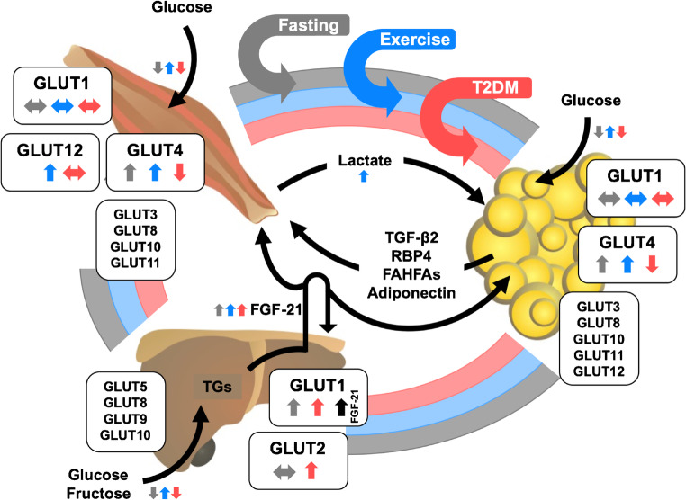
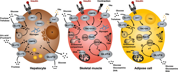
A family of facilitative glucose transporters (GLUTs) is involved in regulating tissue-specific glucose uptake and metabolism in the liver, skeletal muscle, and adipose tissue to ensure homeostatic control of blood glucose levels. Reduced glucose transport activity results in aberrant use of energy substrates and is associated with insulin resistance and type 2 diabetes. It is well established that GLUT2, the main regulator of hepatic hexose flux, and GLUT4, the workhorse in insulin- and contraction-stimulated glucose uptake in skeletal muscle, are critical contributors in the control of whole-body glycemia. However, the molecular mechanism how insulin controls glucose transport across membranes and its relation to impaired glycemic control in type 2 diabetes remains not sufficiently understood. An array of circulating metabolites and hormone-like molecules and potential supplementary glucose transporters play roles in fine-tuning glucose flux between the different organs in response to an altered energy demand.
Overview
The claims and statistics of this paper haven’t been checked yet.
Run the check — free, no account needed. It reads the open-access text and checks the sources it cites.
Integrity signals
Registry
Cites
Statistics
Figures
Verification
Claims the paper makes
The claims and statistics of this paper haven’t been checked yet.
Run the check — free, no account needed. It reads the open-access text and checks the sources it cites.
Claims other papers make about this one
Each is checked against this paper’s own text: does it say what the citing paper says it does?
The cited source says what the claim attributes to it.
In the citing paper
“Beyond GLUT4, skeletal muscle expresses several other glucose transporters, such as GLUT1, GLUT3, GLUT6, GLUT10, and GLUT12, albeit at lower abundance [16].”
From Effects of Skeletal Muscle Hypertrophy on Fat Mass and Glucose Homeostasis in Humans and Animals: A Narrative Review with Systematic Literature Search (2025) · cites this paper as [16]
Self-check
Percentages, sums and reported differences re-computed from the paper’s own numbers.
The self-check needs the paper’s full text.
Citations
Cites
1 retracted
RETRACTED: WNK1 phosphorylation sites in TBC1D1 and TBC1D4 modulate cell surface expression of GLUT1
Henriques, A. F. A., Matos, P., Carvalho, A. S., Azkargorta, M., Elortza, F., Matthiesen, R., & Jordan, P. (2019) · Archives of Biochemistry and Biophysics · doi:10.1016/j.abb.2019.108223 · Cited by 26
RetractedRab proteins as membrane organizers
Zerial, M., & McBride, H. (2001) · Nature Reviews Molecular Cell Biology · doi:10.1038/35052055 · Cited by 3,381
Mechanisms of Insulin Action and Insulin Resistance
Petersen, M. C., & Shulman, G. I. (2018) · Physiological Reviews · doi:10.1152/physrev.00063.2017 · Cited by 3,028
Energy Metabolism in the Liver
Rui, L. (2014) · Comprehensive physiology · doi:10.1002/cphy.c130024 · Cited by 2,401
Inflammatory mechanisms linking obesity and metabolic disease
Saltiel, A. R., & Olefsky, J. M. (2017) · Journal of Clinical Investigation · doi:10.1172/jci92035 · Cited by 2,212 · cited 1×
“, DAGs [202], ceramides [31], and ROS [166], as well as inflammation [201] have been proposed to inhibit insulin signaling towards GLUT4 through interference with phosphorylation events at the level of the insulin receptor (IR), insulin receptor substrate 1 (IRS1), and downstream effectors.”
BackgroundNew genetic loci implicated in fasting glucose homeostasis and their impact on type 2 diabetes risk
DIAGRAM Consortium, Dupuis, J., GIANT Consortium, Global BPgen Consortium, Anders Hamsten on behalf of Procardis Consortium, the MAGIC investigators, Langenberg, C., Prokopenko, I., Saxena, R., Soranzo, N., et al. (2010) · Nature Genetics · doi:10.1038/ng.520 · Cited by 2,166
Serum retinol binding protein 4 contributes to insulin resistance in obesity and type 2 diabetes
Yang, Q., Graham, T. E., Mody, N., Preitner, F., Peroni, O. D., Zabolotny, J. M., Kotani, K., Quadro, L., & Kahn, B. B. (2005) · Nature · doi:10.1038/nature03711 · Cited by 2,086
DeFronzo, R. A., Jacot, E., Jequier, E., Maeder, E., Wahren, J., & Felber, J. P. (1981) · Diabetes · doi:10.2337/diab.30.12.1000 · Cited by 1,770
Adipose tissue dysfunction as determinant of obesity-associated metabolic complications
Longo, M., Zatterale, F., Naderi, J., Parrillo, L., Formisano, P., Raciti, G. A., Beguinot, F., & Miele, C. (2019) · International Journal of Molecular Sciences · doi:10.3390/ijms20092358 · Cited by 1,575
Biology of Human Sodium Glucose Transporters
Wright, E. M., Loo, D. D. F., & Hirayama, B. A. (2011) · Physiological Reviews · doi:10.1152/physrev.00055.2009 · Cited by 1,430
Cited by
Sorted by citations
Papamichail, L., Koch, L. S., Veerman, D., Broersen, K., & van der Meer, A. D. (2025) · Frontiers in Bioengineering and Biotechnology · doi:10.3389/fbioe.2025.1515340 · Cited by 120
Imaging Approaches for the Study of Metabolism in Real Time Using Genetically Encoded Reporters
Chandris, P., Giannouli, C. C., & Panayotou, G. (2022) · Frontiers in Cell and Developmental Biology · doi:10.3389/fcell.2021.725114 · Cited by 19
Havers, T., Held, S., Schönfelder, M., Geisler, S., & Wackerhage, H. (2025) · Sports Medicine · doi:10.1007/s40279-025-02263-w · Cited by 19
“Beyond GLUT4, skeletal muscle expresses several other glucose transporters, such as GLUT1, GLUT3, GLUT6, GLUT10, and GLUT12, albeit at lower abundance [16].”
Santos, H. O., & Penha-Silva, N. (2025) · Nutrition · doi:10.1016/j.nut.2024.112617 · Cited by 11
Huang, Z., Li, L., Dudley, K., Xiao, L., Huang, G., Subramaniam, V. N., Chen, C., Crawford, R., & Xiao, Y. (2025) · BME Frontiers · doi:10.34133/bmef.0181 · Cited by 10
Metadata
- Authors
- Chadt, A., & Al-Hasani, H.
- Affiliations
- Deutsches Diabetes-Zentrum e.V. (DE)
- ORCID
- Chadt, Alexandra
- Venue
- Pflügers Archiv - European Journal of Physiology
- Published
- 2020-06-26
- Volume
- 472
- Issue
- 9
- Pages
- 1273-1298
- ISSN
- 0031-6768 (ISSN-L)
- 1432-2013
- PMC
- PMC7462924
- OpenAlex
- W3038041948
- Cited by
- 510
- References
- 275
- Citations by year
- 2026: 62 · 2025: 89 · 2024: 128 · 2023: 99 · 2022: 84 · 2021: 45 · 2020: 3
- FWCI
- 5.57
- Type
- journal article
- Language
- en
- Open access
- hybrid
- doi.org
- Locations
- Pflügers Archiv - European Journal of Physiology (publishedVersion, cc-by)
- PubMed Central (publishedVersion, cc-by)
- PubMed (publishedVersion)
- Informationszentrum Lebenswissenschaften (ZB MED) (submittedVersion, cc-by)
- Funders
- Deutsche Forschungegemeinschaft (CH1659)
- European Foundation for the Study of Diabetes (EFSD/NovoNordisk)
- Topics
- Life Sciences › Biochemistry, Genetics and Molecular Biology › Molecular Biology › Metabolism, Diabetes, and Cancer
- Health Sciences › Medicine › Physiology (medical) › Adipose Tissue and Metabolism
- Health Sciences › Medicine › Endocrinology, Diabetes and Metabolism › Diabetes, Cardiovascular Risks, and Lipoproteins
- Keywords
- glucose transporters, GLUT2, GLUT4, adipose tissue, liver, skeletal muscle, insulin resistance, type 2 diabetes, glucose uptake, hepatic glucose metabolism, skeletal muscle glucose uptake, blood glucose homeostasis, circulating metabolites, energy metabolism
- MeSH terms
- Adipose Tissue, Animals, Diabetes Mellitus, Humans, Liver, Muscle, Skeletal, Glucose Transport Proteins, Facilitative, Non-alcoholic Fatty Liver Disease
About these checks
Registry, retraction, Cites and statistics results are computed, not judged, with one exception: a printed reference that no index lists is read once by a model before it counts as a citation error. Claims are extracted by a model and kept only when their quote appears verbatim in the text; each cited source is then checked, with a model’s help, for whether it says what the claim relies on. Those verdicts can be wrong — read the claim’s quote and the cited source before relying on one.
Statistics and arithmetic are recomputed, not judged.
Confidence bands are shown only for fields calibrated on our annotated test set. None is calibrated yet, so no band is shown.
What counts as a claim ›How we measure confidence ›How we check this ›
Are you an author, or do you see a problem with this page? Request a review
