Cited by 22

The DOI matches its Crossref record; 3 of the 3 references it cites were found in a registry or an index.

Pharmacologically targeting molecular motor promotes mitochondrial fission for anti-cancer

Qian, Yi, Zhao, Meimei, Han, Qinghua, Wang, Jingkang, Liao, Lixi, Yang, Heng, Liu, Dan, Tu, Pengfei, Liang, Hong, Zeng, Kewu·2021·Acta Pharmaceutica Sinica B

Overview

The claims and statistics of this paper haven’t been checked yet.

Run the check — free, no account needed. It reads the open-access text and checks the sources it cites.

A few free checks a day · papers already verified open instantly · results are public

Integrity signals

Registry

Crossref · 10.1016/j.apsb.2021.01.011

Cites

3 of 3 references verified0 citation errors · 0 not checkable

Statistics

Figures

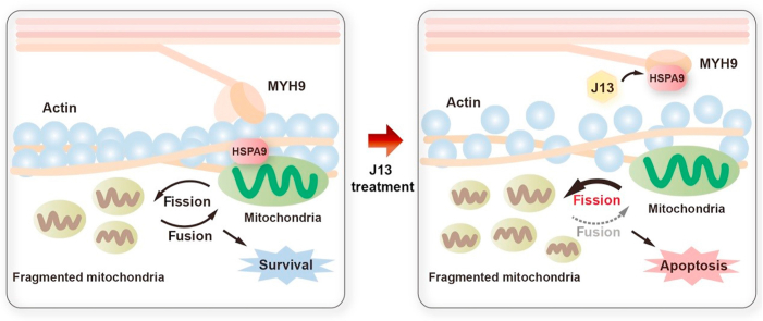
Graphical abstract Graphical abstract Natural-derived small-molecule J13 restrained the interaction between myosin-9 (MYH9) and actin molecular motor via prompting heat-shock protein A9 (HSPA9) attached to MYH9, resulting in excessive mitochondrial fission with implications for human cancer therapy.
Graphical abstract Graphical abstract Natural-derived small-molecule J13 restrained the interaction between myosin-9 (MYH9) and actin molecular motor via prompting heat-shock protein A9 (HSPA9) attached to MYH9, resulting in excessive mitochondrial fission with implications for human cancer therapy.CC BY-NC-ND · PubMed Central
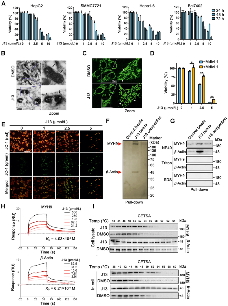
Figure 1 MYH9-actin molecular motor is a direct cellular target of anti-cancer small-molecule J13. (A) J13 inhibited cell viability of HepG2, SMMC7721, Hepa1-6 and Bel7402 cells (n = 6). (B) J13 induced mitochondrial fission in HepG2 cells. Mitochondrial morphology of HepG2 after J13 treatment was detected by transmission electron microscopy scanning (scale bar = 100 nm). (C) J13 caused mitochondrial fragmentation in HepG2 cells, which was visualized by Mito-tracker staining (scale bar = 10 μm). (D) Inhibition of mitochondrial fission by Mdivi-1 markedly reversed J13-dependent anti-cancer effects by MTT assay (n = 6). (E) J13 induced mitochondrial depolarization by JC-1 staining (scale bar = 25 μm). (F) MYH9 and β-actin were identified as J13-binding proteins using pull-down assay, followed by LC–MS/MS analysis with silver staining. (G) Non-covalent bindings of J13 with MYH9 or β-actin were confirmed by immunoblotting assay with different surfactants NP40, Triton, and SDS. (H) Dissociation constants (KD) of J13 with MYH9 or β-actin were detected by surface plasmon resonance (SPR) analysis. (I) and (J) Cellular thermal shift assays (CETSA) were performed to confirm the binding specificity of J13 with MYH9 or β-actin in cell lysates and living cells. Data are presented as mean ± SD from independent experiments; ∗P < 0.05, ∗∗P < 0.01.
Figure 1 MYH9-actin molecular motor is a direct cellular target of anti-cancer small-molecule J13. (A) J13 inhibited cell viability of HepG2, SMMC7721, Hepa1-6 and Bel7402 cells (n = 6). (B) J13 induced mitochondrial fission in HepG2 cells. Mitochondrial morphology of HepG2 after J13 treatment was detected by transmission electron microscopy scanning (scale bar = 100 nm). (C) J13 caused mitochondrial fragmentation in HepG2 cells, which was visualized by Mito-tracker staining (scale bar = 10 μm). (D) Inhibition of mitochondrial fission by Mdivi-1 markedly reversed J13-dependent anti-cancer effects by MTT assay (n = 6). (E) J13 induced mitochondrial depolarization by JC-1 staining (scale bar = 25 μm). (F) MYH9 and β-actin were identified as J13-binding proteins using pull-down assay, followed by LC–MS/MS analysis with silver staining. (G) Non-covalent bindings of J13 with MYH9 or β-actin were confirmed by immunoblotting assay with different surfactants NP40, Triton, and SDS. (H) Dissociation constants (KD) of J13 with MYH9 or β-actin were detected by surface plasmon resonance (SPR) analysis. (I) and (J) Cellular thermal shift assays (CETSA) were performed to confirm the binding specificity of J13 with MYH9 or β-actin in cell lysates and living cells. Data are presented as mean ± SD from independent experiments; ∗P < 0.05, ∗∗P < 0.01.CC BY-NC-ND · PubMed Central
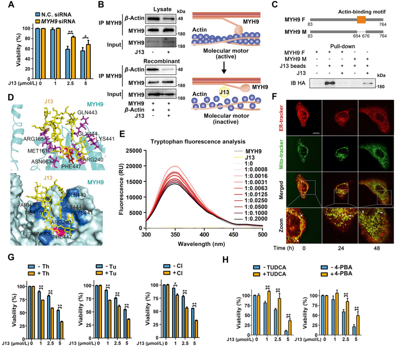
Figure 2 J13 suppresses direct MYH9–actin interaction to promote mitochondrial fission. (A) MYH9 knock-down reversed J13-dependent cell viability decrease in MTT assay. N.C.: knock-down negative control. (B) J13 suppressed the interaction of MYH9 with β-actin by co-IP assay. (C) Pull-down assay revealed that J13 interacted with actin-binding motif of MYH9. MYH9 F and M: full length MYH9 and mutated MYH9 with a deletion of 23 amino acids (654–676). (D) Docking analysis shows the amino acid residues which may form hydrogen bonds with J13 including ARG165, MET161, ARG240, LYS441, GLN443, GLY444, PHE447, and ASN663. (E) J13 induced allosteric effect of MYH9 by tryptophan fluorescence scanning analysis. (F) J13 suppressed the co-location of ER with mitochondria by double fluorescence staining analysis (ER-tracker, red; Mito-tracker, green; scale bar = 10 μm). (G) ER stress inducers including tunicamycin (Tu), thapsigargin (Th), and calcium ionophore III (CI) exacerbated J13-dependent anti-tumor effect by MTT assay. (H) ER stress inhibitor TUDCA and 4-PBA efficiently reversed J13-induced cell viability decrease in HepG2 cells. All data are presented as mean ± SD, n = 6; ∗P < 0.05, ∗∗P < 0.01.
Figure 2 J13 suppresses direct MYH9–actin interaction to promote mitochondrial fission. (A) MYH9 knock-down reversed J13-dependent cell viability decrease in MTT assay. N.C.: knock-down negative control. (B) J13 suppressed the interaction of MYH9 with β-actin by co-IP assay. (C) Pull-down assay revealed that J13 interacted with actin-binding motif of MYH9. MYH9 F and M: full length MYH9 and mutated MYH9 with a deletion of 23 amino acids (654–676). (D) Docking analysis shows the amino acid residues which may form hydrogen bonds with J13 including ARG165, MET161, ARG240, LYS441, GLN443, GLY444, PHE447, and ASN663. (E) J13 induced allosteric effect of MYH9 by tryptophan fluorescence scanning analysis. (F) J13 suppressed the co-location of ER with mitochondria by double fluorescence staining analysis (ER-tracker, red; Mito-tracker, green; scale bar = 10 μm). (G) ER stress inducers including tunicamycin (Tu), thapsigargin (Th), and calcium ionophore III (CI) exacerbated J13-dependent anti-tumor effect by MTT assay. (H) ER stress inhibitor TUDCA and 4-PBA efficiently reversed J13-induced cell viability decrease in HepG2 cells. All data are presented as mean ± SD, n = 6; ∗P < 0.05, ∗∗P < 0.01.CC BY-NC-ND · PubMed Central
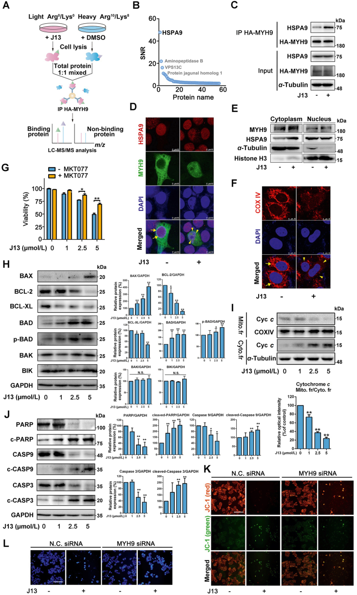
Figure 3 HSPA9 is a crucial MYH9–actin molecular motor substrate protein for controlling mitochondrial dynamics. (A) Schematic illustration of MYH9-binding proteins identification using pull-down coupled with SILAC analysis. (B) HSPA9 was identified as a binding protein of MYH9. (C) J13 promoted the interaction of HSPA9 with MYH9 by co-IP assay. (D) J13 promoted MYH9 interaction with HSPA9 in nucleus. (E) J13 treatment promoted cytosol translocation of HSPA9, and thus increased co-location of MYH9 with HSPA9. (F) J13 induced mitochondrial fragmentation by disrupting COXIV fluorescence distribution. (G) HSPA9 inhibitor MKT-077 (0.5 μmol/L for 24 h) reversed J13-induced cell viability decrease by MTT assay. (H) J13 significantly induced BAX and p-BAD expressions, and inhibited BCL-2 and BCL-XL expressions. (I) J13 induced cytochrome c translocation from mitochondria to cytoplasm. (J) J13 activated caspase-9/3-dependent apoptosis pathway. (K) MYH9 knock-down reversed J13-dependent mitochondrial depolarization (scale bar = 25 μm). (L) MYH9 knock-down rescued J13-dependent cell apoptosis by Hoechst 33258 staining assay (scale bar = 25 μm). All data are presented as mean ± SD from independent experiments (G: n = 6, H–J: n = 3; ∗P < 0.05, ∗∗P < 0.01.
Figure 3 HSPA9 is a crucial MYH9–actin molecular motor substrate protein for controlling mitochondrial dynamics. (A) Schematic illustration of MYH9-binding proteins identification using pull-down coupled with SILAC analysis. (B) HSPA9 was identified as a binding protein of MYH9. (C) J13 promoted the interaction of HSPA9 with MYH9 by co-IP assay. (D) J13 promoted MYH9 interaction with HSPA9 in nucleus. (E) J13 treatment promoted cytosol translocation of HSPA9, and thus increased co-location of MYH9 with HSPA9. (F) J13 induced mitochondrial fragmentation by disrupting COXIV fluorescence distribution. (G) HSPA9 inhibitor MKT-077 (0.5 μmol/L for 24 h) reversed J13-induced cell viability decrease by MTT assay. (H) J13 significantly induced BAX and p-BAD expressions, and inhibited BCL-2 and BCL-XL expressions. (I) J13 induced cytochrome c translocation from mitochondria to cytoplasm. (J) J13 activated caspase-9/3-dependent apoptosis pathway. (K) MYH9 knock-down reversed J13-dependent mitochondrial depolarization (scale bar = 25 μm). (L) MYH9 knock-down rescued J13-dependent cell apoptosis by Hoechst 33258 staining assay (scale bar = 25 μm). All data are presented as mean ± SD from independent experiments (G: n = 6, H–J: n = 3; ∗P < 0.05, ∗∗P < 0.01.CC BY-NC-ND · PubMed Central
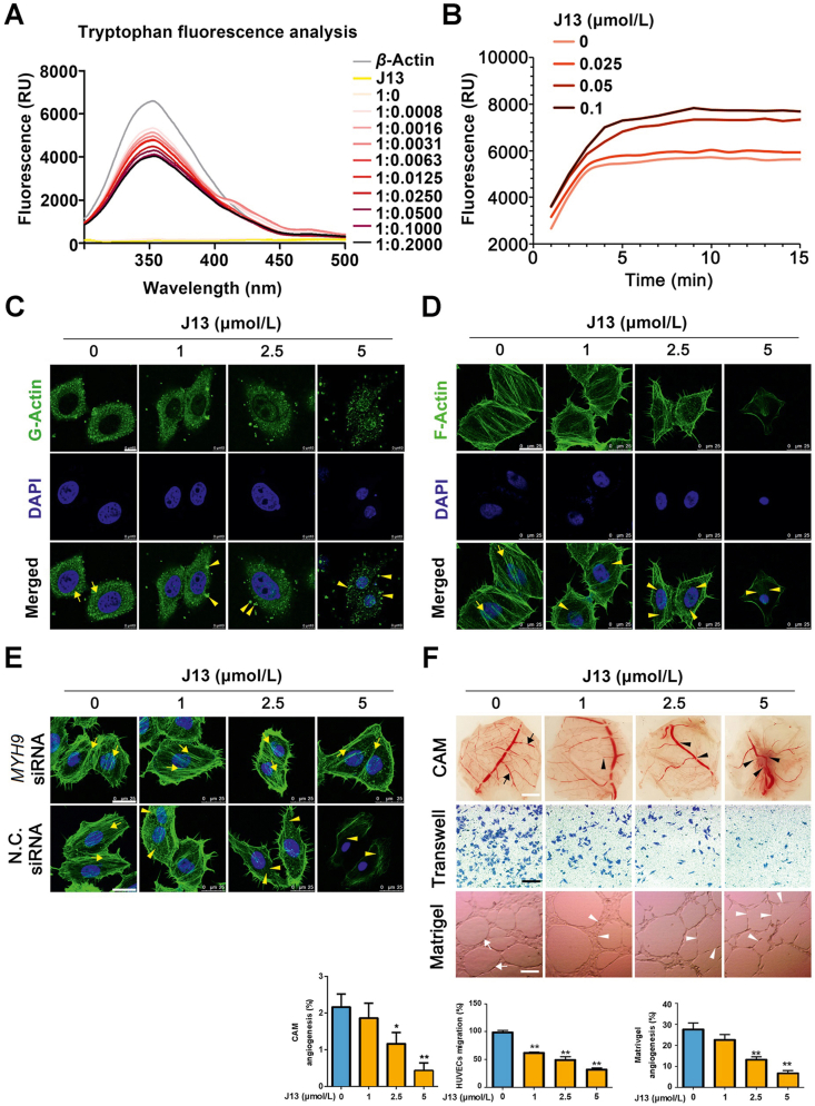
Figure 4 J13 inhibits actin-dependent cell migration and angiogenesis. (A) J13 induced allosteric effect of actin in tryptophan fluorescence scanning analysis. (B) J13 induced excessive G-actin assembly which was detected by G-actin polymerization assay. (C) J13 induced G-actin granulation aggregation in cells which was detected by immunofluorescence staining (scale bar = 10 μm). (D) J13 destroyed F-actin cytoskeleton formation which was detected by fluorescent phalloidin staining (scale bar = 25 μm). (E) MYH9 knock-down reversed J13-dependent F-actin cytoskeleton collapse (scale bar = 25 μm). (F) J13 suppressed CAM microvessels formation, cell migration and capillary-like tube formation. All data are presented as mean ± SD from independent experiments performed in triplicate. ∗P < 0.05, ∗∗P < 0.01.
Figure 4 J13 inhibits actin-dependent cell migration and angiogenesis. (A) J13 induced allosteric effect of actin in tryptophan fluorescence scanning analysis. (B) J13 induced excessive G-actin assembly which was detected by G-actin polymerization assay. (C) J13 induced G-actin granulation aggregation in cells which was detected by immunofluorescence staining (scale bar = 10 μm). (D) J13 destroyed F-actin cytoskeleton formation which was detected by fluorescent phalloidin staining (scale bar = 25 μm). (E) MYH9 knock-down reversed J13-dependent F-actin cytoskeleton collapse (scale bar = 25 μm). (F) J13 suppressed CAM microvessels formation, cell migration and capillary-like tube formation. All data are presented as mean ± SD from independent experiments performed in triplicate. ∗P < 0.05, ∗∗P < 0.01.CC BY-NC-ND · PubMed Central
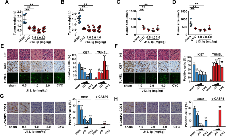
Figure 5 J13 showed anti-tumor effects in vivo. (A) J13 inhibited liver tumor weight growth in H22-bearing ICR mice in vivo by intraperitoneal injection (ip). (B) J13 inhibited liver tumor weight growth of H22-bearing ICR mice in vivo by intragastric administration (ig). (C) J13 inhibited liver tumor size in H22-bearing ICR mice in vivo by intraperitoneal injection (ip). (D) J13 inhibited liver tumor size of H22-bearing ICR mice in vivo by intragastric administration (ig). (E) J13 destroyed cell structure by HE staining (scale bar = 50 μm), decreased Ki67-positive cells number (scale bar = 50 μm), and promoted TUNEL-positive cell apoptosis (scale bar = 20 μm) by ip. (F) J13 destroyed cell structure by HE staining, decreased Ki67-positive cells number, and promoted TUNEL-positive cell apoptosis (scale bar = 20 μm) by ig. (G) J13 reduced CD31-positive cells number and promoted cleaved-caspase 3 (c-CASP3) expression (scale bar = 50 μm) by ip. (H) J13 reduced CD31-positive cells number and promoted c-CASP3 expression (scale bar = 50 μm) by ig. All data are presented as mean ± SD from independent experiments (A–D: n ≥ 8, E–H: n = 3); ∗P < 0.05, ∗∗P < 0.01.
Figure 5 J13 showed anti-tumor effects in vivo. (A) J13 inhibited liver tumor weight growth in H22-bearing ICR mice in vivo by intraperitoneal injection (ip). (B) J13 inhibited liver tumor weight growth of H22-bearing ICR mice in vivo by intragastric administration (ig). (C) J13 inhibited liver tumor size in H22-bearing ICR mice in vivo by intraperitoneal injection (ip). (D) J13 inhibited liver tumor size of H22-bearing ICR mice in vivo by intragastric administration (ig). (E) J13 destroyed cell structure by HE staining (scale bar = 50 μm), decreased Ki67-positive cells number (scale bar = 50 μm), and promoted TUNEL-positive cell apoptosis (scale bar = 20 μm) by ip. (F) J13 destroyed cell structure by HE staining, decreased Ki67-positive cells number, and promoted TUNEL-positive cell apoptosis (scale bar = 20 μm) by ig. (G) J13 reduced CD31-positive cells number and promoted cleaved-caspase 3 (c-CASP3) expression (scale bar = 50 μm) by ip. (H) J13 reduced CD31-positive cells number and promoted c-CASP3 expression (scale bar = 50 μm) by ig. All data are presented as mean ± SD from independent experiments (A–D: n ≥ 8, E–H: n = 3); ∗P < 0.05, ∗∗P < 0.01.CC BY-NC-ND · PubMed Central
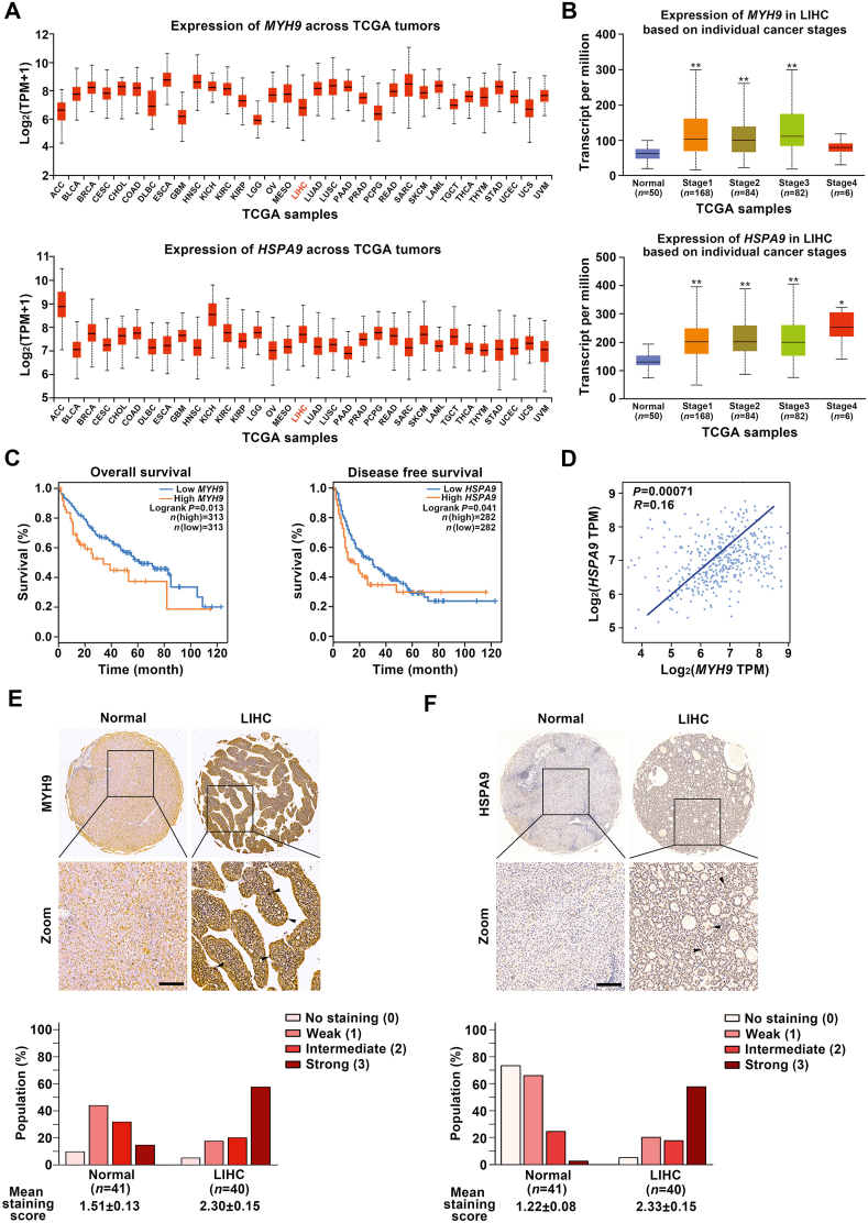
Figure 6 MYH9 and HSPA9 expressions are highly associated with poor survival of liver hepatocellular carcinoma (LIHC) patients. (A) MYH9 and HSPA9 had high expression levels in a variety of malignancies [Log2 (TPM+1) ≥2], including liver hepatocellular carcinoma LIHC from TCGA database. (B) The expression levels of MYH9/HSPA9 in LIHC were positively associated with advanced tumor stage determined by TCGA database. (C) MYH9 expression was positively correlated with LIHC patient overall survival (P = 0.013), while HSPA9 expression was positively related with disease free survival of LIHC patients (P = 0.041) based on GEPIA database. (D) A correlation between MYH9 and HSPA9 mRNA expression levels was shown by scatter plot (P = 0.00071) based on two-tailed Pearson's correlation analysis. (E) and (F) The expression levels of MYH9/HSPA9 were markedly increased in LIHC tissues (n = 40) compared to adjacent normal tissues (n = 41), as shown in representative immunohistochemical images (scale bar = 100 μm) and mean staining scores (P < 0.001, nonparametric ANOVA). ∗P < 0.05, ∗∗P < 0.01.
Figure 6 MYH9 and HSPA9 expressions are highly associated with poor survival of liver hepatocellular carcinoma (LIHC) patients. (A) MYH9 and HSPA9 had high expression levels in a variety of malignancies [Log2 (TPM+1) ≥2], including liver hepatocellular carcinoma LIHC from TCGA database. (B) The expression levels of MYH9/HSPA9 in LIHC were positively associated with advanced tumor stage determined by TCGA database. (C) MYH9 expression was positively correlated with LIHC patient overall survival (P = 0.013), while HSPA9 expression was positively related with disease free survival of LIHC patients (P = 0.041) based on GEPIA database. (D) A correlation between MYH9 and HSPA9 mRNA expression levels was shown by scatter plot (P = 0.00071) based on two-tailed Pearson's correlation analysis. (E) and (F) The expression levels of MYH9/HSPA9 were markedly increased in LIHC tissues (n = 40) compared to adjacent normal tissues (n = 41), as shown in representative immunohistochemical images (scale bar = 100 μm) and mean staining scores (P < 0.001, nonparametric ANOVA). ∗P < 0.05, ∗∗P < 0.01.CC BY-NC-ND · PubMed Central

Verification

Claims the paper makes

The claims and statistics of this paper haven’t been checked yet.

Run the check — free, no account needed. It reads the open-access text and checks the sources it cites.

A few free checks a day · papers already verified open instantly · results are public

Claims other papers make about this one

Each is checked against this paper’s own text: does it say what the citing paper says it does?

Self-check

Percentages, sums and reported differences re-computed from the paper’s own numbers.

The self-check needs the paper’s full text.

Citations

Cites

Show all 3 references

Research works printed but not linked: 55

  1. Chan, 2006, Mitochondria: dynamic organelles in disease, aging, and development, Cell, 125, 1241, 10.1016/j.cell.2006.06.010 · DOI · Paper page
  2. Schapira, 2012, Mitochondrial diseases, Lancet, 379, 1825, 10.1016/S0140-6736(11)61305-6 · DOI
  3. Wei, 2001, Proapoptotic BAX and BAK: a requisite gateway to mitochondrial dysfunction and death, Science, 292, 727, 10.1126/science.1059108 · DOI · Paper page
  4. Lin, 2006, Mitochondrial dysfunction and oxidative stress in neurodegenerative diseases, Nature, 443, 787, 10.1038/nature05292 · DOI · Paper page
  5. Liesa, 2009, Mitochondrial dynamics in mammalian health and disease, Physiol Rev, 89, 799, 10.1152/physrev.00030.2008 · DOI · Paper page
  6. Sabouny, 2020, Reciprocal regulation of mitochondrial fission and fusion, Trends Biochem Sci, 4, 564, 10.1016/j.tibs.2020.03.009 · DOI
  7. Liesa, 2013, Mitochondrial dynamics in the regulation of nutrient utilization and energy expenditure, Cell Metabol, 17, 491, 10.1016/j.cmet.2013.03.002 · DOI · Paper page
  8. Fabian, 2014, Mitochondrial fission and fusion in human diseases, N Engl J Med, 370, 1073, 10.1056/NEJMc1316254 · DOI
  9. Vyas, 2016, Mitochondria and cancer, Cell, 166, 555, 10.1016/j.cell.2016.07.002 · DOI · Paper page
  10. Kashatus, 2015, Erk2 phosphorylation of Drp1 promotes mitochondrial fission and MAPK-driven tumor growth, Mol Cell, 57, 537, 10.1016/j.molcel.2015.01.002 · DOI
  11. Serasinghe, 2015, Mitochondrial division is requisite to RAS-induced transformation and targeted by oncogenic MAPK pathway inhibitors, Mol Cell, 57, 521, 10.1016/j.molcel.2015.01.003 · DOI
  12. Chen, 2017, Mitochondrial dynamics in regulating the unique phenotypes of cancer and stem cells, Cell Metabol, 26, 39, 10.1016/j.cmet.2017.05.016 · DOI
  13. Srinivasan, 2017, Mitochondrial dysfunction and mitochondrial dynamics—the cancer connection, Biochim Biophys Acta Bioenerg, 1858, 602, 10.1016/j.bbabio.2017.01.004 · DOI
  14. Nagdas, 2019, Drp1 promotes KRas-driven metabolic changes to drive pancreatic tumor growth, Cell Rep, 28, 1845, 10.1016/j.celrep.2019.07.031 · DOI
  15. Seo, 2019, Mitochondrial fission factor is a novel Myc-dependent regulator of mitochondrial permeability in cancer, EbioMedicine, 48, 353, 10.1016/j.ebiom.2019.09.017 · DOI
  16. Senft, 2016, Regulators of mitochondrial dynamics in cancer, Curr Opin Cell Biol, 39, 43, 10.1016/j.ceb.2016.02.001 · DOI
  17. Cao, 2019, Matrine has pro-apoptotic effects on liver cancer by triggering mitochondrial fission and activating Mst1-JNK signaling pathways, J Physiol Sci, 69, 185, 10.1007/s12576-018-0634-4 · DOI
  18. Tiku, 2020, Mitochondrial functions in infection and immunity, Trends Cell Biol, 30, 263, 10.1016/j.tcb.2020.01.006 · DOI
  19. Molloy, 2003, Biophysics. Myosin motors walk the walk, Science, 300, 2045, 10.1126/science.1087148 · DOI
  20. Kruppa, 2018, Myosin VI-dependent actin cages encapsulate parkin-positive damaged mitochondria, Dev Cell, 44, 484, 10.1016/j.devcel.2018.01.007 · DOI
  21. Yang, 2019, Ultrastructure and dynamics of the actin–myosin II cytoskeleton during mitochondrial fission, Nat Cell Biol, 21, 603, 10.1038/s41556-019-0313-6 · DOI
  22. Korobova, 2014, A role for myosin II in mammalian mitochondrial fission, Curr Biol, 24, 409, 10.1016/j.cub.2013.12.032 · DOI
  23. Honrath, 2017, Glucose-regulated protein 75 determines ER–mitochondrial coupling and sensitivity to oxidative stress in neuronal cells, Cell Death Discovery, 3, 10.1038/cddiscovery.2017.76 · DOI
  24. Han, 2017, Cytotoxic oleanane triterpenoid saponins from Albizia julibrissin, Fitoterapia, 121, 183, 10.1016/j.fitote.2017.07.015 · DOI
  25. Liao, 2017, Highly selective inhibition of IMPDH2 provides the basis of antineuroinflammation therapy, Proc Natl Acad Sci U S A, 114, 5986, 10.1073/pnas.1706778114 · DOI
  26. Starenki, 2015, Mortalin (GRP75/HSPA9) upregulation promotes survival and proliferation of medullary thyroid carcinoma cells, Oncogene, 34, 4624, 10.1038/onc.2014.392 · DOI
  27. Tang, 2019, GEPIA2: an enhanced web server for large-scale expression profiling and interactive analysis, Nucleic Acids Res, 47, 556, 10.1093/nar/gkz430 · DOI
  28. Consortium, 2013, The genotype-tissue expression (GTEx) project, Nat Genet, 45, 580, 10.1038/ng.2653 · DOI · Paper page
  29. Anderson, 2018, Dysregulation of mitochondrial dynamics proteins are a targetable feature of human tumors, Nat Commun, 9, e1677, 10.1038/s41467-018-04033-x · DOI
  30. He, 2020, The ethnopharmacology, phytochemistry, pharmacology and toxicology of genus Albizia: a review, J Ethnopharmacol, 257, 10.1016/j.jep.2020.112677 · DOI
  31. Feng, 2013, Optimizing extraction process for Yiyuan Yiliu decoction by orthogonal design using multi-index, Chin J Hosp Pharm, 33, 601
  32. Tan, 2013, Study on formation process of “Yiyuan Yiliu Tang” granules, Chin Tradit Pat Med, 35, 2050
  33. Yi, 2014, Investigation of immunity capability of “Yiyuan Yiliu Tang” in H 22 tumor-burdened mice, Chin J Immunol, 30, 205
  34. Zhang, 2006, Julibroside J8-induced HeLa cell apoptosis through caspase pathway, J Asian Nat Prod Res, 8, 457, 10.1080/10286020500173309 · DOI
  35. Qian, 2018, Total saponins of Albiziae Cortex show anti-hepatoma carcinoma effects by inducing S phase arrest and mitochondrial apoptosis pathway activation, J Ethnopharmacol, 221, 20, 10.1016/j.jep.2018.04.015 · DOI
  36. Yu, 2004, Advances in study on bark of Albizzia julibrissin, China J Chin Mater Med, 29, 619
  37. Hua, 2009, Anti-angiogenic activity of julibroside J8, a natural product isolated from Albizia julibrissin, Phytomedicine, 16, 703, 10.1016/j.phymed.2009.01.002 · DOI
  38. Zou, 2005, Diastereoisomeric saponins from Albizia julibrissin, Carbohydr Res, 340, 1329, 10.1016/j.carres.2004.10.027 · DOI
  39. Muller, 2013, Distinct functional interactions between actin isoforms and nonsarcomeric myosins, PLoS One, 8, 10.1371/journal.pone.0070636 · DOI
  40. Makowska, 2015, Specific myosins control actin organization, cell morphology, and migration in prostate cancer cells, Cell Rep, 13, 2118, 10.1016/j.celrep.2015.11.012 · DOI
  41. Yang, 2015, The cap-translation inhibitor 4EGI-1 induces mitochondrial dysfunction via regulation of mitochondrial dynamic proteins in human glioma U251 cells, Neurochem Int, 90, 98, 10.1016/j.neuint.2015.07.019 · DOI
  42. Franco, 2016, Correcting mitochondrial fusion by manipulating mitofusin conformations, Nature, 540, 74, 10.1038/nature20156 · DOI
  43. Erpapazoglou, 2017, From dysfunctional endoplasmic reticulum–mitochondria coupling to neurodegeneration, Neurochem Int, 109, 171, 10.1016/j.neuint.2017.03.021 · DOI
  44. Wu, 2017, Steady-state levels of phosphorylated mitogen-activated protein kinase kinase 1/2 determined by mortalin/HSPA9 and protein phosphatase 1 alpha in KRAS and BRAF tumor cells, Mol Cell Biol, 37, 10.1128/MCB.00061-17 · DOI
  45. Park, 2014, Down-regulation of mortalin exacerbates Abeta-mediated mitochondrial fragmentation and dysfunction, J Biol Chem, 289, 2195, 10.1074/jbc.M113.492587 · DOI
  46. Vander Heiden, 1999, Bcl-2 proteins: regulators of apoptosis or of mitochondrial homeostasis?., Nat Cell Biol, 1, 209, 10.1038/70237 · DOI
  47. Mishra, 2014, Mitochondrial dynamics and inheritance during cell division, development and disease, Nat Rev Mol Cell Biol, 15, 634, 10.1038/nrm3877 · DOI
  48. Westermann, 2010, Mitochondrial fusion and fission in cell life and death, Nat Rev Mol Cell Biol, 11, 872, 10.1038/nrm3013 · DOI · Paper page
  49. Wu, 2013, A mortalin/HSPA9-mediated switch in tumor-suppressive signaling of Raf/MEK/extracellular signal-regulated kinase, Mol Cell Biol, 33, 4051, 10.1128/MCB.00021-13 · DOI
  50. Sane, 2014, Ubiquitin-like (UBX)-domain-containing protein, UBXN2A, promotes cell death by interfering with the p53-Mortalin interactions in colon cancer cells, Cell Death Dis, 5, e1118, 10.1038/cddis.2014.100 · DOI
  51. Altmann, 2008, The class V myosin motor protein, Myo2, plays a major role in mitochondrial motility in Saccharomyces cerevisiae, J Cell Biol, 181, 119, 10.1083/jcb.200709099 · DOI
  52. Hatch, 2014, Novel roles for actin in mitochondrial fission, J Cell Sci, 127, 4549
  53. Boldogh, 2007, Mitochondria on the move, Trends Cell Biol, 17, 502, 10.1016/j.tcb.2007.07.008 · DOI · Paper page
  54. Boldogh, 2006, Interactions of mitochondria with the actin cytoskeleton, Biochim Biophys Acta, 1763, 450, 10.1016/j.bbamcr.2006.02.014 · DOI
  55. Kemp, 2017, Surrogate endpoints in oncology: when are they acceptable for regulatory and clinical decisions, and are they currently overused?., BMC Med, 15, e134, 10.1186/s12916-017-0902-9 · DOI

Cited by

See all 22

Metadata

Authors
Qian, Y., Zhao, M., Han, Q., Wang, J., Liao, L., Yang, H., Liu, D., Tu, P., Liang, H., & Zeng, K.
Venue
Acta Pharmaceutica Sinica B
Publisher
Elsevier BV
Published
2021-07
Volume
11
Issue
7
Pages
1853-1866
ISSN
2211-3835
Cited by
22
References
3
Language
en
Update notices
erratum (2024-05-01)
Funders
  • National Natural Science Foundation of China (81973505, 81773932)
  • National Key Research and Development Program of China (2019YFC1711000, 2019YFC1708902)

About these checks

Registry, retraction, Cites and statistics results are computed, not judged, with one exception: a printed reference that no index lists is read once by a model before it counts as a citation error. Claims are extracted by a model and kept only when their quote appears verbatim in the text; each cited source is then checked, with a model’s help, for whether it says what the claim relies on. Those verdicts can be wrong — read the claim’s quote and the cited source before relying on one.

Statistics and arithmetic are recomputed, not judged.

Confidence bands are shown only for fields calibrated on our annotated test set. None is calibrated yet, so no band is shown.

What counts as a claim ›How we measure confidence ›How we check this ›

Are you an author, or do you see a problem with this page? Request a review

How to cite

Cite CoChat for the checks on this page, and the paper for its own findings.

This page
CoChat. (2026). Citation check: Pharmacologically targeting molecular motor promotes mitochondrial fission for anti-cancer. https://app.cochat.ai/papers/10.1016/j.apsb.2021.01.011
The paper
Qian, Y., Zhao, M., Han, Q., Wang, J., Liao, L., Yang, H., Liu, D., Tu, P., Liang, H., & Zeng, K. (2021). Pharmacologically targeting molecular motor promotes mitochondrial fission for anti-cancer. Acta Pharmaceutica Sinica B. https://doi.org/10.1016/j.apsb.2021.01.011