

The DOI matches its Crossref record; 111 of the 111 references it cites were found in a registry or an index; none of the 114 sources its claims cite is checked for support yet.
Acute neuroendocrine changes after traumatic brain injury
Open accessAbstract
Introduction: Post-traumatic hypopituitarism (PTHP) is a significant, but often neglected consequence of traumatic brain injury (TBI). Research question: We aimed to provide a comprehensive overview of epidemiology, pathophysiology, clinical features and diagnostic approaches of PTHP. Materials and methods: MEDLINE, EMBASE, Cochrane Library and Web of Science were searched. 45 articles of human studies evaluating acute endocrine changes following mild, moderate and severe TBI were selected. Results: Severity of TBI seems to be the most important risk factor of PTHP. Adrenal insufficiency (AI) was present in 10% of TBI patients (prevalence can be as high as 50% after severe TBI), and hypocortisolemia is a predictor of mortality and long-term hypopituitarism. Suppression of the thyroid axis in 2-33% of TBI patients may be an independent predictor of adverse neurological outcome, as well. 9-36% of patients with severe TBI exhibit decreased function of the somatotrophic axis with a divergent effect on the central nervous system. Arginine-Vasopressin (AVP) deficiency is present in 15-51% of patients, associated with increased mortality and unfavorable outcome. Due to shear and injury of the stalk hyperprolactinemia is relatively common (2-50%), but it bears little clinical significance. Sex hormone levels remain within normal values. Discussion and conclusion: PTHP occurs frequently after TBI, affecting various axis and determining patients' outcome. However, evidence is scarce regarding exact epidemiology, diagnosis, and effective clinical application of hormone substitution. Future studies are needed to identify patients at-risk, determine the optimal timing for endocrine testing, and refine diagnostic and treatment approaches to improve outcome.
Overview
CoChat Summary
TL;DR
This systematic review summarizes acute neuroendocrine changes after traumatic brain injury (TBI), focusing on post-traumatic hypopituitarism (PTHP) within 3 months. It finds frequent axis-specific dysfunction, with TBI severity as the most important risk factor, but evidence remains scarce for exact epidemiology, diagnosis, and effective hormone substitution.
Key findings
- Severity of TBI is the most important risk factor for PTHP. Acute adrenal insufficiency was present in 10% of TBI patients, with prevalence up to 50% after severe TBI; hypocortisolemia is a predictor of mortality and long-term hypopituitarism.
- Thyroid axis suppression occurred in 2–33% of TBI patients and may be an independent predictor of adverse neurological outcome; 9–36% of patients with severe TBI showed decreased somatotrophic axis function.
- Arginine-vasopressin deficiency was present in 15–51% of patients and was associated with increased mortality and unfavorable outcome; hyperprolactinemia was relatively common (2–50%) but bore little clinical significance.
- The abstract states sex hormone levels remain within normal values, but the body text reports acute gonadotrophin suppression and low testosterone in several studies.
Methods
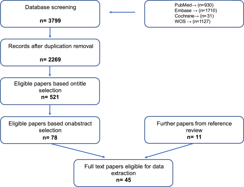
- Systematic review performed according to PRISMA-P. MEDLINE, EMBASE, Cochrane Library, and Web of Science were searched on 12 September 2023 using a TBI-and-endocrine/pituitary search term.
- Inclusion criteria: all TBI types; endocrine disorders assessed in the acute phase (within 3 months); peer-reviewed English full texts. Exclusions: no TBI diagnosis, case presentations, animal data, or publication before 2010. Overall, 45 full texts were included.
- Two independent authors screened titles, abstracts, and full texts; a third author resolved disagreements. References of included texts were reviewed. Data were extracted, critically reviewed, and categorized by endocrine parameters; no quantitative pooling/meta-analysis is described.
Limitations
- Paper states: evidence is scarce regarding exact epidemiology, diagnosis, and effective clinical application of hormone substitution; future studies are needed to identify patients at risk, determine optimal timing of endocrine testing, and refine diagnostic and treatment approaches.
- Paper states: heterogeneity in TBI severity, testing protocols, and diagnostic thresholds limits prevalence estimates; ambiguous nomenclature for adrenal insufficiency, single daily sampling, pulsatile GH secretion, ICU drugs, nutritional/hormonal status, and BMI can affect interpretation.
- Evident from the text: the review synthesizes heterogeneous observational studies with inconsistent findings across axes (e.g., prolactin, GH/IGF-1, thyroid, cortisol outcomes), and it does not report a formal risk-of-bias or study-quality assessment.
Model-written summary. Check it against the paper before relying on it.
Integrity signals
Registry
Cites
Statistics
Figures
Verification
Claims the paper makes
0 Valid · 2 Not valid · 84 Not checked · 32 Own results and interpretations
A result the paper states — it cites no source for it, so there is none to check. The quote shows it is in the paper.
In the paper
“Sex hormone levels remain within normal values.”
§ Acute neuroendocrine changes after traumatic brain injury > Abstract > Results
plain- Asserted: passes — The authors put it forward as true.
- Checkable: passes — It can be checked against evidence.
- Load-bearing: unclear — It is unclear whether the paper’s conclusions depend on it.
- Based: passes — The paper gives a reason why it holds.
- Rests on 2 assumptions not shown here.
None of the sources cited for this claim have been checked yet.
In the paper
“Moreover, a significant correlation was observed between fT4 levels at 30 days and Glasgow Coma Scale scores at discharge.”
§ Acute neuroendocrine changes after traumatic brain injury > Clinical presentation, diagnosis and treatment of TBI-induced neuroendocrine changes > TBI-induced changes of the pituitary-thyroid axis
plainstates an association- Asserted: passes — The authors put it forward as true.
- Checkable: passes — It can be checked against evidence.
- Load-bearing: unclear — It is unclear whether the paper’s conclusions depend on it.
- Based: passes — The paper gives a reason why it holds.
A check of a cited source is scheduled, or we don’t hold its full text yet; the claim is checked once it runs.
In the paper
“Two recent studies have indicated high frequencies (52–78%) of HPA axis dysfunction during the first 10 post-injury days (Hannon et al., 2013; Olivecrona et al., 2013).”
§ Acute neuroendocrine changes after traumatic brain injury > Clinical presentation, diagnosis and treatment of TBI-induced neuroendocrine changes > TBI-induced changes of the hypothalamic-pituitary-adrenal (HPA) axis
What the sources say
hedged- Asserted: passes — The authors put it forward as true.
- Checkable: passes — It can be checked against evidence.
- Load-bearing: unclear — It is unclear whether the paper’s conclusions depend on it.
- Based: passes — The paper gives a reason why it holds.
- Rests on 2 assumptions not shown here.
In the paper
“Similar to adults, a higher incidence of TSH abnormalities was noted in female patients, possibly linked to the natural hormonal fluctuations in female physiology (Aggarwal et al., 2020).”
§ Acute neuroendocrine changes after traumatic brain injury > Clinical presentation, diagnosis and treatment of TBI-induced neuroendocrine changes > TBI-induced changes of the pituitary-thyroid axis
What the sources say
- Not checked yetcites Aggarwal et al., 2020 source
plainstates an association- Asserted: passes — The authors put it forward as true.
- Checkable: passes — It can be checked against evidence.
- Load-bearing: unclear — It is unclear whether the paper’s conclusions depend on it.
- Based: passes — The paper gives a reason why it holds.
In the paper
“The predictive power of cortisol is further supported by the findings of Kumar et al., who demonstrated positive correlation between mean cortisol on day 4 and survival at 3 months (Vishwa Kumar et al., 2021).”
§ Acute neuroendocrine changes after traumatic brain injury > Clinical presentation, diagnosis and treatment of TBI-induced neuroendocrine changes > TBI-induced changes of the hypothalamic-pituitary-adrenal (HPA) axis
What the sources say
- Not checked yetcites Vishwa Kumar et al., 2021 source
boostedstates an association- Asserted: passes — The authors put it forward as true.
- Checkable: passes — It can be checked against evidence.
- Load-bearing: passes — The paper’s conclusions depend on it.
- Based: passes — The paper gives a reason why it holds.
49 more claims
Create a free account to see all 118 claims and to verify the 109 references still open.
- It may state two separate claims as one. 56
- Probably not a substantial claim. 16
- The quote only partly states this claim. 1
Self-check
Percentages, sums and reported differences re-computed from the paper’s own numbers.
Each contradiction between two passages listed here was confirmed by an independent check.
Rated major: it would touch a result — a value a result rests on, reported and recomputed p-values on opposite sides of the significance level, two passages that conflict with no sign which is right, or a cited work retracted before this paper was published.
In the paper
§ Acute neuroendocrine changes after traumatic brain injury > Abstract > Results
“Sex hormone levels remain within normal values.”
§ Acute neuroendocrine changes after traumatic brain injury > Clinical presentation, diagnosis and treatment of TBI-induced neuroendocrine changes > TBI-induced sex hormone deficiency
“Wagner et al. reported low testosterone levels in 100% of the 101 traumatic brain injury patients, which persisted over the observation period (during the first 9 days).”
Rated minor: it would not touch a result — a value no result rests on, a p-value that stays on the same side of the significance level, or a conflict that is clerical, resolved elsewhere in the paper, or outside its own findings.
In the paper
§ Acute neuroendocrine changes after traumatic brain injury > Clinical presentation, diagnosis and treatment of TBI-induced neuroendocrine changes > TBI-induced changes of the hypothalamic-pituitary-adrenal (HPA) axis
“| Alavi et al. (van der Eerden et al., 2010) | 58 | <14 | 0–7 days | 10,3 |”
§ Acute neuroendocrine changes after traumatic brain injury > References
“104. van der Eerden A.W. Should anterior pituitary function be tested during follow-up of all patients presenting at the emergency department because of traumatic brain injury? Eur. J. Endocrinol. 162 1 2010 19 28 19783620”
Rated minor: it would not touch a result — a value no result rests on, a p-value that stays on the same side of the significance level, or a conflict that is clerical, resolved elsewhere in the paper, or outside its own findings.
In the paper
§ Acute neuroendocrine changes after traumatic brain injury > Abstract > Results
“Due to shear and injury of the stalk hyperprolactinemia is relatively common (2–50%), but it bears little clinical significance.”
§ Acute neuroendocrine changes after traumatic brain injury > Clinical presentation, diagnosis and treatment of TBI-induced neuroendocrine changes > TBI-induced changes in prolactin production
“Hyperprolactinemia occurs in more than 50% of TBI survivors in the early, acute phase (Prasanna et al., 2015; Choudhary et al., 2023).”
Citations
Cites
A systematic review of brain injury epidemiology in Europe
Tagliaferri, F., Compagnone, C., Korsic, M., Servadei, F., & Kraus, J. (2006) · Acta Neurochirurgica · doi:10.1007/s00701-005-0651-y · Cited by 1,223
Hormonal Replacement in Hypopituitarism in Adults: An Endocrine Society Clinical Practice Guideline
Fleseriu, M., Hashim, I. A., Karavitaki, N., Melmed, S., Murad, M. H., Salvatori, R., & Samuels, M. H. (2016) · The Journal of Clinical Endocrinology & Metabolism · doi:10.1210/jc.2016-2118 · Cited by 945 · cited 2×
“Additionally, body mass index (BMI) has been noted to influence both GH and IGF-1 levels, with IGF-1 decreasing in cases of very high or low BMI (Fleseriu et al., 2016).”
BackgroundSchneider, H. J., Kreitschmann-Andermahr, I., Ghigo, E., Stalla, G. K., & Agha, A. (2007) · JAMA · doi:10.1001/jama.298.12.1429 · Cited by 549
Metabolic response to the stress of critical illness
Preiser, J.-C., Ichai, C., Orban, J.-C., & Groeneveld, A. B. J. (2014) · British Journal of Anaesthesia · doi:10.1093/bja/aeu187 · Cited by 505
Kelly, D. F., Gaw Gonzalo, I. T., Cohan, P., Berman, N., Swerdloff, R., & Wang, C. (2000) · Journal of Neurosurgery · doi:10.3171/jns.2000.93.5.0743 · Cited by 455
Anterior Pituitary Dysfunction in Survivors of Traumatic Brain Injury
Agha, A., Rogers, B., Sherlock, M., O’Kelly, P., Tormey, W., Phillips, J., & Thompson, C. J. (2004) · The Journal of Clinical Endocrinology & Metabolism · doi:10.1210/jc.2004-0511 · Cited by 390
Occurrence of pituitary dysfunction following traumatic brain injury
Bondanelli, M., De Marinis, L., Ambrosio, M. R., Monesi, M., Valle, D., Zatelli, M. C., Fusco, A., Bianchi, A., Farneti, M., & Degli, E. C. I. (2004) · Journal of Neurotrauma · doi:10.1089/0897715041269713 · Cited by 322
Growth hormone and insulin-like growth factor-1 (IGF-1) and their influence on cognitive aging
Sonntag, W. E., Ramsey, M., & Carter, C. S. (2005) · Ageing Research Reviews · doi:10.1016/j.arr.2005.02.001 · Cited by 258
Neuroprotection by Estrogen and Progesterone in Traumatic Brain Injury and Spinal Cord Injury
Brotfain, E., Gruenbaum, S. E., Boyko, M., Kutz, R., Zlotnik, A. A., & Klein, M. (2016) · Current Neuropharmacology · doi:10.2174/1570159x14666160309123554 · Cited by 229
Hannon, M. J., Crowley, R. K., Behan, L. A., O'Sullivan, E. P., O'Brien, M. M. C., Sherlock, M., Rawluk, D., O'Dwyer, R., Tormey, W., & Thompson, C. J. (2013) · The Journal of Clinical Endocrinology & Metabolism · doi:10.1210/jc.2013-1555 · Cited by 184
Research works printed but not linked: 5
- Ahmed, 2021, Pituitary dysfunction following a traumatic brain injury (TBI) at the desk of a General Practitioner, World Family Medicine, 19, 92
- Dlela, 2020, Acute glucocorticoid defciency following traumatic brain injury, Ann. Intensive Care, 10
- Dutta, 2021, Clinical presentations, hormonal evaluation and imaging abnormalities in patients with multiple pituitary hormone deficiency: a single-centre experience from rural West Bengal, J. Indian Med. Assoc., 119, 13
- Hacioglu, 2019, Neuroendocrine consequences of traumatic brain injury and strategies for its management, Erciyes Medical Journal, 41, 357
- Komoltsev, 2022, Acute glucocorticoid elevation after traumatic brain injury predicts reduces survival rate: a translational study, Eur. J. Neurol., 29, 199
Cited by
An Update on Advances in Hypopituitarism: Etiology, Diagnosis, and Current Management
Iglesias, P. (2024) · Journal of Clinical Medicine · doi:10.3390/jcm13206161 · Cited by 41
Fluid and electrolyte disorders following traumatic brain injury
Tomkins, M., Green, D., O’Reilly, M., & Sherlock, M. (2025) · Best Practice & Research Clinical Endocrinology & Metabolism · doi:10.1016/j.beem.2025.102014 · Cited by 15
Batson, R. D., Goldenberg, J. Z., Wexler, R. S., Wright, T. J., Chua, E., Yuen, A., Freeman, M., Wexler, T., High, W. M., Masel, B. E., et al. (2025) · Best Practice & Research Clinical Endocrinology & Metabolism · doi:10.1016/j.beem.2025.102015 · Cited by 10
Endocrine dysfunction post-traumatic brain injury: challenges and therapeutic approaches
Soltani, Z. E., Nazari, M., Babaei, M., Shakeri, H., Jalali, S., Hajali, V., & Keshavarzi, Z. (2025) · Acta Neurologica Belgica · doi:10.1007/s13760-025-02755-6 · Cited by 9
Magyar-Sümegi, Z. D., Csendes, M., Lendvai-Emmert, D., Sebestyen, G., Tamás, V., Bandi, S. A., Czigler, A., Yabluchanskiy, A., Tarantini, S., Ungvari, Z. I., et al. (2025) · GeroScience · doi:10.1007/s11357-025-01683-w · Cited by 9
Self-citation
Metadata
- Authors
- Magyar-Sumegi, Z. D., Stankovics, L., Lendvai-Emmert, D., Czigler, A., Hegedus, E., Csendes, M., Toth, L., Ungvari, Z., Buki, A., & Toth, P.
- Affiliations
- University of Pecs (HU)
- Semmelweis University (HU)
- Venue
- Brain and Spine
- Publisher
- Elsevier BV
- Published
- 2024
- Volume
- 4
- Pages
- 102830
- ISSN
- 2772-5294 (ISSN-L)
- PMC
- PMC11101905
- OpenAlex
- W4396703655
- Semantic Scholar
- 7a949fb474b5744849648aa5a269e4d1813016d0
- Cited by
- 23
- References
- 111
- Citations by year
- 2026: 9 · 2025: 12 · 2024: 2
- FWCI
- 1.47
- Type
- journal article
- Language
- en
- Open access
- gold
- doi.org
- Licence
- cc-by-nc-nd
- www.elsevier.com/tdm/userlicense/1.0/ (tdm)
- www.elsevier.com/legal/tdmrep-license (tdm)
- creativecommons.org/licenses/by-nc-nd/4.0/ (vor, 126 d)
- Locations
- Brain and Spine (publishedVersion, cc-by-nc-nd)
- PubMed Central (publishedVersion, cc-by-nc-nd)
- PubMed (publishedVersion)
- DOAJ (DOAJ: Directory of Open Access Journals) (submittedVersion, cc-by-sa)
- Funders
- Nemzeti Kutatási Fejlesztési és Innovációs Hivatal (OTKA K-134555)
- Hungarian Scientific Research Fund (FK-146334)
- Nemzeti Kutatási, Fejlesztési és Innovaciós Alap (135784, RRF-2.3.1-21-2022-00003)
- National Research Development and Innovation Office (TKP2021-NKTA-47, RRF-2.3.1-21-2022-00011, 101004093/ EUniWell/EAC-A02-2019 / EAC-A02-2019-1)
- Innovációs és Technológiai Minisztérium
- Topics
- Health Sciences › Medicine › Clinical Neurology › Traumatic Brain Injury and Neurovascular Disturbances
- Health Sciences › Medicine › Endocrinology, Diabetes and Metabolism › Pituitary Gland Disorders and Treatments
- Health Sciences › Medicine › Clinical Neurology › Traumatic Brain Injury Research
- Keywords
- post-traumatic hypopituitarism, traumatic brain injury, adrenal insufficiency, somatotropic axis, central diabetes insipidus, hyperprolactinemia, sex hormones, TBI severity, mortality
About these checks
Registry, retraction, Cites and statistics results are computed, not judged, with one exception: a printed reference that no index lists is read once by a model before it counts as a citation error. Claims are extracted by a model and kept only when their quote appears verbatim in the text; each cited source is then checked, with a model’s help, for whether it says what the claim relies on. Those verdicts can be wrong — read the claim’s quote and the cited source before relying on one.
A language model drew the claims from the paper on 2026-11-14, under version 3 of the claim definition (model deepseek-v4.1-flash, prompt extract-v3).
A second model labelled each claim on 2026-11-14 (model jev-1.13-20260917, definition revision gold-guideline-v0.2).
Statistics and arithmetic are recomputed, not judged.
Confidence bands are shown only for fields calibrated on our annotated test set. None is calibrated yet, so no band is shown.
What counts as a claim ›How we measure confidence ›How we check this ›
Are you an author, or do you see a problem with this page? Request a review
How to cite
Cite CoChat for the checks on this page, and the paper for its own findings.
- This page
- CoChat. (2026). Citation check: Acute neuroendocrine changes after traumatic brain injury. https://app.cochat.ai/papers/10.1016/j.bas.2024.102830
- The paper
- Magyar-Sumegi, Z. D., Stankovics, L., Lendvai-Emmert, D., Czigler, A., Hegedus, E., Csendes, M., Toth, L., Ungvari, Z., Buki, A., & Toth, P. (2024). Acute neuroendocrine changes after traumatic brain injury. Brain and Spine. https://doi.org/10.1016/j.bas.2024.102830
Data sources
Bibliographic data: Semantic Scholar, OpenAlex, Crossref.
Contains information from the Semantic Scholar Academic Graph Datasets, made available under the ODC Attribution License (ODC-BY 1.0).
Semantic Scholar’s data is described in: Kinney, R., et al. (2023). The Semantic Scholar Open Data Platform. arXiv:2301.10140.
