

The DOI matches its Crossref record; 27 of the 27 references it cites were found in a registry or an index.
Encapsulation state of messenger RNA inside lipid nanoparticles
Open accessAbstract
Understanding the structure of messenger RNA (mRNA) lipid nanoparticles, and specifically the microenvironment of the mRNA molecules within these entities, is fundamental to advancing their biomedical potential. Here, we show that a permeating cationic dye, thionine, can serve as a cryogenic electron microscopy contrasting agent by binding selectively to encapsulated mRNA without disturbing lipid nanoparticle morphology. Cryo-electron microscopy images identify the mRNA location, revealing that mRNA may exist within solvent-filled cavities or may be substantially lipid associated.
Overview
The claims and statistics of this paper haven’t been checked yet.
Run the check — free, no account needed. It reads the open-access text and checks the sources it cites.
Integrity signals
Registry
Cites
Statistics
Figures
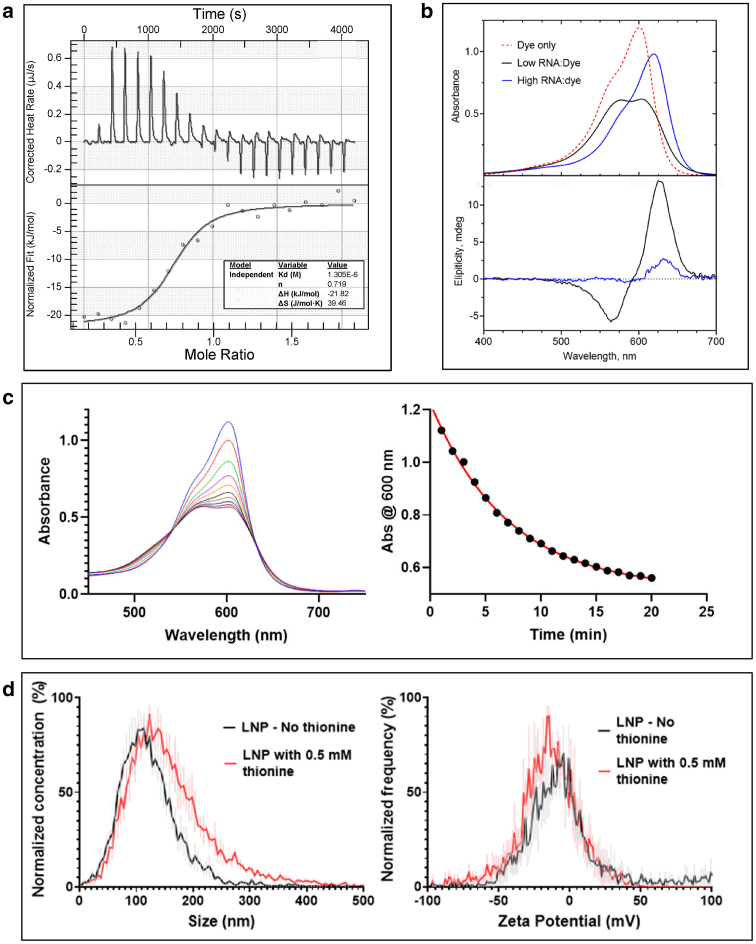
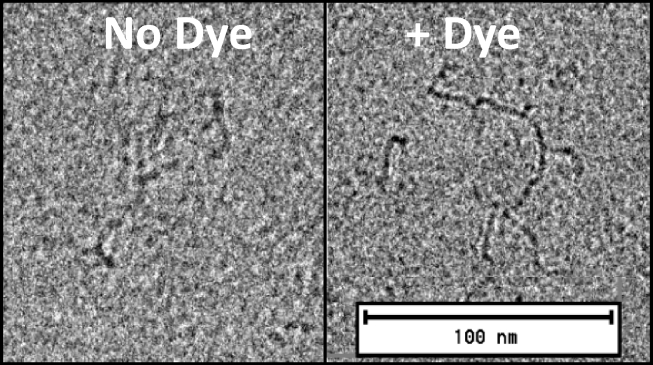
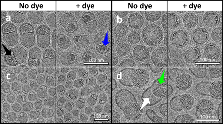
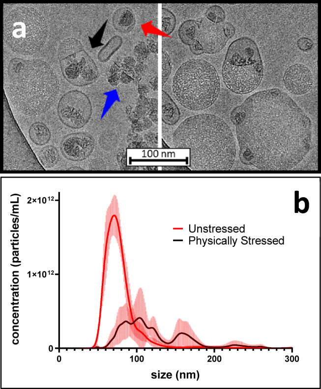
Verification
Claims the paper makes
The claims and statistics of this paper haven’t been checked yet.
Run the check — free, no account needed. It reads the open-access text and checks the sources it cites.
Claims other papers make about this one
Each is checked against this paper’s own text: does it say what the citing paper says it does?
In the citing paper
“A key aspect of LNPs and the characteristic that makes them different from liposomes (spherical vesicles with at least one lipid bilayer and an aqueous core) is the presence of lipids in the core, although data from several studies indicate that water is also present to some extent (Yanez Arteta et al., 2018, Kulkarni et al., 2018, Brader et al., 2021).”
From mRNA-lipid nanoparticle COVID-19 vaccines: structure and stability (2021) · cites this paper as Brader et al., 2021
In the citing paper
“If the LNP shells are permeable to protons –which is likely, as ionized dyes such as 2-(p-toluidino)-6-napthalene sulfonic acid (TNS) and thionine can enter the LNP core (Jayaraman et al., 2012, Brader et al., 2021) – the pH would be similar to the rest of the formulation, around 7 to 8, meaning that most of the ionizable cationic lipids would be uncharged.”
From mRNA-lipid nanoparticle COVID-19 vaccines: structure and stability (2021) · cites this paper as Brader et al., 2021
Self-check
Percentages, sums and reported differences re-computed from the paper’s own numbers.
The self-check needs the paper’s full text.
Citations
Cites
An mRNA Vaccine against SARS-CoV-2 — Preliminary Report
Jackson, L. A., Anderson, E. J., Rouphael, N. G., Roberts, P. C., Makhene, M., Coler, R. N., McCullough, M. P., Chappell, J. D., Denison, M. R., Stevens, L. J., et al. (2020) · New England Journal of Medicine · doi:10.1056/nejmoa2022483 · Cited by 3,237
mRNA-based therapeutics--developing a new class of drugs
Sahin, U., Karikó, K., & Türeci, Ö. (2014) · Nature Reviews Drug Discovery · doi:10.1038/nrd4278 · Cited by 2,474
Rational design of cationic lipids for siRNA delivery
Semple, S. C., Akinc, A., Chen, J., Sandhu, A. P., Mui, B. L., Cho, C. K., Sah, D. W. Y., Stebbing, D., Crosley, E. J., Yaworski, E., et al. (2010) · Nature Biotechnology · doi:10.1038/nbt.1602 · Cited by 1,822
Delivering the Messenger: Advances in Technologies for Therapeutic mRNA Delivery
Kowalski, P. S., Rudra, A., Miao, L., & Anderson, D. G. (2019) · Molecular Therapy · doi:10.1016/j.ymthe.2019.02.012 · Cited by 1,127
Modified mRNA vaccines protect against zika virus infection
Richner, J. M., Himansu, S., Dowd, K. A., Butler, S. L., Salazar, V., Fox, J. M., Julander, J. G., Tang, W. W., Shresta, S., Pierson, T. C., et al. (2017) · Cell · doi:10.1016/j.cell.2017.02.017 · Cited by 909 · cited 1×
“Intriguingly, mRNA-LNPs are known to form both spherical and highly nonspherical morphologies (9,11,16,18,19), but the origins and practical implications of these morphological variants”
BackgroundMiao, L., Li, L., Huang, Y., Delcassian, D., Chahal, J., Han, J., Shi, Y., Sadtler, K., Gao, W., Lin, J., et al. (2019) · Nature Biotechnology · doi:10.1038/s41587-019-0247-3 · Cited by 723 · cited 1×
“Intriguingly, mRNA-LNPs are known to form both spherical and highly nonspherical morphologies (9,11,16,18,19), but the origins and practical implications of these morphological variants”
BackgroundLipid Nanoparticle Assisted mRNA Delivery for Potent Cancer Immunotherapy
Oberli, M. A., Reichmuth, A. M., Dorkin, J. R., Mitchell, M. J., Fenton, O. S., Jaklenec, A., Anderson, D. G., Langer, R., & Blankschtein, D. (2017) · Nano Letters · doi:10.1021/acs.nanolett.6b03329 · Cited by 719
Kulkarni, J. A., Darjuan, M. M., Mercer, J. E., Chen, S., van der Meel, R., Thewalt, J. L., Tam, Y. Y. C., & Cullis, P. R. (2018) · ACS Nano · doi:10.1021/acsnano.8b01516 · Cited by 663 · cited 1×
“In the case of LNPs containing short interfering RNA (siRNA), small angle xray scattering and cryo-electron microscopy (cryo-EM) data have been interpreted as evidence of a multilamellar structure in which the siRNA molecule is sandwiched between bilayer lipid assemblies (10,17).”
BackgroundmRNA vaccine delivery using lipid nanoparticles
Reichmuth, A. M., Oberli, M. A., Jaklenec, A., Langer, R., & Blankschtein, D. (2016) · Therapeutic Delivery · doi:10.4155/tde-2016-0006 · Cited by 624 · cited 1×
“Lipid nanoparticles (LNPs) have emerged as the most promising nonviral delivery vehicle for exogenous mRNA (5,6).”
BackgroundDegradable lipid nanoparticles with predictable in vivo siRNA delivery activity
Whitehead, K. A., Dorkin, J. R., Vegas, A. J., Chang, P. H., Veiseh, O., Matthews, J., Fenton, O. S., Zhang, Y., Olejnik, K. T., Yesilyurt, V., et al. (2014) · Nature Communications · doi:10.1038/ncomms5277 · Cited by 615
Cited by
Sorted by citations
mRNA-lipid nanoparticle COVID-19 vaccines: structure and stability
Schoenmaker, L., Witzigmann, D., Kulkarni, J. A., Verbeke, R., Kersten, G., Jiskoot, W., & Crommelin, D. J. A. (2021) · International Journal of Pharmaceutics · doi:10.1016/j.ijpharm.2021.120586 · Cited by 1,566
“…is likely, as ionized dyes such as 2-(p-toluidino)-6-nap- thalene sulfonic acid (TNS) and thionine can enter the LNP core (Jayaraman et al., 2012; Brader et al., 2021) – the pH would be similar to the rest of the formulation, around 7 to 8, meaning that most of the ionizable cationic lipids…”
InfluentialThe role of lipid components in lipid nanoparticles for vaccines and gene therapy
Gregersen, C. H., Kulkarni, J. A., Witzigmann, D., Lind, M. L., Petersson, K., & Simonsen, J. B. (2022) · Advanced Drug Delivery Reviews · doi:10.1016/j.addr.2022.114416 · Cited by 1,271
“Recent studies have investigated LNP structure/lipid arrangement by using cryo-TEM [94,99,100], molecular dynamics (MD) [101], small-angle neutron scattering (SANS) [54], small-angle x-ray scattering (SAXS) [54,94], fluorescence microscopy [102], and NMR methods [103].”
Chemistry of Lipid Nanoparticles for RNA Delivery
Eygeris, Y., Gupta, M., Kim, J., & Sahay, G. (2021) · Accounts of Chemical Research · doi:10.1021/acs.accounts.1c00544 · Cited by 917
Evolution of drug delivery systems: From 1950 to 2020 and beyond
Park, H., Otte, A., & Park, K. (2021) · Journal of Controlled Release · doi:10.1016/j.jconrel.2021.12.030 · Cited by 507
Payload distribution and capacity of mRNA lipid nanoparticles
Li, S., Hu, Y., Li, A. I., Lin, J., Hsieh, K., Schneiderman, Z., Zhang, P., Zhu, Y., Qiu, C., Kokkoli, E., et al. (2022) · Nature Communications · doi:10.1038/s41467-022-33157-4 · Cited by 299
Cheng, M. H. Y., Leung, J., Zhang, Y., Strong, C., Basha, G., Momeni, A., Chen, Y., Jan, E., Abdolahzadeh, A., Wang, X., et al. (2023) · Advanced Materials · doi:10.1002/adma.202303370 · Cited by 243
Di, J., Du, Z., Wu, K., Jin, S., Wang, X., Li, T., & Xu, Y. (2022) · Pharmaceutical Research · doi:10.1007/s11095-022-03166-5 · Cited by 229
“The single-bilayer LNPs of our small batch was similar to the “blebs” reported (19,20), which was suggested as a bilayer construct with an amorphous electron-dense core doped by occasional electron-lucent cavities (21).”
ResultNanomaterial surface modification toolkit: Principles, components, recipes, and applications
Kaymaz, S. V., Malekzad, H., Sarıgül, H., Soylukan, C., Akyüz, L., & Yüce, M. (2023) · Advances in Colloid and Interface Science · doi:10.1016/j.cis.2023.103035 · Cited by 217
The Storage and In-Use Stability of mRNA Vaccines and Therapeutics: Not A Cold Case
Blenke, E. O., Örnskov, E., Schöneich, C., Nilsson, G. Å., Volkin, D. B., Mastrobattista, E., Almarsson, Ö., & Crommelin, D. J. A. (2022) · Journal of Pharmaceutical Sciences · doi:10.1016/j.xphs.2022.11.001 · Cited by 221
Combinatorial development of nebulized mRNA delivery formulations for the lungs
Jiang, A. Y., Witten, J. J., Raji, I. O., Eweje, F., MacIsaac, C., Meng, S., Oladimeji, F. A., Hu, Y., Manan, R. S., Langer, R., et al. (2023) · Nature Nanotechnology · doi:10.1038/s41565-023-01548-3 · Cited by 215
“Such morphology has been previously reported 78,79 .”
Platforms for Production of Protein-Based Vaccines: From Classical to Next-Generation Strategies
Cid, R., & Bolívar, J. (2021) · Biomolecules · doi:10.3390/biom11081072 · Cited by 176
Kheirolomoom, A., Kare, A. J., Ingham, E. S., Paulmurugan, R., Robinson, E. R., Baikoghli, M., Inayathullah, M., Seo, J. W., Wang, J., Fite, B. Z., et al. (2021) · Biomaterials · doi:10.1016/j.biomaterials.2021.121339 · Cited by 171
Continuous freeze-drying of messenger RNA lipid nanoparticles enables storage at higher temperatures
Meulewaeter, S., Nuytten, G., Cheng, M. H. Y., Smedt, S. C. D., Cullis, P. R., Beer, T. D., Lentacker, I., & Verbeke, R. (2023) · Journal of Controlled Release · doi:10.1016/j.jconrel.2023.03.039 · Cited by 154
“including LNPs containing aqueous compartments (so-called ‘blebs’) which appeared to contain mRNA [40].”
BackgroundResultOptimization of storage conditions for lipid nanoparticle-formulated self-replicating RNA vaccines
Kim, B., Hosn, R. R., Remba, T. K., Yun, D. S., Li, N., Abraham, W. D., Melo, M. B., Cortés, M., Li, B., Zhang, Y., et al. (2022) · Journal of Controlled Release · doi:10.1016/j.jconrel.2022.11.022 · Cited by 157
“While such reactions are not a concern at freezing temperatures, prevention of RNA degradation is a must in refrigeration despite the protective coating the LNPs may offer RNA molecules; thus, it may be possible that the decreased and variable efficacies seen in our LNP-RNA samples kept at 4 C may have been due to suc…”
Tailor made: the art of therapeutic mRNA design
Metkar, M., Pepin, C. S., & Moore, M. J. (2023) · Nature Reviews Drug Discovery · doi:10.1038/s41573-023-00827-x · Cited by 158
Lipid carriers for mRNA delivery
Zhang, W., Jiang, Y., He, Y., Boucetta, H., Wu, J., Chen, Z., & He, W. (2022) · Acta Pharmaceutica Sinica B · doi:10.1016/j.apsb.2022.11.026 · Cited by 134
“For LLPs-mRNA, mRNA is complexed with ionizable cationic lipids in acidic circumstances and loaded in the aqueous cores of LLPs, while mRNA is encapsulated directly in the hydrophilic core of LPs 94 .”
Quality by Design for enabling RNA platform production processes
Daniel, S., Kis, Z., Kontoravdi, C., & Shah, N. (2022) · Trends in biotechnology · doi:10.1016/j.tibtech.2022.03.012 · Cited by 129
Wu, L., Li, X., Qian, X., Wang, S., Liu, J., & Yan, J. (2024) · Vaccines · doi:10.3390/vaccines12020186 · Cited by 137
Takechi‐Haraya, Y., Ohgita, T., Demizu, Y., Saito, H., Izutsu, K., & Sakai‐Kato, K. (2022) · AAPS PharmSciTech · doi:10.1208/s12249-022-02303-y · Cited by 120
“With the current surface evaluation methods, only zeta potential measurement can readily be carried out for such lipid nanoparticles encapsulating mRNA (99).”
BackgroundSimonsen, J. B. (2024) · Journal of Controlled Release · doi:10.1016/j.jconrel.2024.04.022 · Cited by 115
Liposomes as Adjuvants and Vaccine Delivery Systems
Tretiakova, D. S., & Vodovozova, E. L. (2022) · Biochemistry (Moscow) Supplement Series A Membrane and Cell Biology · doi:10.1134/s1990747822020076 · Cited by 109
“At the same time, according to the data of studies using cryo-EM, 4000 nucleotides long mRNA packed in LNPs (approximately equal to the size of mRNA in Pfizer/BioNTech and Moderna vaccines) can be predominantly localized in relatively large water blebs [133].”
BackgroundSzebeni, J., Kiss, B., Bozó, T., Turjeman, K., Levi‐Kalisman, Y., Barenholz, Y., & Kellermayer, M. (2023) · ACS Nano · doi:10.1021/acsnano.2c11904 · Cited by 106
A Biopharmaceutical Perspective on Higher-Order Structure and Thermal Stability of mRNA Vaccines
Kloczewiak, M. A., Banks, J. M., Jin, L., & Brader, M. L. (2022) · Molecular Pharmaceutics · doi:10.1021/acs.molpharmaceut.2c00092 · Cited by 90
Ripoll, M., Bernard, M., Vaure, C., Bazin, E., Commandeur, S., Perkov, V., Lemdani, K., Nicolaï, M., Bonifassi, P., Kichler, A., et al. (2022) · Biomaterials · doi:10.1016/j.biomaterials.2022.121570 · Cited by 89
“2) and the obtained images indicated a majority of bilayer structures forming blebs emanating from small electron dense bodies as already observed by others [44,45].”
TGFβ signaling pathways in human health and disease
Chen, P., Qin, L., & Simons, M. (2023) · Frontiers in Molecular Biosciences · doi:10.3389/fmolb.2023.1113061 · Cited by 89
“These nanoparticles are made of a polyethylene (PEG) lipid, phospholipid, cholesterol, and a cationic lipid and have an average particle size of 70–90 nm and are suitable for delivery of DNA and RNA molecules (Brader et al., 2021; Cui et al., 2022).”
BackgroundSuzuki, Y., Miyazaki, T., Muto, H., Kubara, K., Mukai, Y., Watari, R., Sato, S., Kondo, K., Tsukumo, S., Yasutomo, K., et al. (2022) · Molecular Therapy — Nucleic Acids · doi:10.1016/j.omtn.2022.09.017 · Cited by 83
“Cryoelectron microscopy (cryo-EM) indicated that LNP-CoV mRNA has an electron-dense core with bilayer “ blebs ” 23 (Figure 2B).”
O’Brien, M. N., Costa, A. P., Cebrero, Y. M., Joseph, J., Sarode, A., Zang, N., Kim, L. J., Hofmann, K., Wang, S., Goyon, A., et al. (2023) · Molecular Pharmaceutics · doi:10.1021/acs.molpharmaceut.3c00390 · Cited by 78
Fan, Y., Rigas, D., Kim, L. J., Chang, F.-P., Zang, N., McKee, K., Kemball, C. C., Yu, Z., Winkler, P., Su, W.-C., et al. (2024) · Journal of Controlled Release · doi:10.1016/j.jconrel.2024.07.052 · Cited by 76
Liposomal lipid nanoparticles for extrahepatic delivery of mRNA
Cheng, M. H. Y., Zhang, Y., Fox, K., Leung, J., Strong, C., Kang, E., Chen, Y., Tong, M., Bommadevara, H., Jan, E., et al. (2025) · Nature Communications · doi:10.1038/s41467-025-58523-w · Cited by 77
“The electron dense structures likely re fl ect mRNA complexed with positively charged ionizable lipid as observed elsewhere 24,25 .”
Li, Z., Carter, J., Santos, L., Webster, C. I., van der Walle, C. F., Li, P., Rogers, S. E., & Lu, J. R. (2023) · ACS Nano · doi:10.1021/acsnano.2c06213 · Cited by 76
mRNA – A game changer in regenerative medicine, cell-based therapy and reprogramming strategies
Chabanovska, O., Galow, A.-M., David, R., & Lemcke, H. (2021) · Advanced Drug Delivery Reviews · doi:10.1016/j.addr.2021.114002 · Cited by 81
“Yet, the precise location of these mRNA vesicles remains elusive [166].”
BackgroundA preparation method for mRNA-LNPs with improved properties
Geng, C., Zhou, K., Yan, Y., Li, C., Ni, B., Liu, J., Wang, Y., Zhang, X., Wang, D., Lv, L., et al. (2023) · Journal of Controlled Release · doi:10.1016/j.jconrel.2023.11.017 · Cited by 71
Leveraging Biological Buffers for Efficient Messenger RNA Delivery via Lipid Nanoparticles
Henderson, M. I., Eygeris, Y., Jozić, A., Herrera‐Barrera, M., & Sahay, G. (2022) · Molecular Pharmaceutics · doi:10.1021/acs.molpharmaceut.2c00587 · Cited by 71
A perspective on bleb and empty LNP structures
Simonsen, J. B. (2024) · Journal of Controlled Release · doi:10.1016/j.jconrel.2024.07.046 · Cited by 74
Investigations into mRNA Lipid Nanoparticles Shelf-Life Stability under Nonfrozen Conditions
Reinhart, A.-G., Osterwald, A., Ringler, P., Leiser, Y., Lauer, M. E., Martin, R. E., Ullmer, C., Schumacher, F. F., Korn, C., & Keller, M. (2023) · Molecular Pharmaceutics · doi:10.1021/acs.molpharmaceut.3c00956 · Cited by 67
Structure and Morphology of Lipid Nanoparticles for Nucleic Acid Drug Delivery: A Review
Udepurkar, A. P., Devos, C., Sagmeister, P., Destro, F., Inguva, P., Ahmadi, S., Boulais, É., Quan, Y., Braatz, R. D., & Myerson, A. S. (2025) · ACS Nano · doi:10.1021/acsnano.4c18274 · Cited by 74
Quality assessment of LNP-RNA therapeutics with orthogonal analytical techniques
Parot, J., Méhn, D., Jankevics, H., Markova, N., Carboni, M., Olaisen, C., Hoel, A. D., Sigfúsdóttir, M. S., Meier, F., Drexel, R., et al. (2024) · Journal of Controlled Release · doi:10.1016/j.jconrel.2024.01.037 · Cited by 66
Review of machine learning for lipid nanoparticle formulation and process development
Dorsey, P. J., Lau, C. L., Chang, T., Doerschuk, P. C., & D’Addio, S. M. (2024) · Journal of Pharmaceutical Sciences · doi:10.1016/j.xphs.2024.09.015 · Cited by 74
Nanoparticle-based delivery strategies of multifaceted immunomodulatory RNA for cancer immunotherapy
Yoo, Y. J., Lee, C. H., Park, S. H., & Lim, Y. T. (2022) · Journal of Controlled Release · doi:10.1016/j.jconrel.2022.01.047 · Cited by 62
Intracellular Routing and Recognition of Lipid-Based mRNA Nanoparticles
Delehedde, C., Even, L., Midoux, P., Pichon, C., & Perche, F. (2021) · Pharmaceutics · doi:10.3390/pharmaceutics13070945 · Cited by 60
“Finally, the ionizable lipid ensures the complexation of mRNA in a segregated region of LNPs, protecting it from degradation by extracellular nucleases [35,38].”
BackgroundLipid Nanoparticles for Delivery of CRISPR Gene Editing Components
Wu, F., Li, N., Xiao, Y., Palanki, R., Yamagata, H. M., Mitchell, M. J., & Han, X. (2025) · Small Methods · doi:10.1002/smtd.202401632 · Cited by 64
Rational Design of Lipid Nanoparticles for Enhanced mRNA Vaccine Delivery via Machine Learning
Bae, S., Choi, H., Lee, J., Kang, M., Ahn, S., Lee, Y., Choi, H., Jo, S., Lee, Y., Park, H., et al. (2024) · Small · doi:10.1002/smll.202405618 · Cited by 55
Zeng, S., Chen, Y., Zhou, F., Zhang, T., Fan, X., Chrzanowski, W., Gillies, M. C., & Zhu, L. (2023) · Advanced Drug Delivery Reviews · doi:10.1016/j.addr.2023.114965 · Cited by 51
Oyama, R., Ishigame, H., Tanaka, H., Tateshita, N., Itazawa, M., Imai, R., Nishiumi, N., Kishikawa, J.-I., Kato, T., Anindita, J., et al. (2023) · ACS Nano · doi:10.1021/acsnano.3c02251 · Cited by 49
Analytical techniques for the characterization of nanoparticles for mRNA delivery
Nogueira, S. S., Samaridou, E., Simon, J., Simon, F., Beck‐Broichsitter, M., & Mehta, A. (2024) · European Journal of Pharmaceutics and Biopharmaceutics · doi:10.1016/j.ejpb.2024.114235 · Cited by 57
Advances in the design and delivery of RNA vaccines for infectious diseases
Lokras, A. G., Bobak, T. R., Baghel, S. S., Sebastiani, F., & Foged, C. (2024) · Advanced Drug Delivery Reviews · doi:10.1016/j.addr.2024.115419 · Cited by 52
Markova, N., Cairns, S. A., Jankevics-Jones, H., Kaszuba, M., Caputo, F., & Parot, J. (2021) · Vaccines · doi:10.3390/vaccines10010049 · Cited by 47
“Highly complex, multicomponent, multifunctional viral and non-viral delivery vectors such as mRNA-LNPs and rAAVs require fit-for purpose robust measurement methods for their characterization and the optimization of their stability [17,18], as summarized in Table 1.”
BackgroundDehghani‐Ghahnaviyeh, S., Smith, M., Xia, Y., Dousis, A. D., Grossfield, A. M., & Sur, S. (2023) · The Journal of Physical Chemistry B · doi:10.1021/acs.jpcb.3c01296 · Cited by 47
Morphological Characterization of Self-Amplifying mRNA Lipid Nanoparticles
Thelen, J. L., Leite, W. C., Urban, V. S., O’Neill, H., Grishaev, A., Curtis, J. E., Krueger, S., & Castellanos, M. M. (2024) · ACS Nano · doi:10.1021/acsnano.3c08014 · Cited by 45
The dynamic process of mRNA delivery by lipid nanoparticles in vivo
Ma, R., Li, Y., Wei, Y., Zhou, J., Ma, J., Zhang, M., Tu, J., Jiang, J., Xie, S., Tan, W., et al. (2024) · Nano Today · doi:10.1016/j.nantod.2024.102325 · Cited by 46
Lipid-siRNA Organization Modulates the Intracellular Dynamics of Lipid Nanoparticles
Mo, Y., Keszei, A. F. A., Kothari, S., Liu, H., Pan, A., Kim, P., Bu, J., Kamanzi, A., Dai, D. L., Mazhab‐Jafari, M. T., et al. (2025) · Journal of the American Chemical Society · doi:10.1021/jacs.4c18308 · Cited by 49
“9,12 Lipid-RNA organization may thus serve as a fundamental “fingerprint” property that governs LNP characteristics and intracellular dynamics.”
Transfection Potency of Lipid Nanoparticles Containing mRNA Depends on Relative Loading Levels
Liao, S., Wang, S., Wadhwa, A., Birkenshaw, A., Fox, K., Cheng, M. H. Y., Casmil, I. C., Magaña, A. A., Bathula, N. V., Ho, C.-H., et al. (2024) · ACS Applied Materials & Interfaces · doi:10.1021/acsami.4c20077 · Cited by 47
Atomistic Insights into Organization of RNA-Loaded Lipid Nanoparticles
Paloncýová, M., Šrejber, M., Čechová, P., Kührová, P., Zaoral, F., & Otyepka, M. (2023) · The Journal of Physical Chemistry B · doi:10.1021/acs.jpcb.2c07671 · Cited by 44
Evaluating how cationic lipid affects mRNA-LNP physical properties and biodistribution
Guéguen, C., Chimol, T. B., Briand, M., Renaud, K., Seiler, M., Ziesel, M., Erbacher, P., & Hellal, M. (2023) · European Journal of Pharmaceutics and Biopharmaceutics · doi:10.1016/j.ejpb.2023.08.002 · Cited by 39
Wu, K., Xu, F., Dai, Y., Jin, S., Zheng, A., Zhang, N., & Xu, Y. (2024) · Journal of Controlled Release · doi:10.1016/j.jconrel.2024.11.007 · Cited by 37
Rational design of lipid nanoparticles for enabling gene therapies
Brimacombe, C. A., Kulkarni, J. A., Cheng, M. H. Y., An, K., Witzigmann, D., & Cullis, P. R. (2025) · Molecular Therapy — Methods & Clinical Development · doi:10.1016/j.omtm.2025.101518 · Cited by 45
“184 Both situations appear to drive the ionizable lipid that is not associated with nucleic acid cargo into separate domains at pH 4, leading to more complete separation of mRNA cargo from ioniz-able cationic lipid and improved mRNA stability.”
Why mRNA-ionizable LNPs formulations are so short-lived: causes and way-out
De, A., & Ko, Y. T. (2023) · Expert Opinion on Drug Delivery · doi:10.1080/17425247.2023.2162876 · Cited by 37
Lipid Nanoparticle-Enabled Intracellular Delivery of Prime Editors
Herrera‐Barrera, M., Gautam, M., Lokras, A. G., Vlasova, K. Y., Foged, C., & Sahay, G. (2023) · The AAPS Journal · doi:10.1208/s12248-023-00833-2 · Cited by 39
“5 kb in length, it is likely that only a single transcript can be loaded into the LNPs [39].”
BackgroundLiu, Z., Yang, M., Wang, Y., Wang, T., & Chen, G. (2023) · Chemical Engineering Science · doi:10.1016/j.ces.2023.119052 · Cited by 36
Pratsinis, A., Fan, Y., Portmann, M., Hammel, M., Kou, P., Sarode, A., Ringler, P., Kováčik, Ľ., Lauer, M. E., Lamerz, J., et al. (2023) · International Journal of Pharmaceutics · doi:10.1016/j.ijpharm.2023.122874 · Cited by 35
Li, S., Hu, Y., Lin, J., Schneiderman, Z., Shao, F., Wei, L., Li, A. I., Hsieh, K., Kokkoli, E., Curk, T., et al. (2024) · ACS Nano · doi:10.1021/acsnano.4c02341 · Cited by 37
“…cannot be provided by the mainstream analysis methods such as dynamic light scattering (DLS), 27 cryogenic transmission electron microscopy (cryo-TEM), 25,28,29 nuclear magnetic resonance (NMR), 30 analytical ultracentrifugation, 31,32 and small-angle X-ray and neutron scattering (SAXS and SANS).”
Hardianto, A., Muscifa, Z. S., Widayat, W., Yusuf, M., & Subroto, T. (2023) · Molecules · doi:10.3390/molecules28124836 · Cited by 36
“Encapsulating RNA into LNPs also prevents cleavage by RNase in the blood [3].”
BackgroundVaidya, A. M., Parande, D., Khadse, N., Vargas‐Montoya, N., Agarwal, V., Ortiz, C., Ellis, G., Kaushal, N., Sarode, A., Karve, S., et al. (2024) · Analytical Chemistry · doi:10.1021/acs.analchem.4c00031 · Cited by 37
Vaccine process technology—A decade of progress
Buckland, B. C., Sanyal, G., Ranheim, T., Pollard, D. J., Searles, J. A., Behrens, S., Pluschkell, S. B., Josefsberg, J. O., & Roberts, C. J. (2024) · Biotechnology and Bioengineering · doi:10.1002/bit.28703 · Cited by 35
Brief Comparison of the Efficacy of Cationic and Anionic Liposomes as Nonviral Delivery Systems
Ochoa-Sánchez, C., Rodríguez-León, E., Iñiguez-Palomares, R. A., & Rodríguez-Beas, C. (2024) · ACS Omega · doi:10.1021/acsomega.4c06714 · Cited by 35
Wu, W., Oliveira, L. T., Jain, A., Karpov, Y., Olsen, K., Wu, Y., & Panicker, R. K. G. (2025) · International Journal of Pharmaceutics · doi:10.1016/j.ijpharm.2025.125520 · Cited by 34
Elucidating lipid nanoparticle properties and structure through biophysical analyses
Padilla, M. S., Shepherd, S. J., Hanna, A. R., Kurnik, M., Zhang, X., Chen, M., Byrnes, J., Joseph, R. A., Yamagata, H. M., Ricciardi, A. S., et al. (2025) · Nature Biotechnology · doi:10.1038/s41587-025-02855-x · Cited by 38
Preformed Vesicle Approach to LNP Manufacturing Enhances Retinal mRNA Delivery
Eygeris, Y., Henderson, M. I., Curtis, A. G., Jozić, A., Stoddard, J., Reynaga, R., Chirco, K. R., Su, G., Neuringer, M., Lauer, A. K., et al. (2024) · Small · doi:10.1002/smll.202400815 · Cited by 34
Toward understanding lipid reorganization in RNA lipid nanoparticles in acidic environments
Garaizar, A., Díaz‐Oviedo, C. D., Zablowsky, N., Rissanen, S., Köbberling, J., Sun, J., Steiger, C., Steigemann, P., Mann, F. A., & Meier, K. (2024) · Proceedings of the National Academy of Sciences · doi:10.1073/pnas.2404555121 · Cited by 34
“Such micellar, or even “bleb,” structures at the LNP surface align with previous experimental observations ( 52 ) and are associated with high local concentrations of ionizable lipid ( 49 ).”
Computational Methods for Modeling Lipid-Mediated Active Pharmaceutical Ingredient Delivery
Paloncýová, M., Valério, M., dos Santos, R. N., Kührová, P., Šrejber, M., Čechová, P., Dobchev, D. A., Balsubramani, A., Banáš, P., Agarwal, V., et al. (2025) · Molecular Pharmaceutics · doi:10.1021/acs.molpharmaceut.4c00744 · Cited by 35
mRNA Synthesis and Encapsulation in Ionizable Lipid Nanoparticles
McKenzie, R. E., Minnell, J. J., Ganley, M., Painter, G. F., & Draper, S. L. (2023) · Current Protocols · doi:10.1002/cpz1.898 · Cited by 38
Impact of mixing and shaking on mRNA-LNP drug product quality characteristics
Matthessen, R., Pottelberge, R. V., Goffin, B., & Winter, G. (2024) · Scientific Reports · doi:10.1038/s41598-024-70680-4 · Cited by 32
Laila, U. E., An, W., & Xu, Z.-X. (2024) · Frontiers in Immunology · doi:10.3389/fimmu.2024.1448489 · Cited by 34
Binici, B., Borah, A., Watts, J. A., McLoughlin, D. M., & Perrie, Y. (2024) · International Journal of Pharmaceutics · doi:10.1016/j.ijpharm.2024.124942 · Cited by 34
Walde, P., & Ichikawa, S. (2021) · Applied Sciences · doi:10.3390/app112110345 · Cited by 31
Angle-dependent effects in DLS measurements of polydisperse particles
Jin, L., Jarand, C. W., Brader, M. L., & Reed, W. F. (2021) · Measurement Science and Technology · doi:10.1088/1361-6501/ac42b2 · Cited by 29
“mRNA LNP drug products were produced atModerna (Cambridge,MA) as described previously [20].”
MethodPressure‐dependent fouling behavior during sterile filtration of mRNA‐containing lipid nanoparticles
Messerian, K. O., Зверев, А. И., Kramarczyk, J. F., & Zydney, A. L. (2022) · Biotechnology and Bioengineering · doi:10.1002/bit.28200 · Cited by 28
“All experiments were performed with LNPs whose properties were consistent with those described by Hassett et al. (2021) and Brader et al. (2021).”
BackgroundMethodInfluentialMembrane technology for the purification of RNA and DNA therapeutics
Javidanbardan, A., Messerian, K. O., & Zydney, A. L. (2024) · Trends in biotechnology · doi:10.1016/j.tibtech.2023.11.016 · Cited by 30
Chen, D., Liu, Z., Guo, L., Yang, L., Zhao, Y.-C., & Yang, M. (2025) · Chemical Engineering Journal · doi:10.1016/j.cej.2025.159318 · Cited by 30
Martini 3 Building Blocks for Lipid Nanoparticle Design
Kjølbye, L. R., Valério, M., Paloncýová, M., Borges-Araújo, L., Pestana-Nobles, R., Grünewald, F., Bruininks, B. M. H., Araya‐Osorio, R., Šrejber, M., Mera‐Adasme, R., et al. (2025) · Journal of Chemical Theory and Computation · doi:10.1021/acs.jctc.5c01207 · Cited by 28
Bepperling, A., & Richter, G. M. (2023) · European Biophysics Journal · doi:10.1007/s00249-023-01663-y · Cited by 33
Bader, J., Rüedi, P., Mantella, V., Geisshüsler, S., Brigger, F., Qureshi, B. M., Arroyo, J. O., Montanari, E., & Leroux, J. (2024) · Advanced Science · doi:10.1002/advs.202404860 · Cited by 28
The Novelty of mRNA Viral Vaccines and Potential Harms: A Scoping Review
Halma, M. T. J., Rose, J., Jenks, A., & Lawrie, T. A. (2023) · J — Multidisciplinary Scientific Journal · doi:10.3390/j6020017 · Cited by 25
Unruh, T., Götz, K., Vogel, C. S., Fröhlich, E., Scheurer, A., Porcar, L., & Steiniger, F. (2024) · ACS Nano · doi:10.1021/acsnano.4c02610 · Cited by 25
Goldman, R., Murthy, N. T. V., Trent, N., Balakrishnan, A., Chivukula, S. V., Danz, H., Tibbitts, T. J., Dias, A., Vargas, J., Cooper, D. L., et al. (2023) · Biomaterials · doi:10.1016/j.biomaterials.2023.122243 · Cited by 26
Innovative vaccine platforms against infectious diseases: Under the scope of the COVID-19 pandemic
Tsakiri, M., Naziris, N., & Demetzos, C. (2021) · International Journal of Pharmaceutics · doi:10.1016/j.ijpharm.2021.121212 · Cited by 24
Atomic Force Microscopic Imaging of mRNA-lipid Nanoparticles in Aqueous Medium
Takechi‐Haraya, Y., Usui, A., Izutsu, K., & Abe, Y. (2022) · Journal of Pharmaceutical Sciences · doi:10.1016/j.xphs.2022.11.026 · Cited by 24
Coarse-Grained Simulation of mRNA-Loaded Lipid Nanoparticle Self-Assembly
Grzetic, D. J., Hamilton, N. B., & Shelley, J. C. (2024) · Molecular Pharmaceutics · doi:10.1021/acs.molpharmaceut.4c00216 · Cited by 22
Wang, R., Zhang, Y., Zhong, H., Zang, J., Wang, W., Cheng, H., Chen, Y., & Ouyang, D. (2024) · International Journal of Pharmaceutics · doi:10.1016/j.ijpharm.2024.125114 · Cited by 21
mRNA vaccines: immunogenicity and quality characteristics
Quan, Y., Yang, H., Li, W., & Li, L. X. (2025) · Journal of Nanobiotechnology · doi:10.1186/s12951-025-03800-5 · Cited by 27
“Moderna’s cryo-EM studies have visually demonstrated changes in LNP particle size and morphology under physical stress, showing aggregation and mRNA leakage after freeze-thaw cycles [194].”
McCollum, C. R., Courtney, C. M., O’Connor, N. J., Aunins, T. R., Ding, Y., Jordan, T. X., Rogers, K. L., Brindley, S. M., Brown, J. M., Nagpal, P., et al. (2022) · ACS Biomaterials Science & Engineering · doi:10.1021/acsbiomaterials.2c00510 · Cited by 18
Microfluidic and Turbulent Mixing for mRNA LNP Vaccines
Ahl, P. L. (2025) · Pharmaceutics · doi:10.3390/pharmaceutics17091148 · Cited by 20
“The cationic lipid/mRNA LNP core was also examined with the mRNA binding dye thionine to enhance the contrast of mRNA in cryo-EM images of mRNA LNP [77].”
Lipid nanoparticle steric stabilization roadmap
Cai, X., Zhai, J., Tran, N. L., Mulet, X., & Drummond, C. J. (2022) · Advances in biomembranes and lipid self-assembly · doi:10.1016/bs.abl.2022.05.003 · Cited by 20
On the Influence of Nucleic Acid Backbone Modifications on Lipid Nanoparticle Morphology
An, K., Kurek, D. Z., Mahadeo, M., Zhang, Y., Thewalt, J. L., Cullis, P. R., & Kulkarni, J. A. (2022) · Langmuir · doi:10.1021/acs.langmuir.2c01492 · Cited by 18
Messerian, K. O., Zverev, A., Kramarczyk, J. F., & Zydney, A. L. (2023) · Journal of Membrane Science · doi:10.1016/j.memsci.2023.121896 · Cited by 17
Probing Molecular Packing of Lipid Nanoparticles from 31P Solution and Solid-State NMR
Schroder, R. V., Dorsey, P. J., Vanderburgh, J., Xu, W., D’Addio, S. M., Klein, L. J., Gindy, M. E., & Su, Y. (2024) · Analytical Chemistry · doi:10.1021/acs.analchem.3c04430 · Cited by 17
Delehedde, C., Ciganek, I., Bernard, P.-L., Laroui, N., Silva, C. C. D., Gonçalves, C., Nunès, J. A., Griscelli, A. B., Imeri, J., Huyghe, M., et al. (2024) · Molecular Therapy — Nucleic Acids · doi:10.1016/j.omtn.2024.102263 · Cited by 18
“26,32,40 In contrast with LNPs, mRNA is sandwiched by lipid bilayers in Lx. Imidazolium Lx exposes positive charges on their surface, which likely favor the interaction with the cell plasma membrane.”
Ionizable Drugs Enable Intracellular Delivery of Co‐Formulated siRNA
Slaughter, K. V., Donders, E. N., Jones, M. S., Sabbah, S. G., Elliott, M. J., Shoichet, B. K., Cescon, D. W., & Shoichet, M. S. (2024) · Advanced Materials · doi:10.1002/adma.202403701 · Cited by 17
Mechanistic modeling of lipid nanoparticle formation for the delivery of nucleic acid therapeutics
Inguva, P., Mukherjee, S., Walker, P. J., Tenberg, V., Devos, C., Shin, S., Wu, Y., Santra, S., Wang, J., Singh, S., et al. (2025) · Biotechnology Advances · doi:10.1016/j.biotechadv.2025.108643 · Cited by 19
Elsayed, A., Jaber, N., Al-Remawi, M. M., & Abu–Salah, K. M. (2023) · International Journal of Pharmaceutics · doi:10.1016/j.ijpharm.2023.123360 · Cited by 17
Metadata
- Authors
- Brader, M. L., Williams, S. J., Banks, J. M., Hui, W. H., Zhou, Z. H., & Jin, L.
- Affiliations
- California NanoSystems Institute (US)
- Corresponding author
- Brader, Mark L.
- Venue
- Biophysical Journal
- Publisher
- Elsevier BV
- Published
- 2021-07
- Volume
- 120
- Issue
- 14
- Pages
- 2766-2770
- ISSN
- 0006-3495 (ISSN-L)
- PMC
- PMC8390897
- OpenAlex
- W3139111210
- Cited by
- 236
- References
- 27
- Citations by year
- 2026: 40 · 2025: 59 · 2024: 58 · 2023: 40 · 2022: 27 · 2021: 12
- FWCI
- 10.75
- Type
- journal article
- Language
- en
- Open access
- hybrid
- doi.org
- Licence
- Locations
- Biophysical Journal (publishedVersion, cc-by)
- Europe PMC (PubMed Central) (publishedVersion, other-oa)
- PubMed Central (publishedVersion, cc-by)
- PubMed (publishedVersion)
- eScholarship (California Digital Library) (submittedVersion)
- eScholarship (California Digital Library) (submittedVersion, cc-by)
- Topics
- Life Sciences › Biochemistry, Genetics and Molecular Biology › Molecular Biology › RNA Interference and Gene Delivery
- Physical Sciences › Materials Science › Biomaterials › Nanoparticle-Based Drug Delivery
- Life Sciences › Biochemistry, Genetics and Molecular Biology › Molecular Biology › Lipid Membrane Structure and Behavior
- Keywords
- mRNA lipid nanoparticles, mRNA encapsulation, thionine, mRNA localization
- MeSH terms
- Lipids, RNA, Messenger, Cryoelectron Microscopy, Nanoparticles
About these checks
Registry, retraction, Cites and statistics results are computed, not judged, with one exception: a printed reference that no index lists is read once by a model before it counts as a citation error. Claims are extracted by a model and kept only when their quote appears verbatim in the text; each cited source is then checked, with a model’s help, for whether it says what the claim relies on. Those verdicts can be wrong — read the claim’s quote and the cited source before relying on one.
Statistics and arithmetic are recomputed, not judged.
Confidence bands are shown only for fields calibrated on our annotated test set. None is calibrated yet, so no band is shown.
What counts as a claim ›How we measure confidence ›How we check this ›
Are you an author, or do you see a problem with this page? Request a review
