

The DOI matches its Crossref record; 27 of the 27 references it cites were found in a registry or an index.
Encapsulation state of messenger RNA inside lipid nanoparticles
Open accessAbstract
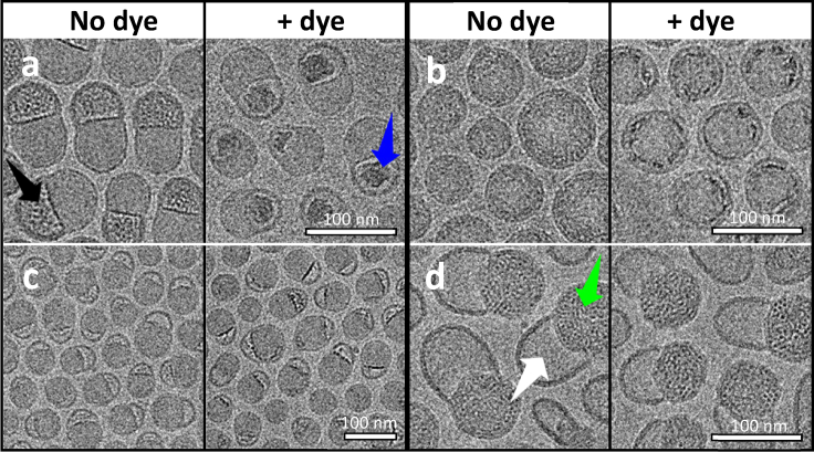
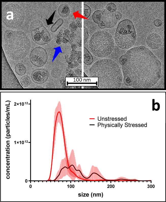
Understanding the structure of messenger RNA (mRNA) lipid nanoparticles, and specifically the microenvironment of the mRNA molecules within these entities, is fundamental to advancing their biomedical potential. Here, we show that a permeating cationic dye, thionine, can serve as a cryogenic electron microscopy contrasting agent by binding selectively to encapsulated mRNA without disturbing lipid nanoparticle morphology. Cryo-electron microscopy images identify the mRNA location, revealing that mRNA may exist within solvent-filled cavities or may be substantially lipid associated.
Overview
The claims and statistics of this paper haven’t been checked yet.
Run the check — free, no account needed. It reads the open-access text and checks the sources it cites.
Integrity signals
Registry
Cites
Statistics
Figures
Verification
Claims the paper makes
The claims and statistics of this paper haven’t been checked yet.
Run the check — free, no account needed. It reads the open-access text and checks the sources it cites.
Claims other papers make about this one
Each is checked against this paper’s own text: does it say what the citing paper says it does?
In the citing paper
“A key aspect of LNPs and the characteristic that makes them different from liposomes (spherical vesicles with at least one lipid bilayer and an aqueous core) is the presence of lipids in the core, although data from several studies indicate that water is also present to some extent (Yanez Arteta et al., 2018, Kulkarni et al., 2018, Brader et al., 2021).”
From mRNA-lipid nanoparticle COVID-19 vaccines: structure and stability (2021) · cites this paper as Brader et al., 2021
In the citing paper
“If the LNP shells are permeable to protons –which is likely, as ionized dyes such as 2-(p-toluidino)-6-napthalene sulfonic acid (TNS) and thionine can enter the LNP core (Jayaraman et al., 2012, Brader et al., 2021) – the pH would be similar to the rest of the formulation, around 7 to 8, meaning that most of the ionizable cationic lipids would be uncharged.”
From mRNA-lipid nanoparticle COVID-19 vaccines: structure and stability (2021) · cites this paper as Brader et al., 2021
Self-check
Percentages, sums and reported differences re-computed from the paper’s own numbers.
The self-check needs the paper’s full text.
Citations
Cites
An mRNA Vaccine against SARS-CoV-2 — Preliminary Report
Jackson, L. A., Anderson, E. J., Rouphael, N. G., Roberts, P. C., Makhene, M., Coler, R. N., McCullough, M. P., Chappell, J. D., Denison, M. R., Stevens, L. J., et al. (2020) · New England Journal of Medicine · doi:10.1056/nejmoa2022483 · Cited by 3,237
mRNA-based therapeutics--developing a new class of drugs
Sahin, U., Karikó, K., & Türeci, Ö. (2014) · Nature Reviews Drug Discovery · doi:10.1038/nrd4278 · Cited by 2,474
Rational design of cationic lipids for siRNA delivery
Semple, S. C., Akinc, A., Chen, J., Sandhu, A. P., Mui, B. L., Cho, C. K., Sah, D. W. Y., Stebbing, D., Crosley, E. J., Yaworski, E., et al. (2010) · Nature Biotechnology · doi:10.1038/nbt.1602 · Cited by 1,822
Delivering the Messenger: Advances in Technologies for Therapeutic mRNA Delivery
Kowalski, P. S., Rudra, A., Miao, L., & Anderson, D. G. (2019) · Molecular Therapy · doi:10.1016/j.ymthe.2019.02.012 · Cited by 1,127
Modified mRNA vaccines protect against zika virus infection
Richner, J. M., Himansu, S., Dowd, K. A., Butler, S. L., Salazar, V., Fox, J. M., Julander, J. G., Tang, W. W., Shresta, S., Pierson, T. C., et al. (2017) · Cell · doi:10.1016/j.cell.2017.02.017 · Cited by 909 · cited 1×
“Intriguingly, mRNA-LNPs are known to form both spherical and highly nonspherical morphologies (9,11,16,18,19), but the origins and practical implications of these morphological variants”
BackgroundMiao, L., Li, L., Huang, Y., Delcassian, D., Chahal, J., Han, J., Shi, Y., Sadtler, K., Gao, W., Lin, J., et al. (2019) · Nature Biotechnology · doi:10.1038/s41587-019-0247-3 · Cited by 723 · cited 1×
“Intriguingly, mRNA-LNPs are known to form both spherical and highly nonspherical morphologies (9,11,16,18,19), but the origins and practical implications of these morphological variants”
BackgroundLipid Nanoparticle Assisted mRNA Delivery for Potent Cancer Immunotherapy
Oberli, M. A., Reichmuth, A. M., Dorkin, J. R., Mitchell, M. J., Fenton, O. S., Jaklenec, A., Anderson, D. G., Langer, R., & Blankschtein, D. (2017) · Nano Letters · doi:10.1021/acs.nanolett.6b03329 · Cited by 719
Kulkarni, J. A., Darjuan, M. M., Mercer, J. E., Chen, S., van der Meel, R., Thewalt, J. L., Tam, Y. Y. C., & Cullis, P. R. (2018) · ACS Nano · doi:10.1021/acsnano.8b01516 · Cited by 663 · cited 1×
“In the case of LNPs containing short interfering RNA (siRNA), small angle xray scattering and cryo-electron microscopy (cryo-EM) data have been interpreted as evidence of a multilamellar structure in which the siRNA molecule is sandwiched between bilayer lipid assemblies (10,17).”
BackgroundmRNA vaccine delivery using lipid nanoparticles
Reichmuth, A. M., Oberli, M. A., Jaklenec, A., Langer, R., & Blankschtein, D. (2016) · Therapeutic Delivery · doi:10.4155/tde-2016-0006 · Cited by 624 · cited 1×
“Lipid nanoparticles (LNPs) have emerged as the most promising nonviral delivery vehicle for exogenous mRNA (5,6).”
BackgroundDegradable lipid nanoparticles with predictable in vivo siRNA delivery activity
Whitehead, K. A., Dorkin, J. R., Vegas, A. J., Chang, P. H., Veiseh, O., Matthews, J., Fenton, O. S., Zhang, Y., Olejnik, K. T., Yesilyurt, V., et al. (2014) · Nature Communications · doi:10.1038/ncomms5277 · Cited by 615
Yanez Arteta, M., Kjellman, T., Bartesaghi, S., Wallin, S., Wu, X., Kvist, A. J., Dabkowska, A., Székely, N., Radulescu, A., Bergenholtz, J., et al. (2018) · Proceedings of the National Academy of Sciences · doi:10.1073/pnas.1720542115 · Cited by 534
Deconvoluting Lipid Nanoparticle Structure for Messenger RNA Delivery
Eygeris, Y., Patel, S., Jozić, A., & Sahay, G. (2020) · Nano Letters · doi:10.1021/acs.nanolett.0c01386 · Cited by 397 · cited 1×
“It has been well established that LNPs with a diversity of morphological features can be generated through both manipulation of rapid precipitation conditions and formulation composition (10,15,26).”
BackgroundNanotechnologies in delivery of mRNA therapeutics using nonviral vector-based delivery systems
Guan, S., & Rosenecker, J. (2017) · Gene Therapy · doi:10.1038/gt.2017.5 · Cited by 389 · cited 1×
“Lipid nanoparticles (LNPs) have emerged as the most promising nonviral delivery vehicle for exogenous mRNA (5,6).”
BackgroundMicrofluidic Mixing: A General Method for Encapsulating Macromolecules in Lipid Nanoparticle Systems
Leung, A. K. K., Tam, Y. Y. C., Chen, S., Hafez, I. M., & Cullis, P. R. (2015) · The Journal of Physical Chemistry B · doi:10.1021/acs.jpcb.5b02891 · Cited by 359 · cited 3×
“This interpretation differs from that of Leung and co-workers (11), who reported a strikingly similar cryo-EM image yet assigned the mRNA location to the body of the particle adjoined to the solvent-filled bleb.”
BackgroundResultLipid nanoparticles for delivery of messenger RNA to the back of the eye
Patel, S., Ryals, R. C., Weller, K. K., Pennesi, M. E., & Sahay, G. (2019) · Journal of Controlled Release · doi:10.1016/j.jconrel.2019.04.015 · Cited by 260 · cited 1×
“Intriguingly, mRNA-LNPs are known to form both spherical and highly nonspherical morphologies (9,11,16,18,19), but the origins and practical implications of these morphological variants”
BackgroundViger-Gravel, J., Schantz, A., Pinon, A. C., Rossini, A. J., Schantz, S., & Emsley, L. (2018) · The Journal of Physical Chemistry B · doi:10.1021/acs.jpcb.7b10795 · Cited by 202
Maeki, M., Fujishima, Y., Sato, Y., Yasui, T., Kaji, N., Ishida, A., Tani, H., Baba, Y., Harashima, H., & Tokeshi, M. (2017) · PLoS ONE · doi:10.1371/journal.pone.0187962 · Cited by 182 · cited 1×
“It has been well established that LNPs with a diversity of morphological features can be generated through both manipulation of rapid precipitation conditions and formulation composition (10,15,26).”
BackgroundFusion-dependent formation of lipid nanoparticles containing macromolecular payloads
Kulkarni, J. A., Witzigmann, D., Leung, J., van der Meel, R., Zaifman, J., Darjuan, M. M., Grisch-Chan, H. M., Thöny, B., Tam, Y. Y. C., & Cullis, P. R. (2019) · Nanoscale · doi:10.1039/c9nr02004g · Cited by 161 · cited 1×
“Intriguingly, mRNA-LNPs are known to form both spherical and highly nonspherical morphologies (9,11,16,18,19), but the origins and practical implications of these morphological variants”
BackgroundVisualizing large RNA molecules in solution.
Gopal, A., Zhou, H. Z., Knobler, C., & Gelbart, W. (2012) · RNA: A publication of the RNA Society · doi:10.1261/rna.027557.111 · Cited by 114 · cited 2×
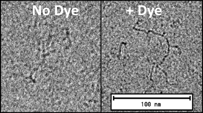
“In the absence of dye (left), the resolution is comparable to published examples (25), whereas in the presence of 0.”
BackgroundVariations on Negative Stain Electron Microscopy Methods: Tools for Tackling Challenging Systems
Scarff, C. A., Fuller, M. J., Thompson, R. F., & Iadanza, M. G. (2018) · Journal of Visualized Experiments · doi:10.3791/57199 · Cited by 110 · cited 1×
“To overcome these limitations, we explored the concept of using an RNA-binding dye to stain LNPs in a somewhat analogous manner to classical cell staining with membrane-permeable dyes (21).”
BackgroundGindy, M. E., DiFelice, K., Kumar, V., Prud’homme, R. K., Celano, R., Haas, R. M., Smith, J. S., & Boardman, D. (2014) · Langmuir · doi:10.1021/la500630h · Cited by 70 · cited 4×
“In the case of LNPs containing short interfering RNA (siRNA), small angle xray scattering and cryo-electron microscopy (cryo-EM) data have been interpreted as evidence of a multilamellar structure in which the siRNA molecule is sandwiched between bilayer lipid assemblies (10,17).”
BackgroundInfluentialIn vitro-Transcribed mRNA Therapeutics: Out of the Shadows and Into the Spotlight
Karikó, K. (2019) · Molecular Therapy · doi:10.1016/j.ymthe.2019.03.009 · Cited by 57
Khan, A. Y., & Kumar, G. S. (2015) · Spectrochimica Acta Part A Molecular and Biomolecular Spectroscopy · doi:10.1016/j.saa.2015.07.091 · Cited by 39
Variations on Negative Stain Electron Microscopy Methods: Tools for Tackling Challenging Systems
Scarff, C. A., Fuller, M. J., Thompson, R. F., & Iadanza, M. G. (2018) · Journal of Visualized Experiments · doi:10.3791/57199-v · Cited by 30
Paul, P., & Kumar, G. S. (2010) · Journal of Hazardous Materials · doi:10.1016/j.jhazmat.2010.08.081 · Cited by 29 · cited 3×
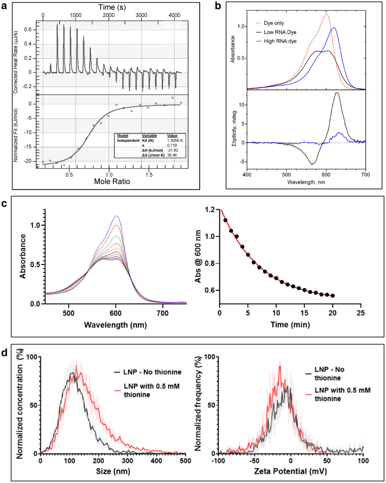
“Intercalation and electrostatic (outside) Biophysical Letter Please cite this article in press as: Brader et al., Encapsulation state of messenger RNA inside lipid nanoparticles, Biophysical Journal (2021), https://doi.org/ 10.1016/j.bpj.2021.03.012 binding modes have been distinguished previously for phenothiazinium…”
BackgroundInfluentialDensity based visualization for molecular simulation
Rozmanov, D. A., Baoukina, S., & Tieleman, D. P. (2014) · Faraday Discussions · doi:10.1039/c3fd00124e · Cited by 29 · cited 1×
“Molecular simulation studies (20) indicate that the LNP structure may involve solvent pockets, and experimental studies have shown nonspherical morphologies to be associated with segregation of the bilayer-forming 1,2-distearoyl-sn-glycero-3-phosphocholine (DSPC) component, resulting in pronounced blebs (11).”
BackgroundInteraction of methylene blue with transfer RNA — a spectroscopic study
Antony, T., Atreyi, M., & Rao, M. V. R. (1995) · Chemico-Biological Interactions · doi:10.1016/0009-2797(95)03616-t · Cited by 22 · cited 1×
“1 b are consistent with the RNA/dye ratio-dependent signatures of these binding modes reported for transfer RNA (24).”
Background
Cited by
Sorted by citations
mRNA-lipid nanoparticle COVID-19 vaccines: structure and stability
Schoenmaker, L., Witzigmann, D., Kulkarni, J. A., Verbeke, R., Kersten, G., Jiskoot, W., & Crommelin, D. J. A. (2021) · International Journal of Pharmaceutics · doi:10.1016/j.ijpharm.2021.120586 · Cited by 1,566
“…is likely, as ionized dyes such as 2-(p-toluidino)-6-nap- thalene sulfonic acid (TNS) and thionine can enter the LNP core (Jayaraman et al., 2012; Brader et al., 2021) – the pH would be similar to the rest of the formulation, around 7 to 8, meaning that most of the ionizable cationic lipids…”
InfluentialThe role of lipid components in lipid nanoparticles for vaccines and gene therapy
Gregersen, C. H., Kulkarni, J. A., Witzigmann, D., Lind, M. L., Petersson, K., & Simonsen, J. B. (2022) · Advanced Drug Delivery Reviews · doi:10.1016/j.addr.2022.114416 · Cited by 1,271
“Recent studies have investigated LNP structure/lipid arrangement by using cryo-TEM [94,99,100], molecular dynamics (MD) [101], small-angle neutron scattering (SANS) [54], small-angle x-ray scattering (SAXS) [54,94], fluorescence microscopy [102], and NMR methods [103].”
Chemistry of Lipid Nanoparticles for RNA Delivery
Eygeris, Y., Gupta, M., Kim, J., & Sahay, G. (2021) · Accounts of Chemical Research · doi:10.1021/acs.accounts.1c00544 · Cited by 917
Evolution of drug delivery systems: From 1950 to 2020 and beyond
Park, H., Otte, A., & Park, K. (2021) · Journal of Controlled Release · doi:10.1016/j.jconrel.2021.12.030 · Cited by 507
Payload distribution and capacity of mRNA lipid nanoparticles
Li, S., Hu, Y., Li, A. I., Lin, J., Hsieh, K., Schneiderman, Z., Zhang, P., Zhu, Y., Qiu, C., Kokkoli, E., et al. (2022) · Nature Communications · doi:10.1038/s41467-022-33157-4 · Cited by 299
Metadata
- Authors
- Brader, M. L., Williams, S. J., Banks, J. M., Hui, W. H., Zhou, Z. H., & Jin, L.
- Affiliations
- California NanoSystems Institute (US)
- Corresponding author
- Brader, Mark L.
- Venue
- Biophysical Journal
- Publisher
- Elsevier BV
- Published
- 2021-07
- Volume
- 120
- Issue
- 14
- Pages
- 2766-2770
- ISSN
- 0006-3495 (ISSN-L)
- PMC
- PMC8390897
- OpenAlex
- W3139111210
- Cited by
- 236
- References
- 27
- Citations by year
- 2026: 40 · 2025: 59 · 2024: 58 · 2023: 40 · 2022: 27 · 2021: 12
- FWCI
- 10.75
- Type
- journal article
- Language
- en
- Open access
- hybrid
- doi.org
- Licence
- Locations
- Biophysical Journal (publishedVersion, cc-by)
- Europe PMC (PubMed Central) (publishedVersion, other-oa)
- PubMed Central (publishedVersion, cc-by)
- PubMed (publishedVersion)
- eScholarship (California Digital Library) (submittedVersion)
- eScholarship (California Digital Library) (submittedVersion, cc-by)
- Topics
- Life Sciences › Biochemistry, Genetics and Molecular Biology › Molecular Biology › RNA Interference and Gene Delivery
- Physical Sciences › Materials Science › Biomaterials › Nanoparticle-Based Drug Delivery
- Life Sciences › Biochemistry, Genetics and Molecular Biology › Molecular Biology › Lipid Membrane Structure and Behavior
- Keywords
- mRNA lipid nanoparticles, mRNA encapsulation, thionine, mRNA localization
- MeSH terms
- Lipids, RNA, Messenger, Cryoelectron Microscopy, Nanoparticles
About these checks
Registry, retraction, Cites and statistics results are computed, not judged, with one exception: a printed reference that no index lists is read once by a model before it counts as a citation error. Claims are extracted by a model and kept only when their quote appears verbatim in the text; each cited source is then checked, with a model’s help, for whether it says what the claim relies on. Those verdicts can be wrong — read the claim’s quote and the cited source before relying on one.
Statistics and arithmetic are recomputed, not judged.
Confidence bands are shown only for fields calibrated on our annotated test set. None is calibrated yet, so no band is shown.
What counts as a claim ›How we measure confidence ›How we check this ›
Are you an author, or do you see a problem with this page? Request a review
