

The DOI matches its Crossref record; 116 of the 116 references it cites were found in a registry or an index.
Elicitation of broadly protective sarbecovirus immunity by receptor-binding domain nanoparticle vaccines
Open accessPublished version of a preprint
Abstract
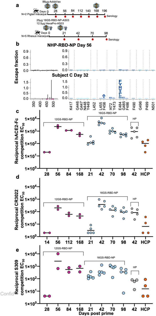
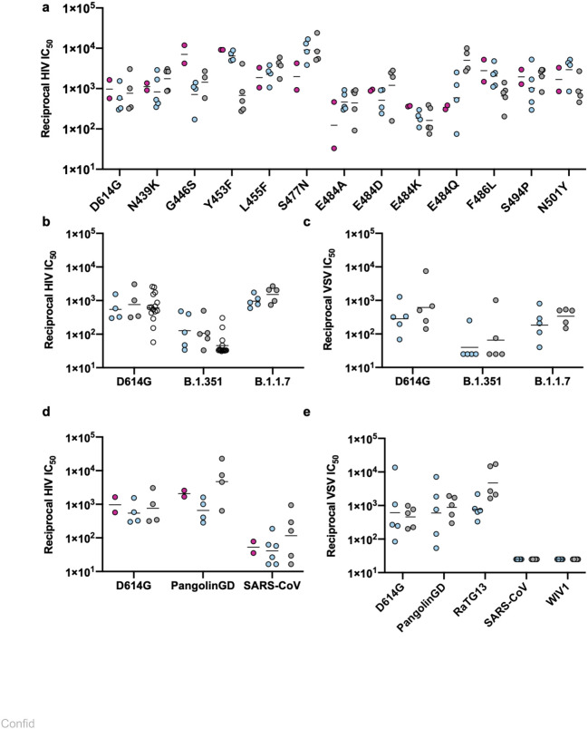
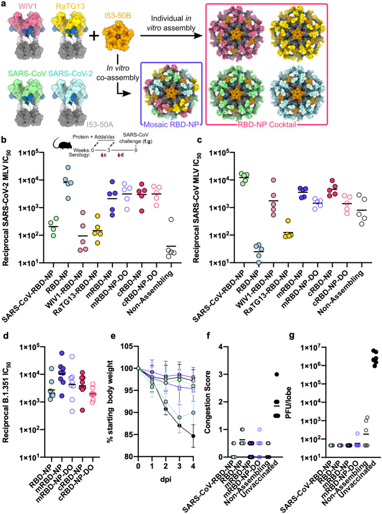
Understanding the ability of SARS-CoV-2 vaccine-elicited antibodies to neutralize and protect against emerging variants of concern and other sarbecoviruses is key for guiding vaccine development decisions and public health policies. We show that a clinical stage multivalent SARS-CoV-2 receptor-binding domain nanoparticle vaccine (SARS-CoV-2 RBD-NP) protects mice from SARS-CoV-2-induced disease after a single shot, indicating that the vaccine could allow dose-sparing. SARS-CoV-2 RBD-NP elicits high antibody titers in two non-human primate (NHP) models against multiple distinct RBD antigenic sites known to be recognized by neutralizing antibodies. We benchmarked NHP serum neutralizing activity elicited by RBD-NP against a lead prefusion-stabilized SARS-CoV-2 spike immunogen using a panel of single-residue spike mutants detected in clinical isolates as well as the B.1.1.7 and B.1.351 variants of concern. Polyclonal antibodies elicited by both vaccines are resilient to most RBD mutations tested, but the E484K substitution has similar negative consequences for neutralization, and exhibit modest but comparable neutralization breadth against distantly related sarbecoviruses. We demonstrate that mosaic and cocktail sarbecovirus RBD-NPs elicit broad sarbecovirus neutralizing activity, including against the SARS-CoV-2 B.1.351 variant, and protect mice against severe SARS-CoV challenge even in the absence of the SARS-CoV RBD in the vaccine. This study provides proof of principle that sarbecovirus RBD-NPs induce heterotypic protection and enables advancement of broadly protective sarbecovirus vaccines to the clinic.
Overview
The claims and statistics of this paper haven’t been checked yet.
Run the check — free, no account needed. It reads the open-access text and checks the sources it cites.
Integrity signals
Registry
Cites
Statistics
Figures
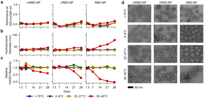
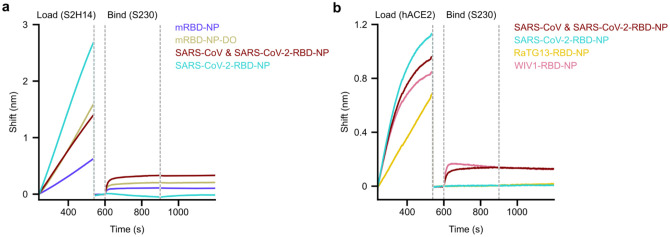
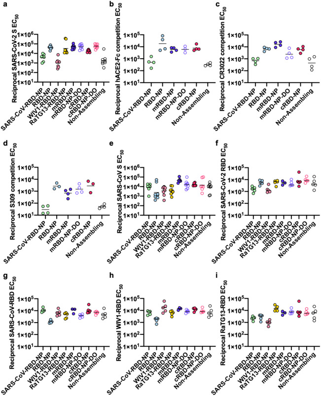
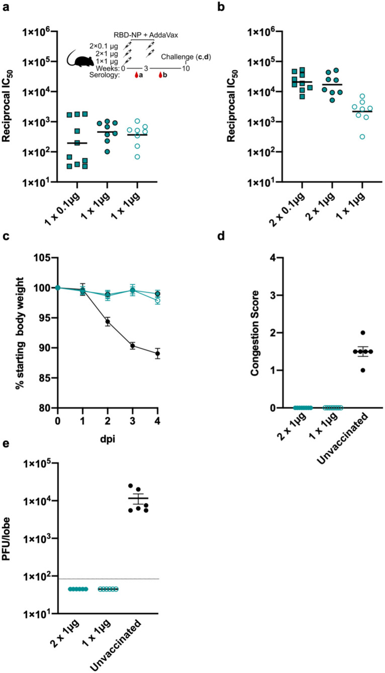
Verification
Claims the paper makes
The claims and statistics of this paper haven’t been checked yet.
Run the check — free, no account needed. It reads the open-access text and checks the sources it cites.
Claims other papers make about this one
Each is checked against this paper’s own text: does it say what the citing paper says it does?
The cited source says what the claim attributes to it.
In the citing paper
“For example, prototypes of multivalent nanoparticle vaccines have been reported to induce broad reactivity against different sarbecoviruses7.”
From Efficacy of an unmodified bivalent mRNA vaccine against SARS-CoV-2 variants in female small animal models (2023) · cites this paper as [7]
Self-check
Percentages, sums and reported differences re-computed from the paper’s own numbers.
The self-check needs the paper’s full text.
Citations
Cites
A pneumonia outbreak associated with a new coronavirus of probable bat origin
Zhou, P., Yang, X.-L., Wang, X.-G., Hu, B., Zhang, L., Zhang, W., Si, H.-R., Zhu, Y., Li, B., Huang, C.-L., et al. (2020) · Nature · doi:10.1038/s41586-020-2012-7 · Cited by 21,969
Hoffmann, M., Kleine-Weber, H., Schroeder, S., Krüger, N., Herrler, T., Erichsen, S., Schiergens, T. S., Herrler, G., Wu, N.-H., Nitsche, A., et al. (2020) · Cell · doi:10.1016/j.cell.2020.02.052 · Cited by 20,286 · cited 1×
“SARS-CoV-2 infects host cells through the attachment of the viral transmembrane spike (S) glycoprotein to angiotensin-converting enzyme 2 (ACE2), followed by fusion of the viral and host membranes (Hoffmann et al., 2020; Lan et al., 2020; Letko et al., 2020; Shang et al., 2020; Walls et al., 2020a; Wang et al., 2020;…”
Safety and Efficacy of the BNT162b2 mRNA Covid-19 Vaccine
Polack, F. P., Thomas, S. J., Kitchin, N., Absalon, J., Gurtman, A., Lockhart, S., Perez, J. L., Pérez Marc, G., Moreira, E. D., Zerbini, C., et al. (2020) · New England Journal of Medicine · doi:10.1056/nejmoa2034577 · Cited by 14,592
Enzymatic assembly of DNA molecules up to several hundred kilobases
Gibson, D. G., Young, L., Chuang, R.-Y., Venter, J. C., Hutchison, C. A., & Smith, H. O. (2009) · Nature Methods · doi:10.1038/nmeth.1318 · Cited by 10,088 · cited 1×
“The RBD-16GS-I53-50A fusion was cloned into pCMV/R using the Xba1 and AvrII restriction sites and Gibson assembly (Gibson et al., 2009).”
Cryo-EM structure of the 2019-nCoV spike in the prefusion conformation
Wrapp, D., Wang, N., Corbett, K. S., Goldsmith, J. A., Hsieh, C.-L., Abiona, O., Graham, B. S., & McLellan, J. S. (2020) · Science · doi:10.1126/science.abb2507 · Cited by 8,897 · cited 2×
“SARS-CoV-2 infects host cells through the attachment of the viral transmembrane spike (S) glycoprotein to angiotensin-converting enzyme 2 (ACE2), followed by fusion of the viral and host membranes (Hoffmann et al., 2020; Lan et al., 2020; Letko et al., 2020; Shang et al., 2020; Walls et al., 2020a; Wang et al., 2020;…”
BackgroundSelf-citationStructure, function and antigenicity of the SARS-CoV-2 spike glycoprotein
Walls, A. C., Park, Y.-J., Tortorici, M. A., Wall, A., McGuire, A. T., & Veesler, D. (2020) · doi:10.1101/2020.02.19.956581 · Cited by 8,586 · cited 5×
“1- with an N-terminal mu-phosphatase signal peptide and a C-terminal octa-histidine tag, flexible linker, and avi tag (GHHHHHHHHGGSSGLNDIFEAQKIEWHE).”
InfluentialSelf-citationStructure of the SARS-CoV-2 spike receptor-binding domain bound to the ACE2 receptor
Lan, J., Ge, J., Yu, J., Shan, S., Zhou, H., Fan, S., Zhang, Q., Shi, X., Wang, Q., Zhang, L., et al. (2020) · Nature · doi:10.1038/s41586-020-2180-5 · Cited by 5,983 · cited 1×
“SARS-CoV-2 infects host cells through the attachment of the viral transmembrane spike (S) glycoprotein to angiotensin-converting enzyme 2 (ACE2), followed by fusion of the viral and host membranes (Hoffmann et al., 2020; Lan et al., 2020; Letko et al., 2020; Shang et al., 2020; Walls et al., 2020a; Wang et al., 2020;…”
BackgroundUCSF ChimeraX: Meeting modern challenges in visualization and analysis
Goddard, T. D., Huang, C. C., Meng, E. C., Pettersen, E. F., Couch, G. S., Morris, J. H., & Ferrin, T. E. (2018) · Protein Science · doi:10.1002/pro.3235 · Cited by 5,267
Structural basis for the recognition of SARS-CoV-2 by full-length human ACE2
Yan, R., Zhang, Y., Li, Y., Xia, L., Guo, Y., & Zhou, Q. (2020) · Science · doi:10.1126/science.abb2762 · Cited by 5,036 · cited 1×
“SARS-CoV-2 infects host cells through the attachment of the viral transmembrane spike (S) glycoprotein to angiotensin-converting enzyme 2 (ACE2), followed by fusion of the viral and host membranes (Hoffmann et al., 2020; Lan et al., 2020; Letko et al., 2020; Shang et al., 2020; Walls et al., 2020a; Wang et al., 2020;…”
Korber, B., Fischer, W. M., Gnanakaran, S., Yoon, H., Theiler, J., Abfalterer, W., Hengartner, N., Giorgi, E. E., Bhattacharya, T., Foley, B., et al. (2020) · Cell · doi:10.1016/j.cell.2020.06.043 · Cited by 4,281
Research works printed but not linked: 1
- Davies, 2020, Estimated transmissibility and severity of novel SARS-CoV-2 Variant of Concern 202012/01 in England, medRxiv
Cited by
Sorted by citations
Broadly neutralizing antibodies overcome SARS-CoV-2 Omicron antigenic shift
Cameroni, E., Bowen, J. E., Rosen, L. E., Saliba, C., Zepeda, S. K., Culap, K., Pinto, D., VanBlargan, L. A., Marco, A. D., di Iulio, J., et al. (2021) · Nature · doi:10.1038/s41586-021-04386-2 · Cited by 1,070
Self-citationStructural basis of SARS-CoV-2 Omicron immune evasion and receptor engagement
McCallum, M., Czudnochowski, N., Rosen, L. E., Zepeda, S. K., Bowen, J. E., Walls, A. C., Hauser, K., Joshi, A., Stewart, C., Dillen, J. R., et al. (2022) · Science · doi:10.1126/science.abn8652 · Cited by 535
“The identification of broadly reactive mAbs that neutralize multiple distinct sarbecoviruses, including SARS-CoV-2 variants, pave the way for designing vaccines eliciting broad sarbecovirus immunity ( 52 – 56 ).”
Self-citationSARS-CoV-2 RBD antibodies that maximize breadth and resistance to escape
Starr, T. N., Czudnochowski, N., Liu, Z., Zatta, F., Park, Y., Addetia, A., Pinto, D., Beltramello, M., Hernandez, P., Greaney, A. J., et al. (2021) · Nature · doi:10.1038/s41586-021-03807-6 · Cited by 503
Self-citationBroad betacoronavirus neutralization by a stem helix–specific human antibody
Pinto, D., Sauer, M. M., Czudnochowski, N., Low, J. S., Tortorici, M. A., Housley, M. P., Noack, J., Walls, A. C., Bowen, J. E., Guarino, B., et al. (2021) · Science · doi:10.1126/science.abj3321 · Cited by 385
Self-citationUnadjuvanted intranasal spike vaccine elicits protective mucosal immunity against sarbecoviruses
Mao, T., Israelow, B., Peña-Hernández, M. A., Suberi, A., Zhou, L., Luyten, S., Reschke, M., Dong, H., Homer, R., Saltzman, W. M., et al. (2022) · Science · doi:10.1126/science.abo2523 · Cited by 356
“Although some recent studies have successfully reported the development of systemic pan-sarbecovirus vaccines (37, 38), P&Sx induces both systemic andmucosal immunity against both SARS-CoV-1 and SARS-CoV-2.”
ResultMolecular basis of immune evasion by the Delta and Kappa SARS-CoV-2 variants
McCallum, M., Walls, A. C., Sprouse, K. R., Bowen, J. E., Rosen, L. E., Dang, H. V., Marco, A. D., Franko, N., Tilles, S. W., Logue, J. K., et al. (2021) · Science · doi:10.1126/science.abl8506 · Cited by 280
Self-citationBroad sarbecovirus neutralization by a human monoclonal antibody
Tortorici, M. A., Czudnochowski, N., Starr, T. N., Marzi, R., Walls, A. C., Zatta, F., Bowen, J. E., Jaconi, S., di Iulio, J., Wang, Z., et al. (2021) · Nature · doi:10.1038/s41586-021-03817-4 · Cited by 255
Self-citationMosaic RBD nanoparticles protect against challenge by diverse sarbecoviruses in animal models
Cohen, A. A., van Doremalen, N., Greaney, A. J., Andersen, H., Sharma, A., Starr, T. N., Keeffe, J. R., Fan, C., Schulz, J. E., Gnanapragasam, P. N. P., et al. (2022) · Science · doi:10.1126/science.abq0839 · Cited by 264
“Target list [bead region] were as follows: T helper: GM-CSF [44], IFN gamma [43], IL-1 beta [18], IL-2 [19], IL-4 [20], IL-5 [21], IL-6 [25], IL-8 (CXCL8) [27], IL-10 [28], IL12p70 [34], IL-13 [35], IL-17A (CTLA-8) [36], IL-18 [66], IL-23 [63], TNF alpha [45] Cytokines: CD40L [74], G-CSF (CSF-3) [42], IFN alpha [48],…”
Self-citationBroadly neutralizing antibodies overcome SARS-CoV-2 Omicron antigenic shift
Cameroni, E., Bowen, J. E., Rosen, L. E., Saliba, C., Zepeda, S. K., Culap, K., Pinto, D., VanBlargan, L. A., Marco, A. D., di Iulio, J., et al. (2021) · Nature · doi:10.1038/d41586-021-03825-4 · Cited by 251
Self-citationImprinted antibody responses against SARS-CoV-2 Omicron sublineages
Park, Y., Pinto, D., Walls, A. C., Liu, Z., Marco, A. D., Benigni, F., Zatta, F., Silacci-Fregni, C., Bassi, J., Sprouse, K. R., et al. (2022) · Science · doi:10.1126/science.adc9127 · Cited by 230
Self-citationSARS-CoV-2 breakthrough infections elicit potent, broad, and durable neutralizing antibody responses
Walls, A. C., Sprouse, K. R., Bowen, J. E., Joshi, A., Franko, N., Navarro, M. J., Stewart, C., Cameroni, E., McCallum, M., Goecker, E. A., et al. (2022) · Cell · doi:10.1016/j.cell.2022.01.011 · Cited by 188
Self-citationThe humoral response and antibodies against SARS-CoV-2 infection
Qi, H., Liu, B., Wang, X., & Zhang, L. (2022) · Nature Immunology · doi:10.1038/s41590-022-01248-5 · Cited by 195
Protein Cages: From Fundamentals to Advanced Applications
Edwardson, T. G. W., Levasseur, M. D., Tetter, S., Steinauer, A., Hori, M., & Hilvert, D. (2022) · Chemical Reviews · doi:10.1021/acs.chemrev.1c00877 · Cited by 171
Targeted modulation of immune cells and tissues using engineered biomaterials
Yousefpour, P., Ni, K., & Irvine, D. J. (2023) · Nature Reviews Bioengineering · doi:10.1038/s44222-022-00016-2 · Cited by 170
Omicron spike function and neutralizing activity elicited by a comprehensive panel of vaccines
Bowen, J. E., Addetia, A., Dang, H. V., Stewart, C., Brown, J. T., Sharkey, W. K., Sprouse, K. R., Walls, A. C., Mazzitelli, I. G., Logue, J. K., et al. (2022) · Science · doi:10.1126/science.abq0203 · Cited by 160
Self-citationKumari, M., Lu, R., Li, M., Huang, J., Hsu, F., Ko, S.-H., Ke, F.-Y., Su, S.-C., Liang, K.-H., Yuan, J. P., et al. (2022) · Journal of Biomedical Science · doi:10.1186/s12929-022-00852-9 · Cited by 166
Nanotechnology-based strategies against SARS-CoV-2 variants
Huang, X., Kon, E., Han, X., Zhang, X., Kong, N., Mitchell, M. J., Peer, D., & Tao, W. (2022) · Nature Nanotechnology · doi:10.1038/s41565-022-01174-5 · Cited by 146
Scientific rationale for developing potent RBD-based vaccines targeting COVID-19
Kleanthous, H., Silverman, J. M., Makar, K. W., Yoon, I.-K., Jackson, N. M., & Vaughn, D. W. (2021) · npj Vaccines · doi:10.1038/s41541-021-00393-6 · Cited by 139
Self-citationAntibody-mediated broad sarbecovirus neutralization through ACE2 molecular mimicry
Park, Y., Marco, A. D., Starr, T. N., Liu, Z., Pinto, D., Walls, A. C., Zatta, F., Zepeda, S. K., Bowen, J. E., Sprouse, K. R., et al. (2022) · Science · doi:10.1126/science.abm8143 · Cited by 135
Self-citationSARS-CoV-2 immune evasion by variant B.1.427/B.1.429Preprint
McCallum, M., Bassi, J., Marco, A. D., Chen, A., Walls, A. C., di Iulio, J., Tortorici, M. A., Navarro, M.-J., Silacci-Fregni, C., Saliba, C., et al. (2021) · bioRxiv (Cold Spring Harbor Laboratory) · doi:10.1101/2021.03.31.437925 · Cited by 128
“429 S pseudotypes were prepared as previously 528 described (62, 63).”
MethodSelf-citation“World in motion” – emulsion adjuvants rising to meet the pandemic challenges
O’Hagan, D. T., der Most, R. G. V., Lodaya, R. N., Coccia, M., & Lofano, G. (2021) · npj Vaccines · doi:10.1038/s41541-021-00418-0 · Cited by 127
Self-citationA broadly cross-reactive antibody neutralizes and protects against sarbecovirus challenge in mice
Martinez, D. R., Schäfer, A., Gobeil, S., Li, D., la Cruz, G. D., Parks, R., Lu, X., Barr, M., Stalls, V., Janowska, K., et al. (2021) · Science Translational Medicine · doi:10.1126/scitranslmed.abj7125 · Cited by 137
Self-citationLiu, Z., Zhou, J., Xu, W., Deng, W., Wang, Y., Wang, M., Wang, Q., Hsieh, M. K., Dong, J., Wang, X., et al. (2022) · Cell Research · doi:10.1038/s41422-022-00612-2 · Cited by 113
“Towards this goal, nanoparticles co-displaying multiple distinct RBDs from beta-coronaviruses could induce cross-nAbs in mice, providing a strategy for simultaneous protection against SARS-CoV-2 and some zoonotic sarbecovirus infections.(67,68) Similarly, nanoparticles displaying the SARS-CoV-2 RBD formulated with 3M-…”
BackgroundRETRACTED: SARS-CoV-2 S2–targeted vaccination elicits broadly neutralizing antibodies
Ng, K. W., Faulkner, N., Finsterbusch, K., Wu, M., Harvey, R., Hussain, S., Greco, M., Liu, Y., Kjær, S., Swanton, C., et al. (2022) · Science Translational Medicine · doi:10.1126/scitranslmed.abn3715 · Cited by 100
RetractedCurrent and future nanoparticle vaccines for COVID-19
Vu, M. N., Kelly, H. G., Kent, S. J., & Wheatley, A. K. (2021) · EBioMedicine · doi:10.1016/j.ebiom.2021.103699 · Cited by 101
“In another approach, the two-component computationally designed protein scaffold I53-50 was constructed to deliver RBDs from four distinct sarbecoviruses - SARS-CoV-1, SARS-CoV-2 and two bat coronaviruses WIV1 and RaTG13 [72].”
Song, J. Y., Choi, W. S., Heo, J. Y., Lee, J. S., Jung, D. S., Kim, S., Park, K., Eom, J. S., Jeong, S. J., Lee, J., et al. (2022) · EClinicalMedicine · doi:10.1016/j.eclinm.2022.101569 · Cited by 94
Self-citationArchitecture and antigenicity of the Nipah virus attachment glycoprotein
Wang, Z., Amaya, M., Addetia, A., Dang, H. V., Reggiano, G., Yan, L., Hickey, A. C., DiMaio, F., Broder, C. C., & Veesler, D. (2022) · Science · doi:10.1126/science.abm5561 · Cited by 94
Self-citationTortorici, M. A., Walls, A. C., Joshi, A., Park, Y., Eguia, R. T., Miranda, M. C., Kepl, E., Dosey, A. M., Stevens‐Ayers, T., Boeckh, M. J., et al. (2022) · Cell · doi:10.1016/j.cell.2022.05.019 · Cited by 89
“…of therapeutics and vaccine development, including monoclonal antibody therapies and vaccines (Baum et al., 2020; Corbett et al., 2020; Corti et al., 2021; Hansen et al., 2020; McCallum et al., 2021a; Piccoli et al., 2020; Pinto et al., 2020; Polack et al., 2020; Walls et al., 2019, 2020b, 2021).”
Self-citationEngineering Self-Assembling Protein Nanoparticles for Therapeutic Delivery
Olshefsky, A., Richardson, C., Pun, S. H., & King, N. P. (2022) · Bioconjugate Chemistry · doi:10.1021/acs.bioconjchem.2c00030 · Cited by 94
Self-citationIn search of a pan-coronavirus vaccine: next-generation vaccine design and immune mechanisms
Cankat, S., Demael, M. U., & Swadling, L. (2023) · Cellular & Molecular Immunology · doi:10.1038/s41423-023-01116-8 · Cited by 84
“Serum from RBD-NP-immunized nonhuman primates strongly cross-reacted with Pangolin-GD and RaTG13 RBDs but had weaker binding to distantly related RmYN02, SARS-CoV-1, WIV16, and ZXC21 RBDs [146].”
BackgroundBianchini, F., Crivelli, V., Abernathy, M. E., Guerra, C., Palus, M., Muri, J., Marcotte, H., Piralla, A., Pedotti, M., Gasparo, R. D., et al. (2023) · Science Immunology · doi:10.1126/sciimmunol.ade0958 · Cited by 88
Ols, S., Lenart, K., Cerveira, R. A., Miranda, M. C., Brunette, N., Kochmann, J., Corcoran, M., Skotheim, R., Philomin, A., Cagigi, A., et al. (2023) · Immunity · doi:10.1016/j.immuni.2023.08.011 · Cited by 80
Self-citationAdvanced technologies for the development of infectious disease vaccines
Gupta, A., Rudra, A., Reed, K., Langer, R., & Anderson, D. G. (2024) · Nature Reviews Drug Discovery · doi:10.1038/s41573-024-01041-z · Cited by 87
Bowen, J. E., Park, Y., Stewart, C., Brown, J. T., Sharkey, W. K., Walls, A. C., Joshi, A., Sprouse, K. R., McCallum, M., Tortorici, M. A., et al. (2022) · Science Immunology · doi:10.1126/sciimmunol.adf1421 · Cited by 81
Self-citationChallenges and developments in universal vaccine design against SARS-CoV-2 variants
Zhao, F., Zai, X., Zhang, Z., Xu, J., & Chen, W. (2022) · npj Vaccines · doi:10.1038/s41541-022-00597-4 · Cited by 83
Immune imprinting and next-generation coronavirus vaccines
Huang, C. Q., Vishwanath, S., Carnell, G., Chan, A. C. Y., & Heeney, J. L. (2023) · Nature Microbiology · doi:10.1038/s41564-023-01505-9 · Cited by 68
“A Delta–Omicron chimaeric RBD dimer was also shown to protect against matched VOCs 127 .”
BackgroundInfluentialAdenovirus-based vaccines—a platform for pandemic preparedness against emerging viral pathogens
Coughlan, L., Kremer, E. J., & Shayakhmetov, D. M. (2022) · Molecular Therapy · doi:10.1016/j.ymthe.2022.01.034 · Cited by 67
“Another innovative vaccine platform is the use of naturally occurring, self-assembling nanoparticles or computationally designed vaccine scaffolds.(34,35) Similar to VLPs, these particles allow for a structurally ordered display of antigen, which, along with their small size, allows them to mimic viruses.”
Proactive vaccination using multiviral Quartet Nanocages to elicit broad anti-coronavirus responses
Hills, R. A., Tan, T. K., Cohen, A. A., Keeffe, J. R., Keeble, A. H., Gnanapragasam, P. N. P., Storm, K. N., Rorick, A. V., West, A. P., Hill, M. L., et al. (2024) · Nature Nanotechnology · doi:10.1038/s41565-024-01655-9 · Cited by 82
“RBDs can be multimerized on VLPs through genetic fusion 15 or isopeptide coupling 12 .”
Imprinted antibody responses against SARS-CoV-2 Omicron sublineages
Park, Y.-J., Pinto, D., Walls, A. C., Liu, Z., De Marco, A., Benigni, F., Zatta, F., Silacci-Fregni, C., Bassi, J., Sprouse, K. R., et al. (2022) · doi:10.1101/2022.05.08.491108 · Cited by 251
Self-citationMao, T., Israelow, B., Suberi, A., Zhou, L., Reschke, M., Peña-Hernández, M. A., Dong, H., Homer, R. J., Saltzman, W., & Iwasaki, A. (2022) · bioRxiv · doi:10.1101/2022.01.24.477597 · Cited by 57
An ultrapotent RBD-targeted biparatopic nanobody neutralizes broad SARS-CoV-2 variants
Chi, X., Zhang, X., Pan, S., Yu, Y., Shi, Y., Lin, T., Duan, H., Liu, X., Chen, W., Yang, X., et al. (2022) · Signal Transduction and Targeted Therapy · doi:10.1038/s41392-022-00912-4 · Cited by 65
The quest for nanoparticle-powered vaccines in cancer immunotherapy
Sun, Z., Zhao, H., Ma, L., Shi, Y., Ji, M., Sun, X., Ma, D., Zhou, W., Huang, T., & Zhang, D. (2024) · Journal of Nanobiotechnology · doi:10.1186/s12951-024-02311-z · Cited by 63
Halfmann, P., Frey, S. J., Loeffler, K., Kuroda, M., Maemura, T., Armbrust, T., Yang, J. E., Hou, Y. J., Baric, R. S., Wright, E., et al. (2022) · EBioMedicine · doi:10.1016/j.ebiom.2022.104341 · Cited by 54
“60 While these results are promising and consistent with an approach that has worked with seasonal in fl u-enza vaccines, concerns include the large number of antigens required to provide broad protection as well as the plasticity of RBD domain and the possible emergence of new vaccine-evading RBD…”
Self-citationCurrent progress in the development of prophylactic and therapeutic vaccines
Li, T., Qian, C., Gu, Y., Zhang, J., Li, S., & Xia, N. (2022) · Science China Life Sciences · doi:10.1007/s11427-022-2230-4 · Cited by 59
“(Walls et al., 2021) and is thus currently undergoing clinical trials (NCT04742738, NCT05007951).”
BackgroundFeng, Y., Yuan, M., Powers, J. M., Hu, M., Munt, J. E., Arunachalam, P. S., Leist, S. R., Bellusci, L., Kim, J., Sprouse, K. R., et al. (2023) · Science Translational Medicine · doi:10.1126/scitranslmed.adg7404 · Cited by 58
Self-citationKang, Y., Sun, C., Sun, J., Xie, C., Zhuang, Z., Xu, H.-Q., Liu, Z., Liu, Y., Peng, S., Yuan, R., et al. (2022) · Nature Communications · doi:10.1038/s41467-022-30222-w · Cited by 56
Arunachalam, P. S., Feng, Y., Ashraf, U., Hu, M., Walls, A. C., Edara, V. V., Zarnitsyna, V. I., Aye, P. P., Golden, N., Miranda, M. C., et al. (2022) · Science Translational Medicine · doi:10.1126/scitranslmed.abq4130 · Cited by 53
“1 (for Omicron) and our previous work (33).”
BackgroundSelf-citationHierarchical design of pseudosymmetric protein nanocages
Dowling, Q. M., Park, Y., Fries, C. N., Gerstenmaier, N., Ols, S., Yang, E. C., Wargacki, A. J., Dosey, A. M., Hsia, Y., Ravichandran, R., et al. (2024) · Nature · doi:10.1038/s41586-024-08360-6 · Cited by 61
Self-citationGrigoryan, L., Lee, A. W. Q., Walls, A. C., Lai, L., Franco, B. D., Arunachalam, P. S., Feng, Y., Luo, W., Vanderheiden, A., Floyd, K. A., et al. (2022) · npj Vaccines · doi:10.1038/s41541-022-00472-2 · Cited by 52
Self-citationAntigen- and scaffold-specific antibody responses to protein nanoparticle immunogens
Kraft, J. C., Pham, M. N., Shehata, L., Brinkkemper, M., Boyoglu-Barnum, S., Sprouse, K. R., Walls, A. C., Cheng, S., Murphy, M., Pettie, D., et al. (2022) · Cell Reports Medicine · doi:10.1016/j.xcrm.2022.100780 · Cited by 50
Self-citationAn antibody class with a common CDRH3 motif broadly neutralizes sarbecoviruses
Liu, L., Iketani, S., Guo, Y., Reddem, E. R., Casner, R. G., Nair, M. S., Yu, J., Chan, J. F., Wang, M., Cerutti, G., et al. (2022) · Science Translational Medicine · doi:10.1126/scitranslmed.abn6859 · Cited by 51
Ellis, D., Brunette, N., Crawford, K. H. D., Walls, A. C., Pham, M. N., Chen, C., Herpoldt, K., Fiala, B., Murphy, M., Pettie, D., et al. (2021) · Frontiers in Immunology · doi:10.3389/fimmu.2021.710263 · Cited by 45
“MLV-Based Pseudovirus Neutralization Assays MLV-based SARS-CoV-2 S pseudotypes were prepared as previously described (10, 25, 50, 70).”
BackgroundMethodSelf-citationMalhi, H., Homad, L. J., Wan, Y.-H., Poudel, B., Fiala, B., Borst, A. J., Wang, J. Y., Walkey, C., Price, J., Wall, A., et al. (2022) · Cell Reports Medicine · doi:10.1016/j.xcrm.2022.100658 · Cited by 48
Self-citationSong, J. Y., Choi, W. S., Heo, J. Y., Kim, E. J., Lee, J. S., Jung, D. S., Kim, S., Park, K., Eom, J. S., Jeong, S. J., et al. (2023) · EClinicalMedicine · doi:10.1016/j.eclinm.2023.102140 · Cited by 50
Self-citationResearch progress on emulsion vaccine adjuvants
Huang, Z., Gong, H., Sun, Q., Yang, J., Yan, X., & Xu, F. (2024) · Heliyon · doi:10.1016/j.heliyon.2024.e24662 · Cited by 51
“Although current preliminary data are encouraging [121], careful evaluation of the immune response induced by emulsion adjuvant vaccines relative to alternative vaccine approaches is needed.”
BackgroundZhang, J., Xu, Y., Chen, M., Huang, Y., Song, T., Yang, C. J., Yang, Y., & Song, Y. (2022) · Journal of the American Chemical Society · doi:10.1021/jacs.2c09229 · Cited by 46
Molecular basis of convergent evolution of ACE2 receptor utilization among HKU5 coronaviruses
Park, Y., Liu, C., Lee, J., Brown, J. T., Ma, C., Liu, P., Gen, R., Xiong, Q., Zepeda, S. K., Stewart, C., et al. (2025) · Cell · doi:10.1016/j.cell.2024.12.032 · Cited by 47
“These findings are reminiscent of the weak cross-clade neutralization observed for sarbecoviruses, 105,106 which harbor RBDs much more similar to each other than merbecovirus RBDs.”
Self-citationBroad-spectrum pan-genus and pan-family virus vaccines
Tan, C. W., Valkenburg, S., Poon, L. L. M., & Wang, L. (2023) · Cell Host & Microbe · doi:10.1016/j.chom.2023.05.017 · Cited by 42
“sarbecoviruses and protect the vaccinated animals from SARSCoV-1 infection.(124) Chimeric spike mRNA vaccine candidates, containing RBD, NTD, and S2 domains from different SARS-”
Rapid and automated design of two-component protein nanomaterials using ProteinMPNN
de Haas, R. J., Brunette, N., Goodson, A., Dauparas, J., Yi, S. Y., Yang, E. C., Dowling, Q., Nguyen, H., Kang, A., Bera, A. K., et al. (2024) · Proceedings of the National Academy of Sciences · doi:10.1073/pnas.2314646121 · Cited by 42
“In vitro assembly has been used in the manufacturing processes for several designed protein nanoparticle vaccines in clinical trials (35, 49) (NCT05664334 and NCT04750343), including a mosaic nanoparticle vaccine for influenza that codisplays four distinct hemagglutinin antigens on the same…”
Self-citationOmicron BA.1 and BA.2 neutralizing activity elicited by a comprehensive panel of human vaccinesPreprint
Bowen, J. E., Sprouse, K. R., Walls, A. C., Mazzitelli, I. G., Logue, J. K., Franko, N., Ahmed, K., Ansari, A. S., Cameroni, E., Gori, A., et al. (2022) · bioRxiv (Cold Spring Harbor Laboratory) · doi:10.1101/2022.03.15.484542 · Cited by 38
Self-citationVishwanath, S., Carnell, G., Ferrari, M., Asbach, B., Billmeier, M., George, C. L., Sans, M. S., Nadesalingam, A., Huang, C. Q., Paloniemi, M., et al. (2023) · Nature Biomedical Engineering · doi:10.1038/s41551-023-01094-2 · Cited by 40
“Strategies to achieve pan-sarbecovirus and pan-betacoronavirus protection include synthetic mRNAs expressing chimaeric versions of different spike proteins 29 , and mosaic or cocktail nanoparticles expressing RBDs derived from different coronaviruses 30 .”
Antigen spacing on protein nanoparticles influences antibody responses to vaccination
Ellis, D., Dosey, A. M., Boyoglu-Barnum, S., Park, Y., Gillespie, R. A., Syeda, H., Hutchinson, G. B., Tsybovsky, Y., Murphy, M., Pettie, D., et al. (2023) · Cell Reports · doi:10.1016/j.celrep.2023.113552 · Cited by 42
“…include Epstein-Barr virus gp350 89 and human cytomegalovirus glycoprotein B. 90 The strategy has also been combined with antigen stabilization, 86,91,92 germline targeting, 93–95 and mosaic nanoparticle display 34,85,96,97 to further increase the potency and breadth of vaccine-elicited antibodies.”
MethodSelf-citationDesign of a mutation-integrated trimeric RBD with broad protection against SARS-CoV-2
Liang, Y., Zhang, J., Yuan, R. Y., Wang, M. Y., He, P., Su, J. G., Han, Z. B., Jin, Y. Q., Hou, J. W., Zhang, H., et al. (2022) · Cell Discovery · doi:10.1038/s41421-022-00383-5 · Cited by 42
Martinez, D. R., Schaefer, A., Gobeil, S., Li, D., la Cruz, G. D., Parks, R., Lu, X., Barr, M., Manne, K., Mansouri, K., et al. (2021) · bioRxiv (Cold Spring Harbor Laboratory) · doi:10.1101/2021.04.27.441655 · Cited by 37
Self-citationSpySwitch enables pH- or heat-responsive capture and release for plug-and-display nanoassembly
Vester, S. K., Rahikainen, R., Anuar, I. N. A. K., Hills, R. A., Tan, T. K., & Howarth, M. R. (2022) · Nature Communications · doi:10.1038/s41467-022-31193-8 · Cited by 35
Host range, transmissibility and antigenicity of a pangolin coronavirus
Hou, Y. J., Chiba, S., Leist, S. R., Meganck, R. M., Martinez, D. R., Schäfer, A., Catanzaro, N., Sontake, V., West, A., Edwards, C. E., et al. (2023) · Nature Microbiology · doi:10.1038/s41564-023-01476-x · Cited by 36
Self-citationBroad receptor tropism and immunogenicity of a clade 3 sarbecovirus
Lee, J., Zepeda, S. K., Park, Y., Taylor, A. L., Quispe, J. D., Stewart, C., Leaf, E. M., Treichel, C., Corti, D., King, N. P., et al. (2023) · Cell Host & Microbe · doi:10.1016/j.chom.2023.10.018 · Cited by 33
Self-citationMaltseva, M., Keeshan, A., Cooper, C., & Langlois, M. (2024) · Human Vaccines & Immunotherapeutics · doi:10.1080/21645515.2024.2384192 · Cited by 34
Zhang, Y.-N., Paynter, J., Sou, C., Fourfouris, T., Wang, Y., Abraham, C., Ngo, T., Zhang, Y., He, L., & Zhu, J. (2021) · Science Advances · doi:10.1126/sciadv.abj3107 · Cited by 35
Broadly neutralizing antibodies to SARS-related viruses can be readily induced in rhesus macaques
He, W., Yuan, M., Callaghan, S., Musharrafieh, R. G., Song, G., Silva, M., Beutler, N., Lee, W.-H., Yong, P., Torres, J. L., et al. (2022) · Science Translational Medicine · doi:10.1126/scitranslmed.abl9605 · Cited by 32
Rationally designed immunogens enable immune focusing following SARS-CoV-2 spike imprinting
Hauser, B. M., Sangesland, M., Denis, K. S., Lam, E. C., Case, J. B., Windsor, I. W., Feldman, J. E., Caradonna, T. M., Kannegieter, T., Diamond, M., et al. (2022) · Cell Reports · doi:10.1016/j.celrep.2022.110561 · Cited by 38
“Although protein- and mRNA-based immunization strategies elicit SARS-2 and broad sarbecovirus immunity (Cohen et al., 2021; Martinez et al., 2021; Walls et al., 2021), it remains unknownwhether these vaccine candidates can successfully focus immunity toward conserved crossneutralizing epitopes in the context of SARS-2…”
Wang, H., Xue, Q., Zhang, H. C., Yuan, G., Wang, X., Sheng, K., Li, C., Cai, J., Sun, Y., Zhao, J., et al. (2023) · Emerging Microbes & Infections · doi:10.1080/22221751.2023.2249121 · Cited by 30
“There are also two backbones pseudovirus systems that simultaneously used in the neutralization assays for SARS-CoV-2 [38,39].”
MethodZhou, D., Cheng, R., Yao, Y., Zhang, G., Li, X., Wang, B., Wang, Y., Yu, F., Yang, S., Liu, H., et al. (2024) · npj Vaccines · doi:10.1038/s41541-024-00954-5 · Cited by 29
“Duetotheseadvantages,thisdeliverysystemhasbeen extensively utilized in vaccine developments against diverse infectious pathogens and tumors, including in fl uenza vaccines 35,36 , Epstein-Barr virus vaccines 37,38 , and COVID-19 vaccines 39 – 42 .”
BackgroundMethodHutchinson, G. B., Abiona, O. M., Ziwawo, C. T., Werner, A. P., Ellis, D., Tsybovsky, Y., Leist, S. R., Palandjian, C., West, A., Fritch, E. J., et al. (2023) · Nature Communications · doi:10.1038/s41467-023-41661-4 · Cited by 29
“Other studies have reported intrasubgroup cross-reactivity through natural infection 7 or via nanoparticle-based immunogens using mosaic co-display of RBDs from diverse SARS-like CoV S proteins 9,10 .”
BackgroundSelf-citationAdvances in SARS-CoV-2 receptor-binding domain-based COVID-19 vaccines
Guan, X., Yang, Y., & Du, L. (2023) · Expert Review of Vaccines · doi:10.1080/14760584.2023.2211153 · Cited by 29
“Such approaches have also been used to design S-based mosaic nanoparticle vaccines that target different SARS-CoV-2 variants, as well as RBD-based universal mosaic nanoparticle vaccines against both SARS-CoV-2 and other Sarbecoviruses [114,116–118].”
BackgroundMethodS glycoprotein diversity of the Omicron variantPreprint
Sarkar, R., Lo, M., Saha, R., Dutta, S., & Chawla‐Sarkar, M. (2021) · medRxiv · doi:10.1101/2021.12.04.21267284 · Cited by 26
“The E484Q has been shown to reduce serum neutralizing antibody titers [33-35].”
BackgroundInducing broad-based immunity against viruses with pandemic potential
Sette, A., & Saphire, E. O. (2022) · Immunity · doi:10.1016/j.immuni.2022.04.010 · Cited by 25
Moore, K. A., Leighton, T., Ostrowsky, J. T., Anderson, C. J., Danila, R. N., Ulrich, A. K., Lackritz, E. M., Mehr, A. J., Baric, R. S., Baylor, N. W., et al. (2023) · Vaccine · doi:10.1016/j.vaccine.2023.02.032 · Cited by 25
Self-citationLenart, K., Cerveira, R. A., Hellgren, F., Ols, S., Sheward, D. J., Kim, C., Cagigi, A., Gagné, M., Davis, B. J., Germosen, D., et al. (2024) · npj Vaccines · doi:10.1038/s41541-024-00806-2 · Cited by 26
“Elicitation of broadly neutralizing B cell responses is therefore critical for developing highly ef fi cacious vaccine immunity to past, present, and future SARS-CoV-2 variants 36,37 .”
Self-citationProtein Nanoparticles as Vaccine Platforms for Human and Zoonotic Viruses
Pandey, K. K., Sahoo, B. R., & Pattnaik, A. K. (2024) · Viruses · doi:10.3390/v16060936 · Cited by 28
“In these studies, either the full-length S [142], mosaic S from several variants of concern [143], the RBD [139,141], or the mosaic RBD from multiple sarbecoviruses [144] was displayed as trimeric subunits on the surface of the I53-50 through genetic fusion or tag coupling.”
BackgroundAntibody-mediated immunity to SARS-CoV-2 spike
Errico, J. M., Adams, L. J., & Fremont, D. H. (2022) · Advances in immunology · doi:10.1016/bs.ai.2022.07.001 · Cited by 22
“Although SARS-CoV cross-reactive RBM (ACE2-blocking) antibodies exist in polyclonal settings (Walls et al., 2021; Wec et al., 2020), examples of true class 1 or 2 broad sarbecovirus neutralizing mAbs like S2K146 are rare; S2K146 is itself unlike other class I RBM antibodies in that it displays a…”
Brinkkemper, M., Veth, T. S., Brouwer, P. J. M., Turner, H. L., Poniman, M., Burger, J. A., Bouhuijs, J. H., Olijhoek, W., Bontjer, I., Snitselaar, J. L., et al. (2022) · iScience · doi:10.1016/j.isci.2022.105649 · Cited by 25
“Similar approaches, using RBDs from S proteins of multiple SARS and SARS-like strains, have demonstrated potential in mice and non-human primates.(51,52) Future mosaic NP approaches may exclude SARS-CoV2 S to specifically boost cross-reactive responses.”
Self-citationAddetia, A., Piccoli, L., Case, J. B., Park, Y., Beltramello, M., Guarino, B., Dang, H. V., Pinto, D., Scheaffer, S. M., Sprouse, K. R., et al. (2023) · bioRxiv (Cold Spring Harbor Laboratory) · doi:10.1101/2023.01.17.523798 · Cited by 25
Self-citationMosaic RBD nanoparticle elicits immunodominant antibody responses across sarbecoviruses
Liu, C., Xu, S., Zheng, Y., Xie, Y., Xu, K., Chai, Y., Luo, T., Dai, L., & Gao, G. F. (2024) · Cell Reports · doi:10.1016/j.celrep.2024.114235 · Cited by 24
Lamerton, R. E., Marcial‐Juárez, E., Faustini, S. E., Pérez-Toledo, M., Goodall, M. D., Jossi, S. E., Newby, M. L., Chapple, I., Dietrich, T., Veenith, T., et al. (2022) · Frontiers in Immunology · doi:10.3389/fimmu.2022.838780 · Cited by 20
Zhou, J., Liu, Z., Zhang, G., Xu, W., Xing, L., Lu, L., Wang, Q., & Jiang, S. (2022) · Journal of Medical Virology · doi:10.1002/jmv.28172 · Cited by 24
“For example, the vaccine RBD‐NP utilized a two‐component icosahedral protein nanoparticle (I53‐50) to display the SARS‐CoV‐2 RBD protein.(113) The vaccine's efficacy in different animal models was confirmed (Table 2).”
BackgroundMethodInfluentialHendricks, G. G., Grigoryan, L., Navarro, M. J., Catanzaro, N., Hubbard, M. L., Powers, J. M., Mattocks, M. D., Treichel, C., Walls, A. C., Lee, J., et al. (2025) · Science Translational Medicine · doi:10.1126/scitranslmed.adu2085 · Cited by 24
“Second, as shown previously (20, 33, 44–48), multivalent display of RBDs on self-assembling protein nanoparticles improves immunogenicity, particularly post-prime, as indicated by the enhanced immunogenicity of the adjuvanted protein RBD nanoparticles compared with non-assembling RBD trimers at…”
Self-citationLee, D. B., Kim, H., Jeong, J. H., Jang, U. S., Jang, Y., Roh, S., Jeon, H., Kim, E. J., Han, S. Y., Maeng, J. Y., et al. (2023) · Proceedings of the National Academy of Sciences · doi:10.1073/pnas.2208425120 · Cited by 23
“In contrast, mosaic nanoparticles achieved by mixing heterologous β-CoV RBDs provide an avidity advantage to elicit superior cross-reactive B cells than an admixture of nanoparticles (24, 25).”
BackgroundJacob-Dolan, C., Yu, J., McMahan, K., Giffin, V. M., Chandrashekar, A., Martinot, A. J., Anioke, T., Powers, O. C., Hall, K., Hope, D. L., et al. (2023) · npj Vaccines · doi:10.1038/s41541-023-00622-0 · Cited by 23
Self-citationWang, D., Yuan, Y., Liu, B., Epstein, N. D., & Yang, Y. (2023) · International Journal of Biological Macromolecules · doi:10.1016/j.ijbiomac.2023.123979 · Cited by 23
Corleis, B., Hoffmann, D., Rauch, S., Fricke, C., Roth, N., Gergen, J., Kovacikova, K., Schlottau, K., Halwe, N. J., Ulrich, L., et al. (2023) · Nature Communications · doi:10.1038/s41467-023-36110-1 · Cited by 22
Computational design of vaccine immunogens
Castro, K. M., Scheck, A., Xiao, S., & Correia, B. E. (2022) · Current Opinion in Biotechnology · doi:10.1016/j.copbio.2022.102821 · Cited by 22
Dosey, A. M., Ellis, D., Boyoglu-Barnum, S., Syeda, H., Saunders, M., Watson, M. J., Kraft, J. C., Pham, M. N., Guttman, M., Lee, K. K., et al. (2023) · Cell Reports · doi:10.1016/j.celrep.2023.113553 · Cited by 22
Self-citationMartinez, D. R., Moreira, F. R., Catanzaro, N., Diefenbacher, M. V., Zweigart, M. R., Gully, K. L., la Cruz, G. D., Brown, A. J., Adams, L. E., Yount, B. L., et al. (2024) · Science Translational Medicine · doi:10.1126/scitranslmed.adj4504 · Cited by 35
Self-citationAddetia, A., Stewart, C., Seo, A. J., Sprouse, K. R., Asiri, A. Y., Al-Mozaini, M., Memish, Z. A., Alshukairi, A. N., & Veesler, D. (2024) · Cell Reports · doi:10.1016/j.celrep.2024.114530 · Cited by 19
“As such, betacoronavirus RBD-based vaccines will likely confer high neutralizing antibody titers and robust protection across this genera and could become an universal strategy to develop betacoronavirus vaccines, as successfully implemented with the SKYcovione COVID-19 vaccine 67,68 .”
Self-citationGlycosylation and Serological Reactivity of an Expression-enhanced SARS-CoV-2 Viral Spike Mimetic
Chawla, H., Jossi, S. E., Faustini, S. E., Samsudin, F., Allen, J. D., Watanabe, Y., Newby, M. L., Marcial‐Juárez, E., Lamerton, R. E., McLellan, J. S., et al. (2021) · Journal of Molecular Biology · doi:10.1016/j.jmb.2021.167332 · Cited by 19
Self-citationStructural basis for broad sarbecovirus neutralization by a human monoclonal antibodyPreprint
Tortorici, M. A., Czudnochowski, N., Starr, T. N., Marzi, R., Walls, A. C., Zatta, F., Bowen, J. E., Jaconi, S., di Iulio, J., Wang, Z., et al. (2021) · bioRxiv (Cold Spring Harbor Laboratory) · doi:10.1101/2021.04.07.438818 · Cited by 22
Self-citationSARS-CoV-2 spike conformation determines plasma neutralizing activityPreprint
Bowen, J. E., Walls, A. C., Joshi, A., Sprouse, K. R., Stewart, C., Tortorici, M. A., Franko, N., Logue, J. K., Mazzitelli, I. G., Tilles, S. W., et al. (2021) · bioRxiv (Cold Spring Harbor Laboratory) · doi:10.1101/2021.12.19.473391 · Cited by 21
Self-citationZhang, J., Xu, Y., Chen, M., Wang, S., Lin, G., Huang, Y., Yang, C. J., Yang, Y., & Song, Y. (2024) · Angewandte Chemie International Edition · doi:10.1002/anie.202412294 · Cited by 20
Vaccine-mediated protection against Merbecovirus and Sarbecovirus challenge in mice
Martinez, D. R., Schäfer, A., Gavitt, T. D., Mallory, M. L., Lee, E., Catanzaro, N., Chen, H., Gully, K. L., Scobey, T., Korategere, P., et al. (2023) · Cell Reports · doi:10.1016/j.celrep.2023.113248 · Cited by 19
Self-citation
Metadata
- Authors
- Walls, A. C., Miranda, M. C., Schäfer, A., Pham, M. N., Greaney, A., Arunachalam, P. S., Navarro, M.-J., Tortorici, M. A., Rogers, K., O’Connor, M. A., Shirreff, L., Ferrell, D. E., Bowen, J., Brunette, N., Kepl, E., Zepeda, S. K., Starr, T., Hsieh, C.-L., Fiala, B., Wrenn, S., Pettie, D., Sydeman, C., Sprouse, K. R., Johnson, M., Blackstone, A., Ravichandran, R., Ogohara, C., Carter, L., Tilles, S. W., Rappuoli, R., Leist, S. R., Martinez, D. R., Clark, M., Tisch, R., O’Hagan, D. T., Van Der Most, R., Van Voorhis, W. C., Corti, D., McLellan, J. S., Kleanthous, H., Sheahan, T. P., Smith, K. D., Fuller, D. H., Villinger, F., Bloom, J., Pulendran, B., Baric, R. S., King, N. P., & Veesler, D.
- Affiliations
- University of Washington (US)
- University of North Carolina at Chapel Hill (US)
- Stanford University (US)
- Centre National de la Recherche Scientifique (FR)
- University of Louisiana at Lafayette (US)
- Fred Hutch Cancer Center (US)
- The University of Texas at Austin (US)
- GlaxoSmithKline (Italy) (IT)
- Vir Biotechnology (Switzerland) (CH)
- Gates Foundation (US)
- Corresponding author
- King, Neil P.
- Veesler, David
- ORCID
- Walls, Alexandra C.
- Miranda, Marcos C.
- Schäfer, Alexandra
- Pham, Minh N.
- Greaney, Allison
- Arunachalam, Prabhu S.
- Navarro, Mary-Jane
- Tortorici, M. Alejandra
- Rogers, Kenneth
- O’Connor, Megan A.
- Shirreff, Lisa
- Ferrell, Douglas E.
- Bowen, John
- Brunette, Natalie
- Kepl, Elizabeth
- Zepeda, Samantha K.
- Starr, Tyler
- Hsieh, Ching-Lin
- Fiala, Brooke
- Wrenn, Samuel
- Pettie, Deleah
- Sprouse, Kaitlin R.
- Johnson, Max
- Ravichandran, Rashmi
- Carter, Lauren
- Tilles, Sasha W.
- Rappuoli, Rino
- Leist, Sarah R.
- Martinez, David R.
- Clark, Matthew
- Tisch, Roland
- Corti, Davide
- McLellan, Jason S.
- Kleanthous, Harry
- Sheahan, Timothy P.
- Smith, Kelly D.
- Fuller, Deborah H.
- Villinger, Francois
- Bloom, Jesse
- Pulendran, Bali
- Baric, Ralph S.
- King, Neil P.
- Veesler, David
- Venue
- Cell
- Publisher
- Elsevier BV
- Published
- 2021-10
- Volume
- 184
- Issue
- 21
- Pages
- 5432-5447.e16
- ISSN
- 0092-8674 (ISSN-L)
- PMC
- PMC7986998
- OpenAlex
- W3199882158
- Cited by
- 185
- References
- 116
- Citations by year
- 2026: 15 · 2025: 24 · 2024: 27 · 2023: 35 · 2022: 59 · 2021: 25
- FWCI
- 11.55
- Type
- journal article
- Language
- en
- Open access
- hybrid
- doi.org
- Licence
- cc-by
- www.elsevier.com/tdm/userlicense/1.0/ (tdm)
- www.elsevier.com/legal/tdmrep-license (tdm)
- www.elsevier.com/open-access/userlicense/1.0/ (vor, 378 d)
- doi.org/10.15223/policy-017 (stm-asf)
- doi.org/10.15223/policy-037 (stm-asf)
- doi.org/10.15223/policy-012 (stm-asf)
- doi.org/10.15223/policy-029 (stm-asf)
- doi.org/10.15223/policy-004 (stm-asf)
- Locations
- Cell (publishedVersion, cc-by)
- bioRxiv (Cold Spring Harbor Laboratory) (acceptedVersion, cc-by-nc-nd)
- Europe PMC (PubMed Central) (publishedVersion)
- PubMed Central (publishedVersion)
- PubMed (publishedVersion)
- PubMed Central (submittedVersion, cc-by-nc-nd)
- Funders
- Burroughs Wellcome Fund (U01 AI149644, HHSN272201700036I-75N93020F00001)
- Howard Hughes Medical Institute
- Defense Threat Reduction Agency (HDTRA1-18-1-0001)
- National Institute of General Medical Sciences (R01GM120553)
- Bill and Melinda Gates Foundation (017592, OPP1156262, INV\u2013017592)
- Pfizer
- University of Washington
- Gilead Sciences
- GlaxoSmithKline (63/032,502)
- North Carolina General Assembly
- National Institute of Allergy and Infectious Diseases (DP1AI158186, F32 AI152296, T32 AI007151, R01-AI127521, 5T32GM008268-32, HHSN272201700059C)
- Office of Research Infrastructure Programs, National Institutes of Health (P51-OD010425)
- Topics
- Health Sciences › Medicine › Infectious Diseases › SARS-CoV-2 and COVID-19 Research
- Life Sciences › Agricultural and Biological Sciences › Animal Science and Zoology › Animal Virus Infections Studies
- Life Sciences › Biochemistry, Genetics and Molecular Biology › Molecular Biology › vaccines and immunoinformatics approaches
- Keywords
- Sarbecovirus, SARS-CoV-2, receptor binding domain, nanoparticle vaccine, multivalent vaccine, heterologous protection, neutralizing antibodies, SARS-CoV, spike protein mutations, mosaic vaccine, dose sparing, non-human primates
About these checks
Registry, retraction, Cites and statistics results are computed, not judged, with one exception: a printed reference that no index lists is read once by a model before it counts as a citation error. Claims are extracted by a model and kept only when their quote appears verbatim in the text; each cited source is then checked, with a model’s help, for whether it says what the claim relies on. Those verdicts can be wrong — read the claim’s quote and the cited source before relying on one.
Statistics and arithmetic are recomputed, not judged.
Confidence bands are shown only for fields calibrated on our annotated test set. None is calibrated yet, so no band is shown.
What counts as a claim ›How we measure confidence ›How we check this ›
Are you an author, or do you see a problem with this page? Request a review
