

The DOI matches its Crossref record; 437 of the 439 references it cites were found in a registry or an index.
Transcranial magnetic stimulation of the brain: What is stimulated? - A consensus and critical position paper
Open accessAbstract
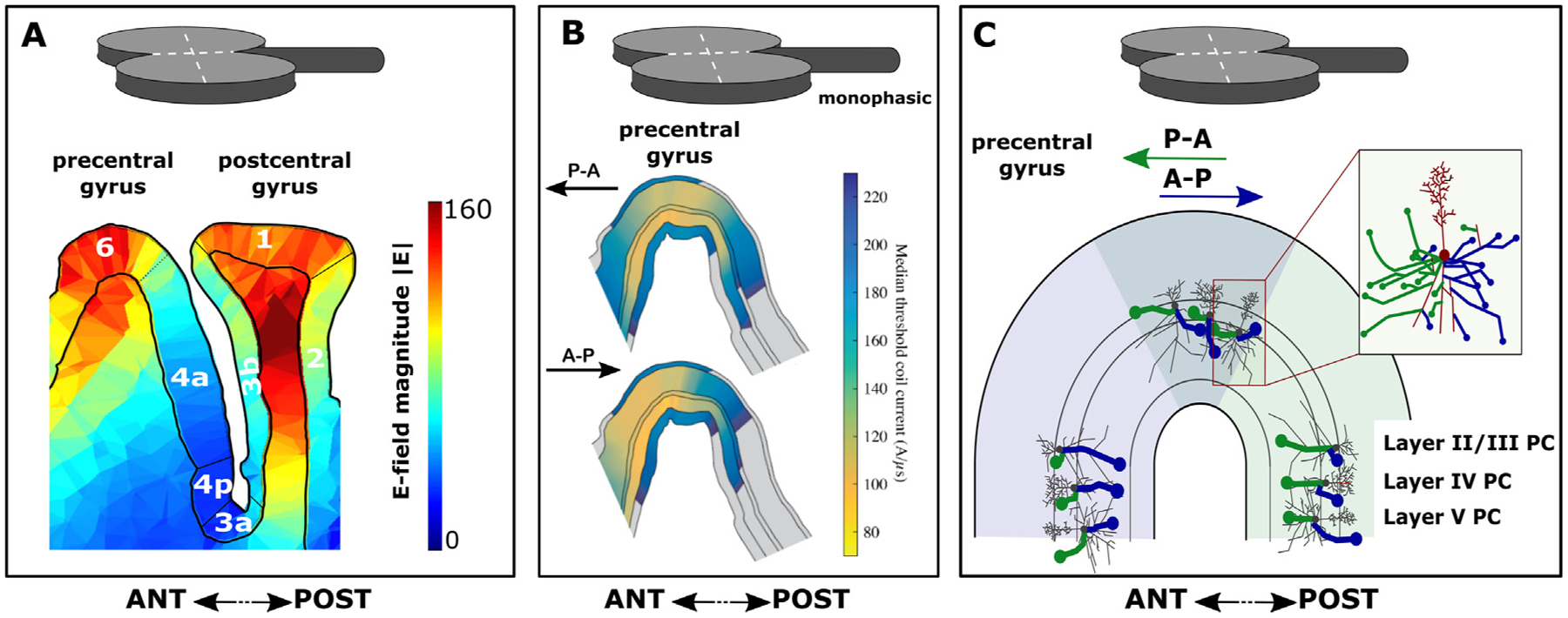
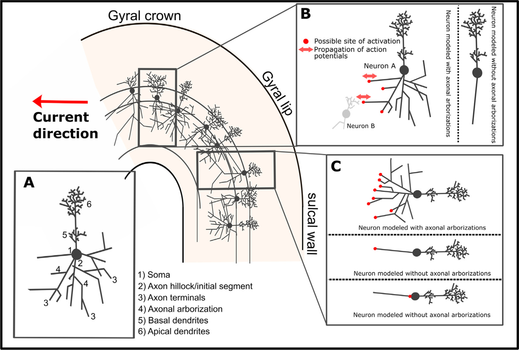
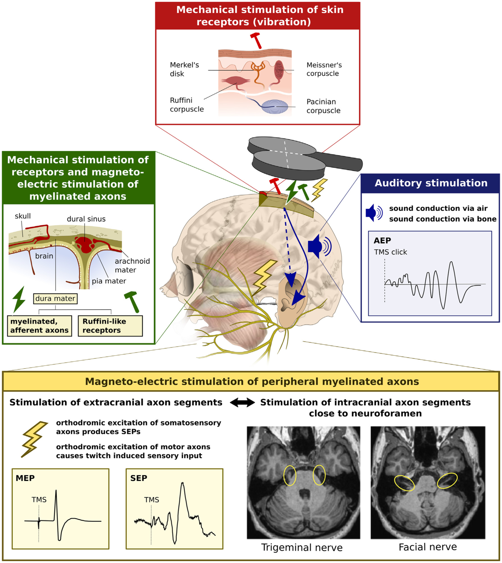
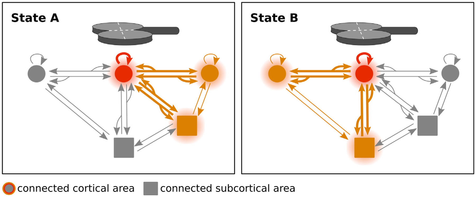
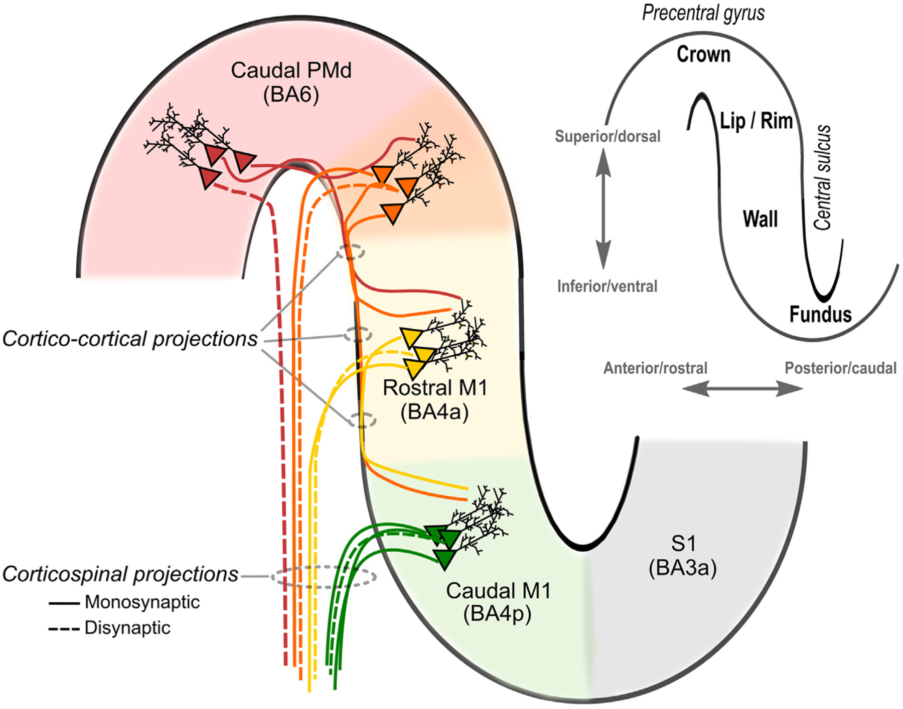
Transcranial (electro)magnetic stimulation (TMS) is currently the method of choice to non-invasively induce neural activity in the human brain. A single transcranial stimulus induces a time-varying electric field in the brain that may evoke action potentials in cortical neurons. The spatial relationship between the locally induced electric field and the stimulated neurons determines axonal depolarization. The induced electric field is influenced by the conductive properties of the tissue compartments and is strongest in the superficial parts of the targeted cortical gyri and underlying white matter. TMS likely targets axons of both excitatory and inhibitory neurons. The propensity of individual axons to fire an action potential in response to TMS depends on their geometry, myelination and spatial relation to the imposed electric field and the physiological state of the neuron. The latter is determined by its transsynaptic dendritic and somatic inputs, intrinsic membrane potential and firing rate. Modeling work suggests that the primary target of TMS is axonal terminals in the crown top and lip regions of cortical gyri. The induced electric field may additionally excite bends of myelinated axons in the juxtacortical white matter below the gyral crown. Neuronal excitation spreads ortho- and antidromically along the stimulated axons and causes secondary excitation of connected neuronal populations within local intracortical microcircuits in the target area. Axonal and transsynaptic spread of excitation also occurs along cortico-cortical and cortico-subcortical connections, impacting on neuronal activity in the targeted network. Both local and remote neural excitation depend critically on the functional state of the stimulated target area and network. TMS also causes substantial direct co-stimulation of the peripheral nervous system. Peripheral co-excitation propagates centrally in auditory and somatosensory networks, but also produces brain responses in other networks subserving multisensory integration, orienting or arousal. The complexity of the response to TMS warrants cautious interpretation of its physiological and behavioural consequences, and a deeper understanding of the mechanistic underpinnings of TMS will be critical for advancing it as a scientific and therapeutic tool.
Overview
The claims and statistics of this paper haven’t been checked yet.
Run the check — free, no account needed. It reads the open-access text and checks the sources it cites.
Integrity signals
Registry
Cites
Statistics
Figures
Verification
Claims the paper makes
The claims and statistics of this paper haven’t been checked yet.
Run the check — free, no account needed. It reads the open-access text and checks the sources it cites.
Claims other papers make about this one
Each is checked against this paper’s own text: does it say what the citing paper says it does?
The cited source says what the claim attributes to it.
In the citing paper
“studies suggest that cerebellar modulation is possible with TMS (Hardwick et al., 2014; Siebner et al., 2022)”
From A double-blind, randomized, sham-controlled clinical trial of cerebellar intermittent theta burst stimulation for bipolar disorder: Safety, tolerability, mood and cognitive effects (2025) · cites this paper as Siebner et al., 2022
Self-check
Percentages, sums and reported differences re-computed from the paper’s own numbers.
The self-check needs the paper’s full text.
Citations
Cites
NON-INVASIVE MAGNETIC STIMULATION OF HUMAN MOTOR CORTEX
Barker, A. T., Jalinous, R., & Freeston, I. L. (1985) · The Lancet · doi:10.1016/s0140-6736(85)92413-4 · Cited by 4,466
Theta Burst Stimulation of the Human Motor Cortex
Huang, Y.-Z., Edwards, M. J., Rounis, E., Bhatia, K. P., & Rothwell, J. C. (2005) · Neuron · doi:10.1016/j.neuron.2004.12.033 · Cited by 4,158
Self-citationIsolated Hand Paresis: A Case Series
Alstadhaug, K. B., & Sjulstad, A. S. (2013) · Cerebrovascular Diseases Extra · doi:10.1159/000350708 · Cited by 4,090
Corticocortical inhibition in human motor cortex.
Kujirai, T., Caramia, M. D., Rothwell, J. C., Day, B. L., Thompson, P. D., Ferbert, A., Wroe, S. J., Asselman, P., & MARSDEN, C. D. (1993) · The Journal of Physiology · doi:10.1113/jphysiol.1993.sp019912 · Cited by 3,132
Self-citationLocalization of the motor hand area to a knob on the precentral gyrus. A new landmark
Yousry, T. A. (1997) · Brain · doi:10.1093/brain/120.1.141 · Cited by 1,840
Dynamics of Sparsely Connected Networks of Excitatory and Inhibitory Spiking Neurons
Brunel, N. (2000) · Journal of Computational Neuroscience · doi:10.1023/a:1008925309027 · Cited by 1,737
Breakdown of cortical effective connectivity during sleep
Massimini, M., Ferrarelli, F., Huber, R., Esser, S. K., Singh, H., & Tononi, G. (2005) · Science · doi:10.1126/science.1117256 · Cited by 1,534
Interhemispheric inhibition of the human motor cortex.
Ferbert, A., Priori, A., Rothwell, J. C., Day, B. L., Colebatch, J. G., & MARSDEN, C. D. (1992) · The Journal of Physiology · doi:10.1113/jphysiol.1992.sp019243 · Cited by 1,490
Self-citationGABAergic cell subtypes and their synaptic connections in rat frontal cortex
Kawaguchi, Y. (1997) · Cerebral Cortex · doi:10.1093/cercor/7.6.476 · Cited by 1,416
Rizzolatti, G., & Luppino, G. (2001) · Neuron · doi:10.1016/s0896-6273(01)00423-8 · Cited by 1,356
Research works printed but not linked: 3
- Draganski, 2008, Evidence for segregated and integrative connectivity patterns in the human Basal Ganglia, J Neurosci, 28, 7143, 10.1523/JNEUROSCI.1486-08.2008 · DOI
- Edgley SA, Eyre JA, RN, L, Miller S. Direct and indirect activation of corticospinal neurons by electrical and magnetic stimulation in the anesthetized macaque monkey. J Physiol (Lond). 1992; 446, 224.
- Ziemann, 1996, Interaction between intracortical inhibition and facilitation in human motor cortex, J Physiol, 496, 873, 10.1113/jphysiol.1996.sp021734 · DOI
Cited by
Sorted by citations
Items 401–500 of 565
Histologically Informed Multiscale Modeling of the Neuronal Elements Activated by TMSPreprint
Worbs, T., Nielsen, J. D., Wang, B., Hansen, J. G., Grill, W. M., Peterchev, A. V., & Thielscher, A. (2026) · bioRxiv (Cold Spring Harbor Laboratory) · doi:10.64898/2026.06.05.730288 · Cited by 1
Self-citationEditorial: Brain stimulation: From basic research to clinical use
Benali, A., Tsutsui, K., Sekino, M., & Pfeiffer, F. (2022) · Frontiers in Human Neuroscience · doi:10.3389/fnhum.2022.1092165
Bando, H. (2022) · International Physical Medicine & Rehabilitation Journal · doi:10.15406/ipmrj.2022.07.00304
Transcranial Magnetic Stimulation in Bipolar II Disorder Treatment: A Case Report
Tran, N. N., Hutto, S., Thompson, L. R., & Hawkins, A. (2023) · Cureus · doi:10.7759/cureus.45918
Li, Q., Luo, C., Deng, Y., Tang, X., Shao, H., Saeed, A., Han, X., Huang, X., & Guo, F. (2023) · Research Square · doi:10.21203/rs.3.rs-3418988/v1
Alteration of Interhemispheric Inhibition in Patients With Lateral Epicondylalgia
Sato, Y., Takanaka, S., & Izumi, S. (2023) · Journal of Pain · doi:10.1016/j.jpain.2023.11.023
Decoding auditory working memory content from EEG aftereffects of auditory-cortical TMSPreprint
Uluç, I., Daneshzand, M., Jas, M., Kotlarz, P. L., Lankinen, K., Fiedler, J. L., Mamashli, F., Pajankar, N., Turpin, T., de Lara, L. I. N., et al. (2024) · bioRxiv (Cold Spring Harbor Laboratory) · doi:10.1101/2024.03.04.583379
Zadeh, A. K., Raghuram, H., Shrestha, S., Kibreab, M., Kathol, I., Martino, D., Pike, G. B., Pichardo, S., & Monchi, O. (2024) · SSRN Electronic Journal · doi:10.2139/ssrn.4769614
İşler, C., Taşkıran, E., Baş, G., Türk, B. G., Özkara, Ç., & Uzan, M. (2024) · Research Square · doi:10.21203/rs.3.rs-4270427/v1
Together towards more accessible, standardized TMS protocols: Reply to Sorkhabi and Leedham
Wang, Y., Nummenmaa, A. R., & Kimberley, T. J. (2024) · Brain stimulation · doi:10.1016/j.brs.2024.04.008
Neuromodulation of disrupted brain networks
Klooster, D., Leunissen, I., Tik, M., Baeken, C., & Sack, A. T. (2024) · Elsevier eBooks · doi:10.1016/b978-0-12-820480-1.00190-x
Ugbenin, B. E., & Mgbeanuli, C. C. (2024) · South Asian Journal of Social Studies and Economics · doi:10.9734/sajsse/2024/v21i8869
Li, X., Dong, B., & Wang, J. (2024) · Journal of Neurorestoratology · doi:10.1016/j.jnrt.2024.100147
Biomarkers from bench to bedside
Voetterl, H. (2024) · Research Publications (Maastricht University) · doi:10.26481/dis.20240625hv
Barua, D. R., Bhowmik, S., & Datta, S. (2024) · Advances in medical diagnosis, treatment, and care (AMDTC) book series · doi:10.4018/979-8-3693-2089-1.ch005
Gimbutaitė, G., Stočkūnaitė, P., & Petrusevičienė, L. (2024) · Reabilitacijos mokslai slauga kineziterapija ergoterapija · doi:10.33607/rmske.v2i31.1539
Memory modulation through brain stimulation
Rose, N. S., Fragetta, J., & Reinhart, R. M. G. (2024) · Elsevier eBooks · doi:10.1016/b978-0-443-15754-7.00061-4
Music Medicine for Neurological Disorders
Chauhan, N. B., Kale, M., Gujar-Kale, P., Desai, M., Jain, D. K., & Iyer, A. (2024) · Integrated science · doi:10.1007/978-3-031-73061-0_26
Corominas-Teruel, X., Bracco, M., Lohof, A. M., Sherrard, R. M., Colomina, M. T., Mahon, S., Charpier, S., & Valero‐Cabré, A. (2025) · bioRxiv (Cold Spring Harbor Laboratory) · doi:10.1101/2025.02.04.636500
Ramírez-Guerrero, J. J., Narganes-Pineda, C., Martín-Signes, M., & Chica, A. B. (2025) · NeuroImage · doi:10.1016/j.neuroimage.2025.121137
Role of transcranial magnetic stimulation in the management of trichotillomania: A systematic review
Kar, S. K., Kumari, B., Joseph, R., Chatterjee, S., Joshi, M., & Agrawal, A. (2025) · World Journal of Meta-Analysis · doi:10.13105/wjma.v13.i1.98933
Bi-modal microwave neuromodulation via thermal and nonthermal mechanismsPreprint
Marar, C., Yu, F., Chen, G., Lin, J., Yang, C., & Cheng, J. (2025) · bioRxiv (Cold Spring Harbor Laboratory) · doi:10.1101/2025.03.26.645466
Zhong, X., Wu, Y., Jiang, H., Wang, Z., & Jiles, D. C. (2025) · IEEE Transactions on Magnetics · doi:10.1109/tmag.2025.3555973
Modulating Interhemispheric Connectivity Through Chemogenetic Stimulation: A fMRI-fMRS StudyPreprint
Just, N. (2025) · Research Square · doi:10.21203/rs.3.rs-6369938/v1
MagnEspScope - DIY microscope with integrated magnetic system for magnetogenetic studiesPreprint
Минин, А. С., Uimin, M. A., Belousova, A. V., & Zubarev, I. V. (2025) · bioRxiv (Cold Spring Harbor Laboratory) · doi:10.1101/2025.05.10.653253
Zhang, X., Hancock, R., & Santaniello, S. (2025) · Journal of Neural Engineering · doi:10.1088/1741-2552/ade18b
The Application of Repetitive Transcranial Magnetic Stimulation in Post-Stroke Rehabilitation
闫, 雯. (2025) · Advances in Clinical Medicine · doi:10.12677/acm.2025.1561785
Auer, G. A., Pruewasser, L., Scharinger, C., Laczkovics, C., Zesch, E., Plener, P. L., Antal, A., Koenig, J., & Konicar, L. (2025) · Neuroscience & Biobehavioral Reviews · doi:10.1016/j.neubiorev.2025.106258
Self-citationSalemi, M. H., & Harirchian, M. H. (2025) · Neurodegenerative Disease Management · doi:10.1080/17582024.2025.2520716
Malderen, S. V., Hehl, M., Nuyts, M., Verstraelen, S., Swinnen, S. P., & Cuypers, K. (2025) · The Cerebellum · doi:10.1007/s12311-025-01871-6
Shen, X. Y., Zhao, Z., Huang, W. Y., & Zhong, J. G. (2025) · Cureus · doi:10.7759/cureus.87672
TMS for Mood Disorders: An Ethical Perspective
Poorganji, M., Boyd, E. C., Nagra, D. S., & Appelbaum, L. G. (2025) · Advances in neuroethics · doi:10.1007/978-3-031-92401-9_10
Chen, Y., Hu, Y., & Zhang, J. (2025) · Physiotherapy Theory and Practice · doi:10.1080/09593985.2025.2532571
Sela, O. W. B., Geva, S. O., Pell, G. S., Roth, Y., Friedman, J., Muhana, A., Frenkel‐Toledo, S., & Soroker, N. (2025) · doi:10.36227/techrxiv.175356536.62169355/v1
Wischnewski, M., & Zhao, Z. (2025) · Physics of Life Reviews · doi:10.1016/j.plrev.2025.07.019
Neurophysiological signatures of Stanford Neuromodulation Therapy in treatment resistant depression
Momi, D., Buchanan, D. M., Wada, M., Geoly, A. D., Johnson, N. D., Cole, E. J., Maron‐Katz, A., Tischler, C., Cline, C. C., Pizzagalli, D. A., et al. (2026) · Molecular Psychiatry · doi:10.1038/s41380-026-03837-4 · Cited by 1
Agboada, D., Rethwilm, R., Kuder, M., Mack, W., & Seiberl, W. (2025) · SSRN Electronic Journal · doi:10.2139/ssrn.5412680
Agrawal, B., Feuermann, Y., Panov, J., & Kaphzan, H. (2025) · International Journal of Molecular Sciences · doi:10.3390/ijms26178634
Technology-assisted interventions for neuropsychiatric disorders
Fliti, T., Shhaytli, A., Serhal, A., Takesh, Z., & Chokor, M. (2025) · Progress in brain research · doi:10.1016/bs.pbr.2025.08.017
Phosphenes and the effects of charged particles on the visual system
Carozzo, S., & Sannita, W. G. (2025) · Frontiers in Neurology · doi:10.3389/fneur.2025.1621659
Neuroscience Methods for Investigating Brain Plasticity
Verra, L., Renz, F. M., & Schuck, N. W. (2025) · Oxford University Press eBooks · doi:10.1093/oxfordhb/9780197677131.013.24 · Cited by 1
İşler, C., Tanrıverdi, T., Taşkıran, E., Baş, G., Türk, B. G., Özkara, Ç., & Uzan, M. (2025) · Journal of Clinical Neuroscience · doi:10.1016/j.jocn.2025.111650
Madsen, M. A. J., Christiansen, L., & Siebner, H. R. (2025) · Journal of Neurophysiology · doi:10.1152/jn.00411.2025
Self-citationLi, L., Li, H., & Fan, Q. (2025) · Sensors · doi:10.3390/s25196241
Falato, E., Ranieri, F., Musumeci, G., D’Alonzo, M., Capone, F., Pino, G. D., Pilato, F., & Lazzaro, V. D. (2025) · Clinical Neurophysiology · doi:10.1016/j.clinph.2025.2111381
Abnormal inter-hemispheric functional cooperation in blepharospasm
Liu, J.-P., Gu, Y., Shi, Y., Wang, S.-F., Song, C.-M., Liu, B.-Q., Chen, Y., & Li, H. (2025) · Frontiers in Neurology · doi:10.3389/fneur.2025.1660039
Arrington, C. N., Bledniak, E., Kinnie, B., Farkas, E. B., Morris, R. D., & Hoeft, F. (2025) · Neurobiology of Language · doi:10.1162/nol.a.217
Repetitive Transcranial Magnetic Stimulation in Dementia
Mikellides, G., Jomeh, A. A. E., Roy, E., & Kyriazis, M. (2025) · Current Aging Science · doi:10.2174/0118746098392580251003064940
Changes in corticospinal excitability in response to mediolateral gait instability
Huiberts, R. O., Bruijn, S. M., & Davies, J. L. (2025) · Experimental Physiology · doi:10.1113/ep093199
Fitzsimmons, S., Coomans, C., Breedt, L. C., Batelaan, N. M., van der Werf, Y. D., van den Heuvel, O. A., Douw, L., & Vriend, C. (2025) · medRxiv · doi:10.1101/2025.11.12.25340097
Raffin, E., Salamanca-Giron, R., Huxlin, K. R., Reynaud, O., Mattera, L., Martuzzi, R., & Hummel, F. C. (2025) · The Journal of Physiology · doi:10.1113/jp289699 · Cited by 1
A Multiscale Integrated Approach to the Study of the Nervous System in Health and Disease
Cavaliere, C., Esposito, F., Papa, M., & Cirillo, G. (2025) · Neuroimaging · doi:10.3390/neuroimaging1010002
Sugarmann, L. R., Hasbrouck, A., Hsu, J. L., Owate-Chujor, O., & Fridley, J. (2025) · Current Physical Medicine and Rehabilitation Reports · doi:10.1007/s40141-025-00519-0
How to design a NIBS study in the cognitive domain: Common pitfalls and recommendations
Hartwigsen, G., Bergmann, T. O., O’Shea, J., Silvanto, J., & Sack, A. T. (2025) · Handbook of behavioral neuroscience · doi:10.1016/b978-0-443-26602-7.00008-7
dsGrid: a dual-site TMS grid-search method for personalized targeting of motor network connectivity
Deluisi, J. A., Goldenkoff, E. R., Lee, T. G., Brissenden, J. A., Taylor, S. F., Polk, T. A., Wittenberg, G. F., & Vesia, M. (2025) · Brain stimulation · doi:10.1016/j.brs.2025.102998
A Multi-Target Transcranial Magnetic Stimulation System With Coupled Modeling Control
Hu, L., Xu, Y., Lv, H., & Zhao, C. (2025) · IEEE Transactions on Magnetics · doi:10.1109/tmag.2025.3644394
Webler, R. D., Carrasco, C., Cooper, S., Chen, M., Hunt, C., Hennessy, S., Cao, L., Lam, C., Chiu, A., Differding, C., et al. (2023) · Research Square · doi:10.21203/rs.3.rs-2850235/v1
Kolmos, M., Madsen, M. J., Liu, M. L., Karabanov, A., Johansen, K. L., Thielscher, A., Gandrup, K. L., Lundell, H., Fuglsang, S., Thade, E., et al. (2024) · Figshare · doi:10.6084/m9.figshare.26577095
Self-citationKolmos, M., Madsen, M. J., Liu, M. L., Karabanov, A., Johansen, K. L., Thielscher, A., Gandrup, K. L., Lundell, H., Fuglsang, S., Thade, E., et al. (2024) · Figshare · doi:10.6084/m9.figshare.26577092
Self-citationKolmos, M., Madsen, M. J., Liu, M. L., Karabanov, A., Johansen, K. L., Thielscher, A., Gandrup, K. L., Lundell, H., Fuglsang, S., Thade, E., et al. (2024) · Figshare · doi:10.6084/m9.figshare.26577089
Self-citationKolmos, M., Madsen, M. J., Liu, M. L., Karabanov, A., Johansen, K. L., Thielscher, A., Gandrup, K. L., Lundell, H., Fuglsang, S., Thade, E., et al. (2024) · Figshare · doi:10.6084/m9.figshare.26577098
Self-citationNavigated rTMS for the Treatment of Pain
Lefaucheur, J.-P., & Bardel, B. (2025) · Springer eBooks · doi:10.1007/978-3-031-97155-6_16
Martín-López, E., Ruiz-Ramos, A., Rosario-Lozano, M. P., & Blasco‐Amaro, J. A. (2025) · doi:10.52766/cuvk5385
Xia, Z., Hoeft, F., Arrington, C. N., & Krishnamurthy, L. C. (2025) · doi:10.17605/osf.io/2tdj4
Rasheed, L. S. A. (2025) · Applied Neuropsychology Child · doi:10.1080/21622965.2025.2608127
Arheix-Parras, S., Moore, S., & Desai, R. H. (2026) · Brain Science · doi:10.3390/brainsci16010117
Matsugi, A., Okada, Y., Mori, N., & Hosomi, K. (2026) · BMC Research Notes · doi:10.1186/s13104-026-07673-1 · Cited by 1
Snow, N. J., Mohamed, B., Duraid, S., & Ploughman, M. (2026) · Multiple Sclerosis and Related Disorders · doi:10.1016/j.msard.2026.107035
Normative Deviations Reveal Task-Evoked and Clinical Network ReorganizationPreprint
Kröll, J., Abdelmotaleb, M., Kocataş, H., Müller, V. I., Oliveros, L. K. P., Meinzer, M., Flöel, A., Eickhoff, S. B., & Patil, K. R. (2026) · bioRxiv (Cold Spring Harbor Laboratory) · doi:10.64898/2026.02.04.703503
Bruera, A., & Hartwigsen, G. (2026) · Neurobiology of Language · doi:10.1162/nol.a.244
Accelerated DMN-Targeted cTBS Improves Processing Speed Deficits in SchizophreniaPreprint
Connolly, J. G., Blyth, S. H., Yildiz, G., Rogers, B. P., Vandekar, S., Halko, M. A., Brady, R. O., & Ward, H. B. (2026) · medRxiv · doi:10.64898/2026.02.11.26346103
Temporo-Occipital and Medial Temporal Networks Underlying Object-Location LearningPreprint
Abdelmotaleb, M., Kocataş, H., Guevara, L. M. C., Niemann, F., Shahbabaie, A., Kuhnke, P., Malinowski, R., Antonenko, D., Hartwigsen, G., Meinzer, M., et al. (2026) · bioRxiv (Cold Spring Harbor Laboratory) · doi:10.64898/2026.02.13.705708
Insight into the impact of focal stimulation on large-scale network dynamics
Anaraki, A. K., Dhami, P., Chatterjee, R., Blumberger, D. M., Daskalakis, Z. J., Moreno, S., & Farzan, F. (2026) · Journal of Neural Engineering · doi:10.1088/1741-2552/ae4a4f
Sela, O. W. B., Ofir, S., Pell, G. S., Roth, Y., Friedman, J., Muhana, A., Frenkel‐Toledo, S., & Soroker, N. (2026) · PLoS ONE · doi:10.1371/journal.pone.0343725
Personalizing neuromodulation for chronic pain: A connectivity-guided randomized trialPreprint
Martino, E. D., Bach, M. M., Couto, B. A., Jakobsen, A., Martins, P. N., Ingemann-Molden, S., Casali, A. G., Graven‐Nielsen, T., & de Andrade, D. C. (2026) · medRxiv · doi:10.64898/2026.03.02.26347430
Cerebrum functional network alterations following posterior fossa surgery in Children
Zhu, R., Hu, B., Zhai, H., Hu, C., Han, X., Lu, Q., Ni, J., Guan, X., & Gong, J. (2026) · Child s Nervous System · doi:10.1007/s00381-026-07187-y
Brain-Computer Interfaces for Vision Recovery in Precortical Vision Loss
Yang, C., Guo, A., & Lin, K. (2026) · Eye and Brain · doi:10.2147/eb.s561691
Durner, G., Colbus, G., Marschal, V., Sestito, G., Kesseler, H.-G., Sannwald, L. W., Grübel, N., Wirtz, C. R., Paľa, A., & Becker, R. (2026) · Brain and Behavior · doi:10.1002/brb3.71313
Liu, S., Huang, Z., Tan, J., & Zhao, N. (2026) · Frontiers in Neurology · doi:10.3389/fneur.2026.1723901
Dămășaru, C., Roșu, G., Tuţă, L., Cernian, A. S., & Rus, M. (2026) · Bioengineering · doi:10.3390/bioengineering13040395
Wang, Y., Li, Y., Wang, H., Wang, C., & Qie, S. (2026) · Journal of Integrative Neuroscience · doi:10.31083/jin47371
Ritter, P., Freigang, S., Valentı́n, A., Zaar, K., Reishofer, G., Jehna, M., Michenthaler, M. C., Karakaya, S., Moser, P., Frank, L., et al. (2026) · Journal of Clinical Medicine · doi:10.3390/jcm15072644
Gao, H., Fukuda, S., Futamura, M., Kawasaki, T., Hamada, N., Mori, N., Hosomi, K., & Hiraoka, K. (2026) · The Cerebellum · doi:10.1007/s12311-026-01992-6
Constantin, D.-A., Cristina, D. B., Leaşu, F., Nacu, A.-G., & Rogozea, L. M. (2026) · Brain Sciences · doi:10.3390/brainsci16040392
Ethical risks and considerations of brain-controlled and neuromodulation technologies
Huang, K., Yang, H., Zhu, S., Chen, Y., Li, T., Zhao, L., Gong, A., Nan, W., Xu, J., & Fu, Y. (2026) · Cognitive Neurodynamics · doi:10.1007/s11571-026-10445-z
Bakulin, I. S., Poydasheva, A. G., Sinitsyn, D. O., Sergeeva, A. N., Zabirova, A. K., Lagoda, D. Y., Sergeev, D. V., Suponeva, N. A., & Пирадов, М. А. (2026) · Annals of Clinical and Experimental Neurology · doi:10.17816/acen.1465
Carta, D., Buono, V. L., Culicetto, L., Bertino, S., Terranova, C., Formica, C., Quartarone, A., & Marino, S. (2026) · Neurological Sciences · doi:10.1007/s10072-026-09011-6
Pople, C. B., Vasileiadi, M., Zaidi, A., Silver, D., Musa, L., Nyman, A. J., Baskaran, A., Lin, F., Lin, F., Cash, R. F. H., et al. (2026) · medRxiv · doi:10.64898/2026.04.09.26350062
Self-citationLi, Z., Gurjanov, X., Lemberg, A., Sinitsa, P., Sack, A. T., & Duecker, F. (2026) · Neuromodulation Technology at the Neural Interface · doi:10.1016/j.neurom.2026.02.008
Gazquez, J., Cadena, C. C., He, W., Yamada, E., Alteköster, C., Soyka, F., Laakso, I., Hirata, A., Joseph, W., Tarnaud, T., et al. (2026) · Physics in Medicine and Biology · doi:10.1088/1361-6560/aea302
Microwaves stimulate and inhibit neurons via modulation of TRPV and TREK channels
Marar, C., Yu, F., Chen, G., Gong, X., Lin, J., Yang, C., & Cheng, J. (2026) · Journal of Neural Engineering · doi:10.1088/1741-2552/ae62a7
Macías-García, P., Rashid-López, R., Cruz-Gómez, Á. J., Sánchez-Fernández, F. L., Sarrias-Arrabal, E., Cano-Cano, F., Lozano-Soto, E., Sanmartino, F., Espinosa‐Rosso, R., & González-Rosa, J. J. (2026) · Neurotherapeutics · doi:10.1016/j.neurot.2026.e00911
Nikolenko, V., Zharova, N., Mikliaeva, E., Pomelenok, A., Vostrikova, I., Ershova, E., Raikh, N., Zharikova, T. M., Pontes-Silva, A., & Zharikov, Y. O. (2026) · Aktuelle Rheumatologie · doi:10.1055/a-2839-9811
Metadata
- Authors
- Siebner, H. R., Funke, K., Aberra, A. S., Antal, A., Bestmann, S., Chen, R., Classen, J., Davare, M., Di Lazzaro, V., Fox, P. T., Hallett, M., Karabanov, A. N., Kesselheim, J., Beck, M. M., Koch, G., Liebetanz, D., Meunier, S., Miniussi, C., Paulus, W., Peterchev, A. V., Popa, T., Ridding, M. C., Thielscher, A., Ziemann, U., Rothwell, J. C., & Ugawa, Y.
- Affiliations
- University of Copenhagen (DK)
- Ruhr University Bochum (DE)
- Duke University (US)
- Universitätsmedizin Göttingen (DE)
- Wellcome Centre for Human Neuroimaging (GB)
- University Health Network (CA)
- Leipzig University (DE)
- King's College London (GB)
- The University of Texas at San Antonio Health Science Center (US)
- National Institutes of Health (US)
- Hvidovre Hospital (DK)
- University of Ferrara (IT)
- Centre National de la Recherche Scientifique (FR)
- University of Trento (IT)
- Fukushima Medical University (JP)
- University of South Australia (AU)
- Hertie Institute for Clinical Brain Research (DE)
- UCL Queen Square Institute of Neurology (GB)
- Corresponding author
- Siebner, Hartwig R.
- ORCID
- Siebner, Hartwig R.
- Funke, Klaus
- Aberra, Aman S.
- Antal, Andrea
- Bestmann, Sven
- Chen, Robert
- Classen, Joseph
- Davare, Marco
- Fox, Peter T.
- Hallett, Mark
- Karabanov, Anke N.
- Kesselheim, Janine
- Beck, Mikkel M.
- Koch, Giacomo
- Liebetanz, David
- Meunier, Sabine
- Miniussi, Carlo
- Paulus, Walter
- Peterchev, Angel V.
- Popa, Traian
- Ridding, Michael C.
- Thielscher, Axel
- Ziemann, Ulf
- Rothwell, John C.
- Ugawa, Yoshikazu
- Venue
- Clinical Neurophysiology
- Publisher
- Elsevier BV
- Published
- 2022-08
- Volume
- 140
- Pages
- 59-97
- ISSN
- 1388-2457 (ISSN-L)
- PMC
- PMC9753778
- OpenAlex
- W4280629063
- Cited by
- 566
- References
- 439
- Citations by year
- 2026: 138 · 2025: 179 · 2024: 138 · 2023: 90 · 2022: 20
- FWCI
- 37.47
- Type
- journal article
- Language
- en
- Open access
- hybrid
- doi.org
- Licence
- Locations
- Clinical Neurophysiology (publishedVersion, cc-by)
- PubMed (publishedVersion)
- Research at the University of Copenhagen (University of Copenhagen) (publishedVersion, cc-by)
- PubMed Central (acceptedVersion, cc-by)
- UniSA Research Outputs Repository (University of South Australia) (submittedVersion, other-oa)
- Brunel University Research Archive (BURA) (Brunel University London) (submittedVersion, cc-by)
- UCL Discovery (University College London) (submittedVersion)
- Infoscience (Ecole Polytechnique Fédérale de Lausanne) (submittedVersion, cc-by-nc-nd)
- Institutional Research Information System University of Ferrara (University of Ferrara) (submittedVersion)
- Institutional Research Information System (Università degli Studi di Trento) (submittedVersion, cc-by-nc-nd)
- Research Portal (King's College London) (submittedVersion, cc-by)
- Technical University of Denmark, DTU Orbit (Technical University of Denmark, DTU) (submittedVersion, cc-by)
- Institutional Research Information System University of Ferrara (University of Ferrara) (submittedVersion, cc-by)
- Funders
- National Science Foundation (1106401)
- Niedersächsische Ministerium für Wissenschaft und Kultur
- Deutsche Forschungsgemeinschaft (FU256/3-2, SFB 874, 122679504)
- Institut National de la Santé et de la Recherche Médicale
- Ministry of Education, Culture, Sports, Science and Technology (16H05322, 20K07866, 19H01091, 15H05881)
- Schweizerischer Nationalfonds zur Förderung der Wissenschaftlichen Forschung
- Bundesministerium für Bildung und Forschung (01EE1403B, 01GP2124B)
- Lundbeckfonden (R118‐A11308, R186-2015-2138, R244‐2017‐196, R313–2019–622, R186-2015)
- Novo Nordisk
- Fondazione Cassa Di Risparmio Di Trento E Rovereto
- Novo Nordisk Fonden (NNF14OC001)
- H. Lundbeck A/S (R244-2017-196, R186-2015-2138)
- Fondation Defitech
- National Institutes of Health (1106401, H2020)
- Horizon 2020 Framework Programme (101017716)
- Biotechnology and Biological Sciences Research Council
- National Institute of Mental Health (R01MH111865, RF1MH114268, R01MH127104)
- National Institute of Neurological Disorders and Stroke (R01NS117405, R01NS088674)
- Topics
- Life Sciences › Neuroscience › Neurology › Transcranial Magnetic Stimulation Studies
- Physical Sciences › Engineering › Biomedical Engineering › Ultrasound and Hyperthermia Applications
- Life Sciences › Neuroscience › Cellular and Molecular Neuroscience › Neuroscience and Neuropharmacology Research
- Keywords
- transcranial magnetic stimulation, induced electric field, axonal depolarization, cortical gyri, myelinated axons, multisensory integration
- MeSH terms
- Action Potentials, Brain, Humans, Neurons, Evoked Potentials, Motor, Consensus, Transcranial Magnetic Stimulation
About these checks
Registry, retraction, Cites and statistics results are computed, not judged, with one exception: a printed reference that no index lists is read once by a model before it counts as a citation error. Claims are extracted by a model and kept only when their quote appears verbatim in the text; each cited source is then checked, with a model’s help, for whether it says what the claim relies on. Those verdicts can be wrong — read the claim’s quote and the cited source before relying on one.
Statistics and arithmetic are recomputed, not judged.
Confidence bands are shown only for fields calibrated on our annotated test set. None is calibrated yet, so no band is shown.
What counts as a claim ›How we measure confidence ›How we check this ›
Are you an author, or do you see a problem with this page? Request a review
