

The DOI matches its Crossref record; 5 of the 5 references it cites were found in a registry or an index.
Rational design of monomeric IL37 variants guided by stability and dynamical analyses of IL37 dimers
Open accessPublished version of a preprint
Abstract
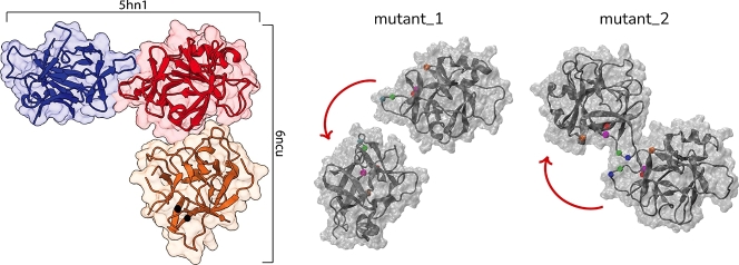
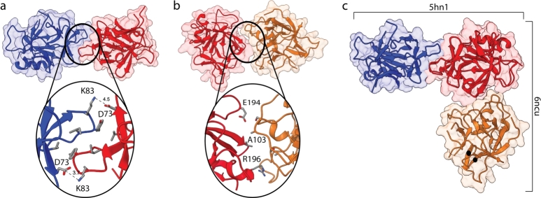
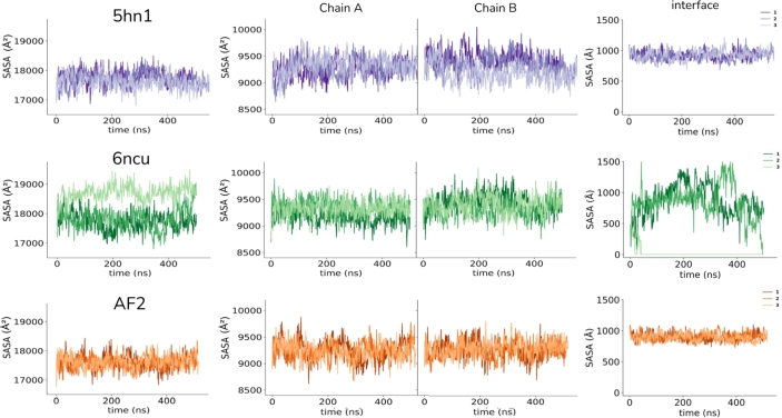
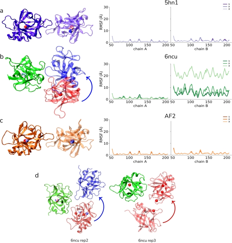
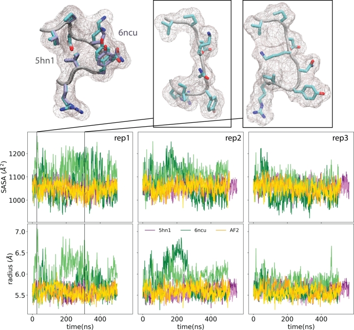
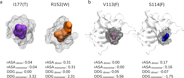
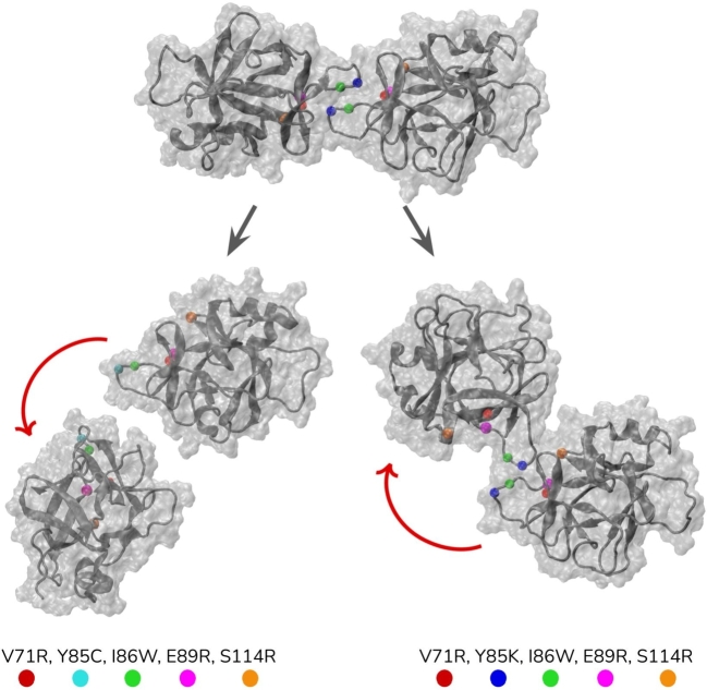
IL37 plays important roles in the regulation of innate immunity and its oligomeric status is critical to these roles. In its monomeric state, IL37 can effectively inhibit the inflammatory response of IL18 by binding to IL18R α , a capacity lost in its dimeric form, underlining the pivotal role of the oligomeric status of IL37 in its anti-inflammatory action. Until now, two IL37 dimer structures have been deposited in PDB, reflecting a substantial difference in their dimer interfaces. Given this discrepancy, we analyzed the PDB structures of the IL37 dimer (PDB IDs: 6ncu , 5hn1 ) along with a AF2-multimer prediction by molecular dynamics (MD) simulations. Results showed that the 5hn1 and AF2-predicted dimers have the same interface and stably maintained their conformations throughout simulations, while the recent IL37 dimer (PDB ID: 6ncu ) with a different interface did not, proposing a possible issue with the recent IL37 dimer structure ( 6ncu ). Next, focusing on the stable dimer structures, we have identified five critical positions of V71/Y85/I86/E89/S114, three new positions compared to the literature, that would reduce dimer stability without affecting the monomer structure. Two quintuple mutants were tested by MD simulations and showed partial or complete dissociation of the dimer. Overall, the insights gained from this study reinforce the validity of the 5hn1 and AF2 multimer structures, while also advancing our understanding of the IL37 dimer interface through the generation of monomer-locked IL37 variants.
Overview
The claims and statistics of this paper haven’t been checked yet.
Run the check — free, no account needed. It reads the open-access text and checks the sources it cites.
Integrity signals
Registry
Cites
Statistics
Figures
Verification
Claims the paper makes
The claims and statistics of this paper haven’t been checked yet.
Run the check — free, no account needed. It reads the open-access text and checks the sources it cites.
Self-check
Percentages, sums and reported differences re-computed from the paper’s own numbers.
The self-check needs the paper’s full text.
Citations
Cites
Highly accurate protein structure prediction with AlphaFold
Jumper, J., Evans, R., Pritzel, A., Green, T., Figurnov, M., Ronneberger, O., Tunyasuvunakool, K., Bates, R., Žídek, A., Potapenko, A., et al. (2021) · Nature · doi:10.1038/s41586-021-03819-2 · Cited by 43,764
Accurate prediction of protein structures and interactions using a 3-track network
Baek, M., DiMaio, F., Anishchenko, I., Dauparas, J., Ovchinnikov, S., Lee, G. R., Wang, J., Cong, Q., Kinch, L. N., Schaeffer, R. D., et al. (2021) · doi:10.1101/2021.06.14.448402 · Cited by 5,549
A genomic mutational constraint map using variation in 76,156 human genomes
Chen, S., Francioli, L. C., Goodrich, J. K., Collins, R. L., Kanai, M., Wang, Q., Alföldi, J., Watts, N. A., Vittal, C., Gauthier, L. D., et al. (2024) · Nature · doi:10.1038/s41586-023-06045-0 · Cited by 1,727
Mariani, V., Biasini, M., Barbato, A., & Schwede, T. (2013) · Bioinformatics · doi:10.1093/bioinformatics/btt473 · Cited by 1,138
Benchmarking AlphaFold for protein complex modeling reveals accuracy determinants
Yin, R., Feng, B. Y., Varshney, A., & Pierce, B. G. (2021) · doi:10.1101/2021.10.23.465575 · Cited by 335
Research works printed but not linked: 66
- 10.1111/imr.12621 · DOI · Paper page
- van de VeerdonkFrank L.StoeckmanAngela K.WuGoupingBoeckermannAaron N.AzamTaniaNeteaMihai G.Il-38 binds to the il-36 receptor and has biological effects on immune cells similar to il-36 receptor antagonistProc Natl Acad Sci109820123001300522315422 · DOI
- RudloffInaGodsellJackNold-PetryClaudia A.HarrisJamesHoiAlbertaMorandEric F.Brief report: interleukin-38 exerts antiinflammatory functions and is associated with disease activity in systemic lupus erythematosusArthritis Rheumatol671220153219322526314375 · DOI
- DinarelloCharlesArendWilliamSimsJohnSmithDirkBlumbergHalO'NeillLukeIl-1 family nomenclatureNat Immunol1111201097320959797 · DOI
- 10.1073/pnas.1819672116 · DOI
- Ellisdon Andrew M. Nold-Petry Claudia A. D'Andrea Laura Cho Steven X. Lao Jason C. Rudloff Ina Homodimerization attenuates the anti-inflammatory activity of interleukin-37 Sci Immunol 2 8 2017 eaaj1548
- Zeng Huiqiong Zhou Kaixia Ye Zhizhong Biology of interleukin-37 and its role in autoimmune diseases Exp Ther Med 24 2 2022 1 10
- 10.1016/j.chembiol.2021.10.004 · DOI
- Sánchez-FernándezAlbaZandeeStephanieAmo-AparicioJesúsCharabatiMarcPratAlexandreGarlandaCeciliaIl-37 exerts therapeutic effects in experimental autoimmune encephalomyelitis through the receptor complex il-1r5/il-1r8Theranostics1112021133391457 · DOI
- 10.1007/s13238-017-0408-4 · DOI
- 10.1021/acs.jcim.2c00054 · DOI
- 10.1093/bioinformatics/btn268 · DOI
- 10.1126/science.2471267 · DOI
- 10.1534/genetics.117.300064 · DOI
- 10.1016/j.csbj.2023.06.021 · DOI
- ŠaliAndrejBlundellTom L.Comparative protein modelling by satisfaction of spatial restraintsJ Mol Biol234319937798158254673 · DOI · Paper page
- 10.1110/ps.062416606 · DOI
- 10.1126/science.abj8754 · DOI · Paper page
- Evans Richard O'Neill Michael Pritzel Alexander Antropova Natasha Senior Andrew Green Tim Protein complex prediction with alphafold-multimer bioRxiv 2021 2021-10
- 10.1038/s41592-022-01488-1 · DOI · Paper page
- MirditaMilotSteineggerMartinSödingJohannesMmseqs2 desktop and local web server app for fast, interactive sequence searchesBioinformatics351620192856285830615063 · DOI · Paper page
- 10.1038/nbt.3988 · DOI · Paper page
- Yang Zhenyu Zeng Xiaoxi Zhao Yi Chen Runsheng Alphafold2 and its applications in the fields of biology and medicine Signal Transduct Targeted Ther 8 1 2023 115 · DOI
- 10.1002/jcc.20945 · DOI · Paper page
- 10.1002/jcc.21287 · DOI
- Lee Jumin Cheng Xi Swails Jason M. Yeom Min Sun Eastman Peter K. Lemkul Justin A. Charmm-gui input generator for namd, gromacs, amber, openmm, and charmm/openmm simulations using the charmm36 additive force field Biophys J 110 3 2016 641a
- DolinskyTodd J.NielsenJens E.McCammonJ. AndrewBakerNathan A.Pdb2pqr: an automated pipeline for the setup of Poisson–Boltzmann electrostatics calculationsNucleic Acids Res32suppl_22004W665W66715215472 · DOI
- 10.1002/pro.3280 · DOI
- 10.1002/jcc.20289 · DOI · Paper page
- MacKerell A.D. Jr. Bashford D. Bellott M. Dunbrack R.L. Jr. Evanseck J.D. Field M.J. All-atom empirical potential for molecular modeling and dynamics studies of proteins J Phys Chem B 102 18 1998 3568 3616 · DOI
- HuangJingMacKerellAlexander D.Jr.Charmm36 all-atom additive protein force field: validation based on comparison to nmr dataJ Comput Chem342520132135214523832629 · DOI
- 10.1063/1.445869 · DOI
- 10.1063/1.464397 · DOI
- Martyna Glenn J. Tobias Douglas J. Klein Michael L. Constant pressure molecular dynamics algorithms J Chem Phys 101 5 1994 4177 4189 · DOI
- Feller Scott E. Zhang Yuhong Pastor Richard W. Brooks Bernard R. Constant pressure molecular dynamics simulation: the Langevin piston method J Chem Phys 103 11 1995 4613 4621 · DOI
- Pastor Richard W. Brooks Bernard R. Szabo Attila An analysis of the accuracy of Langevin and molecular dynamics algorithms Mol Phys 65 6 1988 1409 1419 · DOI
- Gowers Richard J. Linke Max Barnoud Jonathan Reddy Tyler JE Melo Manuel N. Seyler Sean L. Mdanalysis: a python package for the rapid analysis of molecular dynamics simulations Proceedings of the 15th python in science conference vol. 98 2016 SciPy Austin TX 105 · DOI
- Michaud-AgrawalNaveenDenningElizabeth J.WoolfThomas B.BecksteinOliverMdanalysis: a toolkit for the analysis of molecular dynamics simulationsJ Comput Chem321020112319232721500218 · DOI
- HumphreyWilliamDalkeAndrewSchultenKlausVmd: visual molecular dynamicsJ Mol Graph141199633388744570 · DOI · Paper page
- 10.1002/pro.3943 · DOI
- 10.1002/pro.3235 · DOI · Paper page
- Meng Elaine C. Goddard Thomas D. Pettersen Eric F. Couch Greg S. Pearson Zach J. Morris John H. Ucsf chimerax: tools for structure building and analysis Protein Sci 2023 e4792
- GrantBarry J.SkjaervenLarsYaoXin-QiuThe bio3d packages for structural bioinformaticsProtein Sci3012021203032734663 · DOI
- SchymkowitzJoostBorgJesperStricherFrancoisNysRobbyRousseauFredericSerranoLuisThe foldx web server: an online force fieldNucleic Acids Res33suppl_22005W382W38815980494 · DOI · Paper page
- BillR.MillerI.I.I.Dwight McGeeT.Jr.SwailsJason M.HomeyerNadineGohlkeHolgerMmpbsa. py: an efficient program for end-state free energy calculationsJ Chem Theory Comput8920123314332126605738 · DOI
- CaseDavid A.AktulgaHasan MetinBelfonKellonCeruttiDavid S.Andrés CisnerosG.WilianViníciusAmbertoolsJ Chem Inf Model632020236183619137805934 · DOI
- 10.1016/j.csbj.2021.09.003 · DOI
- OliveiraNuno F.B.RodriguesFilipe E.P.VitorinoJoão N.M.FaíscaPatrícia F.N.MachuqueiroMiguelInterfacial dynamics and growth modes of β2-microglobulin dimersJ Chem Inf Model631420234447445737132512 · DOI
- Zhang Zinan Z. Zhang Yu He Tingyan Sweeney Colin L. Baris Safa Karakoc-Aydiner Elif Homozygous il37 mutation associated with infantile inflammatory bowel disease Proc Natl Acad Sci 118 10 2021 e2009217118
- Chen Yuting Lu Haoyu Zhang Ning Zhu Zefeng Wang Shuqin Li Minghui Premps: predicting the effects of single mutations on protein stability bioRxiv 2020 2020-04
- Dang Dung Thanh Molecular approaches to protein dimerization: opportunities for supramolecular chemistry Front Chem 10 2022 829312
- 10.1016/j.tibs.2004.09.006 · DOI
- PlaxcoKevin W.GrossMichaelProtein complexes: the evolution of symmetryCurr Biol1912009R25R2619138586 · DOI
- KrissinelEvgenyHenrickKimInference of macromolecular assemblies from crystalline stateJ Mol Biol3723200777479717681537 · DOI
- 10.1073/pnas.1810054115 · DOI
- 10.1111/febs.15717 · DOI
- DahanayakeJayangika N.GautamDevaki N.VermaRajniMitchell-KochKatie R.To keep or not to keep? The question of crystallographic waters for enzyme simulations in organic solventMol Simul421220161001101327403032 · DOI
- 10.1111/j.1742-4658.2007.06178.x · DOI
- 10.1110/ps.073360508 · DOI
- XuQifangDunbrackRoland L.The protein common interface database (protcid)—a comprehensive database of interactions of homologous proteins in multiple crystal formsNucleic Acids Res39suppl_12010D761D77021036862
- BaskaranKumaranDuarteJose M.BiyaniNikhilBlivenSpencerCapitaniGuidoA pdb-wide, evolution-based assessment of protein-protein interfacesBMC Struct Biol141201411124410837
- 10.1107/S2052252514005442 · DOI
- LevyEmmanuel D.Piqsi: protein quaternary structure investigationStructure151120071364136717997962 · DOI
- LiWenyuSrinivasulaSrinivasa M.ChaiJijieLiPingweiWuJia-WeiZhangZhiJiaStructural insights into the pro-apoptotic function of mitochondrial serine protease htra2/omiNat Struct Biol96200243644111967569 · DOI
- Timucin Emel Ugur Sezerman O. The conserved lid tryptophan, w211, potentiates thermostability and thermoactivity in bacterial thermoalkalophilic lipases PLoS ONE 8 12 2013 e85186
- GenhedenSamuelRydeUlfThe mm/pbsa and mm/gbsa methods to estimate ligand-binding affinitiesExpert Opin Drug Discov105201544946125835573 · DOI
Metadata
- Authors
- Sardag, I., Duvenci, Z. S., Belkaya, S., & Timucin, E.
- Affiliations
- Boğaziçi University (TR)
- Acibadem Mehmet Ali Aydinlar University, Institute of Health Sciences, Department of Biostatistics and Bioinformatics, Istanbul 34752, Turkey
- Bilkent University (TR)
- Corresponding author
- Belkaya, Serkan
- Venue
- Computational and Structural Biotechnology Journal
- Published
- 2024-04-22
- Volume
- 23
- Pages
- 1854-1863
- ISSN
- 2001-0370 (ISSN-L)
- PMC
- PMC11177541
- OpenAlex
- W4394996705
- Cited by
- 4
- References
- 5
- Citations by year
- 2026: 1 · 2025: 3
- FWCI
- 0.54
- Type
- journal article
- Language
- en
- Open access
- gold
- doi.org
- Licence
- Locations
- Computational and Structural Biotechnology Journal (publishedVersion, cc-by)
- bioRxiv (Cold Spring Harbor Laboratory) (acceptedVersion, cc-by)
- PubMed Central (publishedVersion, cc-by)
- PubMed (publishedVersion)
- DOAJ (DOAJ: Directory of Open Access Journals) (submittedVersion, cc-by-sa)
- Bilkent University Institutional Repository (Bilkent University) (submittedVersion, cc-by)
- Funders
- Türkiye Bilimsel ve Teknolojik Araştırma Kurumu
- EMBO (IG 4727)
- Topics
- Life Sciences › Biochemistry, Genetics and Molecular Biology › Molecular Biology › Protein Structure and Dynamics
- Life Sciences › Immunology and Microbiology › Immunology › Cytokine Signaling Pathways and Interactions
- Life Sciences › Biochemistry, Genetics and Molecular Biology › Molecular Biology › Receptor Mechanisms and Signaling
- Keywords
- IL-37, dimer stability, dimer interface, oligomeric state, molecular dynamics simulation, dimer dissociation, critical residues
About these checks
Registry, retraction, Cites and statistics results are computed, not judged, with one exception: a printed reference that no index lists is read once by a model before it counts as a citation error. Claims are extracted by a model and kept only when their quote appears verbatim in the text; each cited source is then checked, with a model’s help, for whether it says what the claim relies on. Those verdicts can be wrong — read the claim’s quote and the cited source before relying on one.
Statistics and arithmetic are recomputed, not judged.
Confidence bands are shown only for fields calibrated on our annotated test set. None is calibrated yet, so no band is shown.
What counts as a claim ›How we measure confidence ›How we check this ›
Are you an author, or do you see a problem with this page? Request a review
How to cite
Cite CoChat for the checks on this page, and the paper for its own findings.
- This page
- CoChat. (2026). Citation check: Rational design of monomeric IL37 variants guided by stability and dynamical analyses of IL37 dimers. https://app.cochat.ai/papers/10.1016/j.csbj.2024.04.037
- The paper
- Sardag, I., Duvenci, Z. S., Belkaya, S., & Timucin, E. (2024). Rational design of monomeric IL37 variants guided by stability and dynamical analyses of IL37 dimers. Computational and Structural Biotechnology Journal. https://doi.org/10.1016/j.csbj.2024.04.037
