

The DOI matches its Crossref record; 2 of the 2 references it cites were found in a registry or an index.
Advances in drug delivery systems, challenges and future directions
Overview
The claims and statistics of this paper haven’t been checked yet.
Run the check — free, no account needed. It reads the open-access text and checks the sources it cites.
Integrity signals
Registry
Cites
Statistics
Figures
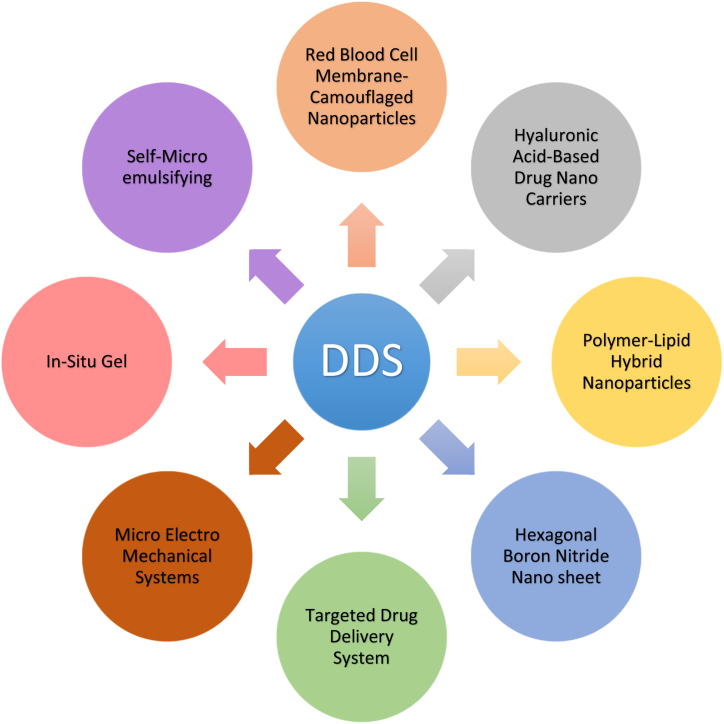
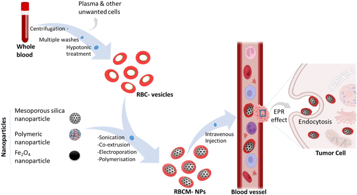
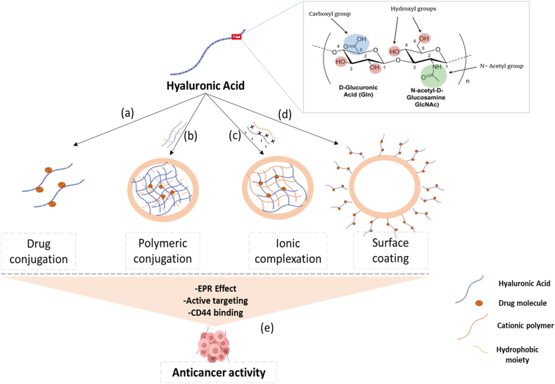
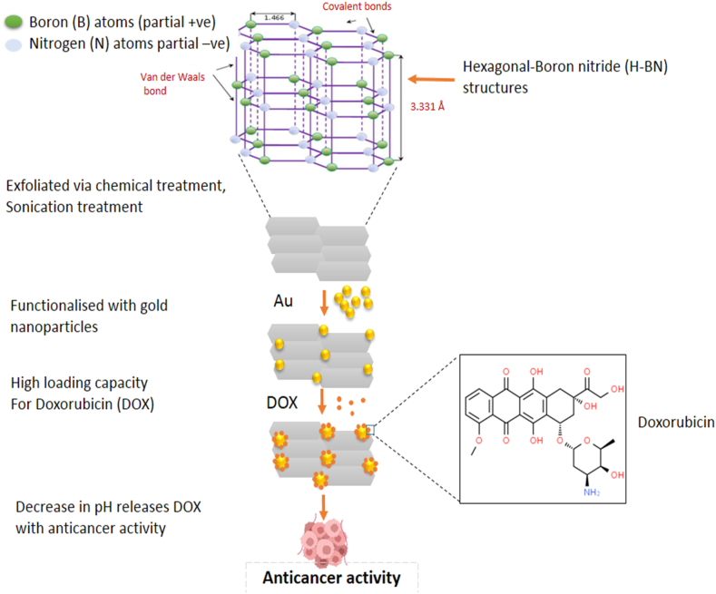
Verification
Claims the paper makes
The claims and statistics of this paper haven’t been checked yet.
Run the check — free, no account needed. It reads the open-access text and checks the sources it cites.
Claims other papers make about this one
Each is checked against this paper’s own text: does it say what the citing paper says it does?
In the citing paper
“DDS have been specifically established to tackle these issues, as it utilizes these issues as means of targeting tumor sites, which provides efficiency, safety, and site-specific drug delivery [28]. It brings about high local drug concentrations, extended drug retention on tumors, and minimal off-target side effects.”
From Advances in green-synthesized magnetic nanoparticles for targeted cancer therapy: mechanisms, applications, and future perspectives (2026) · cites this paper as [26]
Self-check
Percentages, sums and reported differences re-computed from the paper’s own numbers.
The self-check needs the paper’s full text.
Citations
Cites
The evolution of commercial drug delivery technologies
Vargason, A. M., Anselmo, A. C., & Mitragotri, S. (2021) · Nature Biomedical Engineering · doi:10.1038/s41551-021-00698-w · Cited by 1,361 · cited 2×
“Furthermore, it improves drug solubility, target site accumulation, efficacy, pharmacological activity, pharmacokinetic properties, patient acceptance, and compliance, and reduces drug toxicity [2].”
BackgroundCombination therapy based on nano codelivery for overcoming cancer drug resistance
Wang, H., & Huang, Y. (2020) · Medicine in Drug Discovery · doi:10.1016/j.medidd.2020.100024 · Cited by 115
Research works printed but not linked: 123
- Rayaprolu, 2018, Excipients in parenteral formulations: selection considerations and effective utilization with small molecules and biologics, Drug Dev. Ind. Pharm., 44, 1565, 10.1080/03639045.2018.1483392 · DOI
- Alqahtani, 2021, Advances in oral drug delivery, Front. Pharmacol., 12, 10.3389/fphar.2021.618411 · DOI
- Chauhan, 2022, Recent Advances in Ophthalmic Drug Delivery, 1
- Thirunavukkarasu, 2022, Transdermal drug delivery systems for the effective management of type 2 diabetes mellitus: a review, Diabetes Res. Clin. Pract., 10.1016/j.diabres.2022.109996 · DOI
- Sharma, 2022, Subcutaneous drug delivery: a review of the state-of-the-art modelling and experimental techniques, J. Biomech. Eng.
- Misbah Ul Haq, 2021, Rectal drug delivery system: an overview, Clin. Pharmacol. Biopharm., 10
- Mahant, 2022, Emerging trends and potential prospects in vaginal drug delivery, Curr. Drug Deliv.
- Cho, 2018, Biochemical and Biophysical Research Communications the immunotherapeutic effects of recombinant Bacillus rin resistant to antimicrobial peptides on Calmette-Gu e bladder cancer cells, Biochem. Biophys. Res. Commun.
- Palugan, 2021, International Journal of Pharmaceutics : X Intravesical drug delivery approaches for improved therapy of urinary bladder diseases, Int. J. Pharm. X., 3
- Keraliya, 2012, Osmotic drug delivery system as a part of modified release dosage form, ISRN Pharm
- Mattos, 2017, Biogenic silica nanoparticles loaded with neem bark extract as green, slow-release biocide, 142, 4206
- Ding, 2017, A review of drug release mechanisms from nanocarrier systems, Mater. Sci. Eng. C., 76, 1440, 10.1016/j.msec.2017.03.130 · DOI
- Danhier, 2015, To exploit the tumor microenvironment : passive and active tumor targeting of nanocarriers for anti-cancer drug delivery, J. Contr. Release, 148, 135, 10.1016/j.jconrel.2010.08.027 · DOI
- Patra, 2018, Nano based drug delivery systems : recent developments and future prospects, J. Nanobiotechnol., 1
- Malm, 1951, Cellulose acetate phthalate as an enteric coating material, J. Am. Pharm. Assoc. (Scientific Ed.)., 40, 520, 10.1002/jps.3030401014 · DOI
- Yun, 2014, Controlled drug delivery systems: the next 30 years, Front. Chem. Sci. Eng., 8, 276, 10.1007/s11705-014-1426-x · DOI
- Zhang, 2017, Big data analysis of global advances in pharmaceutics and drug delivery 1980-2014, Drug Discov. Today, 22, 1201, 10.1016/j.drudis.2017.05.012 · DOI
- Bangham, 1964, Negative staining of phospholipids and their structural modification by surface-active agents as observed in the electron microscope, J. Mol. Biol., 8, 10.1016/S0022-2836(64)80115-7 · DOI
- Bangham, 1965, Diffusion of univalent ions across the lamellae of swollen phospholipids, J. Mol. Biol., 13, 10.1016/S0022-2836(65)80093-6 · DOI · Paper page
- Birrenbach, 1976, Polymerized micelles and their use as adjuvants in immunology, J. Pharm. Sci., 65, 1763, 10.1002/jps.2600651217 · DOI
- Couvreur, 1977, Nanocapsules: a new type of lysosomotropic carrier, FEBS Lett., 84, 323, 10.1016/0014-5793(77)80717-5 · DOI
- Park, 2016, Drug delivery of the future: chasing the invisible gorilla, J. Contr. Release, 240, 2, 10.1016/j.jconrel.2015.10.048 · DOI
- Ye, 2010, Issues in long-term protein delivery using biodegradable microparticles, J. Control. Release Off. J. Control. Release Soc., 146, 241, 10.1016/j.jconrel.2010.05.011 · DOI · Paper page
- Park, 2014, Controlled drug delivery systems: past forward and future back, J. Contr. Release, 190, 3, 10.1016/j.jconrel.2014.03.054 · DOI
- Yun, 2015, Controlled drug delivery: historical perspective for the next generation, J. Contr. Release, 219, 2, 10.1016/j.jconrel.2015.10.005 · DOI
- Rahman, 2020, Novel drug delivery systems for loading of natural plant extracts and their biomedical applications, Int. J. Nanomed., 15, 2439, 10.2147/IJN.S227805 · DOI
- Vincy, 2022, Recent progress in red blood cells-derived particles as novel bioinspired drug delivery systems: challenges and strategies for clinical translation, Front. Chem., 10, 10.3389/fchem.2022.905256 · DOI
- Patil, 2019, A review on topical gels as drug delivery system, J. Drug Deliv. Therapeut., 9
- Hanley, 2021, Phototheranostics using erythrocyte-based particles, Biomolecules, 11, 10.3390/biom11050729 · DOI
- Wang C, 2014, Multifunctional theranostic red blood cells for magnetic-field-enhanced in vivo combination therapy of cancer, Adv. Mater., 26, 4794, 10.1002/adma.201400158 · DOI
- Jiawei Chen, 2020, Nanoscale delivery system for nutraceuticals: preparation, application, characterization, safety, and future trends, Food Eng. Rev., 12, 14, 10.1007/s12393-019-09208-w · DOI
- Zinger, 2021, Biomimetic nano drug delivery carriers for treating cardiovascular diseases, Nanomed. Nanotechnol. Biol. Med., 33, 10.1016/j.nano.2021.102360 · DOI
- Gao, 2020, Neuron tau-targeting biomimetic nanoparticles for curcumin delivery to delay progression of Alzheimer's disease, J. Nanobiotechnol., 18, 71, 10.1186/s12951-020-00626-1 · DOI
- Mamo, 2010, Emerging nanotechnology approaches for HIV/AIDS treatment and prevention, Nanomedicine, 5, 269, 10.2217/nnm.10.1 · DOI
- Moss, 2013, HIV/AIDS review, Radiol. Technol., 84, 247
- Alaniz, 2002, Interaction of CD44 with different forms of hyaluronic acid. Its role in adhesion and migration of tumor cells, Cell Commun. Adhes., 9, 117, 10.1080/15419060214522 · DOI
- Kang, 2015, Nanocarrier-mediated co-delivery of chemotherapeutic drugs and gene agents for cancer treatment, Acta Pharm. Sin. B., 5, 169, 10.1016/j.apsb.2015.03.001 · DOI
- Rizvi, 2018, Applications of nanoparticle systems in drug delivery technology, Saudi Pharmaceut. J., 26, 64, 10.1016/j.jsps.2017.10.012 · DOI
- Yu, 2020, Development of a hyaluronic acid-based nanocarrier incorporating doxorubicin and cisplatin as a pH-sensitive and CD44-targeted anti-breast cancer drug delivery system, Front. Pharmacol., 11, 10.3389/fphar.2020.532457 · DOI · Paper page
- Jia, 2022, Hyaluronic acid-based nano drug delivery systems for breast cancer treatment: recent advances, Front. Bioeng. Biotechnol., 10, 10.3389/fbioe.2022.990145 · DOI
- Huang, 2018, Application of hyaluronic acid as carriers in drug delivery, Drug Deliv., 25, 766, 10.1080/10717544.2018.1450910 · DOI
- Sharker, 2019, Hexagonal boron nitrides (white graphene): a promising method for cancer drug delivery, Int. J. Nanomed., 14, 9983, 10.2147/IJN.S205095 · DOI
- Weng, 2016, Functionalized hexagonal boron nitride nanomaterials: emerging properties and applications, Chem. Soc. Rev., 45, 3989, 10.1039/C5CS00869G · DOI
- Liu, 2019, Functionalized h-BN nanosheets as a theranostic platform for SERS real-time monitoring of MicroRNA and photodynamic therapy, Angew. Chem. Int. Ed. Engl., 58, 7757, 10.1002/anie.201902776 · DOI
- Jose, 2018, Polymeric lipid hybrid nanoparticles: properties and therapeutic applications, Crit. Rev. Ther. Drug Carrier Syst., 35, 555, 10.1615/CritRevTherDrugCarrierSyst.2018024751 · DOI
- Pérez-Herrero, 2015, Advanced targeted therapies in cancer: drug nanocarriers, the future of chemotherapy, Eur. J. Pharm. Biopharm. Off. J. Arbeitsgemeinschaft Fur Pharm. Verfahrenstechnik e.V., 93, 52
- Choudhury, 2019, Strategizing biodegradable polymeric nanoparticles to cross the biological barriers for cancer targeting, Int. J. Pharm., 565, 509, 10.1016/j.ijpharm.2019.05.042 · DOI
- Rizwanullah, 2020, Polymer-lipid hybrid nanoparticles: a next-generation nanocarrier for targeted treatment of solid tumors, Curr. Pharmaceut. Des., 26, 1206, 10.2174/1381612826666200116150426 · DOI
- Mohanty, 2020, Utilization of polymer-lipid hybrid nanoparticles for targeted anti-cancer therapy, Molecules, 25, 10.3390/molecules25194377 · DOI · Paper page
- Pouton, 2006, Formulation of poorly water-soluble drugs for oral administration: physicochemical and physiological issues and the lipid formulation classification system, Eur. J. Pharm. Sci. Off. J. Eur. Fed. Pharm. Sci., 29, 278
- Savjani, 2012, Drug solubility: importance and enhancement techniques, ISRN Pharm
- Vinarov, 2021, Impact of gastrointestinal tract variability on oral drug absorption and pharmacokinetics: an UNGAP review, Eur. J. Pharm. Sci. Off. J. Eur. Fed. Pharm. Sci., 162
- Tang, 2008, Development of solid self-emulsifying drug delivery systems: preparation techniques and dosage forms, Drug Discov. Today, 13, 606, 10.1016/j.drudis.2008.04.006 · DOI
- Rajpoot, 2019, Self-microemulsifying drug-delivery system: ongoing challenges and future ahead, in, 393
- Gursoy, 2004, Self-emulsifying drug delivery systems (SEDDS) for improved oral delivery of lipophilic drugs, Biomed. Pharmacother., 58, 173, 10.1016/j.biopha.2004.02.001 · DOI
- Mehanna, 2021, Formulation and applications of lipid-based nanovehicles: spotlight on self-emulsifying systems, Adv. Pharmaceut. Bull., 11, 56, 10.34172/apb.2021.006 · DOI
- Baloch, 2019, Self-nanoemulsifying drug delivery system (SNEDDS) for improved oral bioavailability of chlorpromazine: in vitro and in vivo evaluation, Medicina (Kaunas), 55
- Dokania, 2015, Self-microemulsifying drug delivery system (SMEDDS)--challenges and road ahead, Drug Deliv., 22, 675, 10.3109/10717544.2014.896058 · DOI
- Lokhande, 2018, An update review on recent advancements in multiple emulsion, Int. J. Res. Sci. Innov., 5, 90
- Parmar, 2011, Study of cosurfactant effect on nanoemulsifying area and development of lercanidipine loaded (SNEDDS) self nanoemulsifying drug delivery system, Colloids Surf. B Biointerfaces, 86, 327, 10.1016/j.colsurfb.2011.04.016 · DOI
- Rahman, 2013, Role of excipients in successful development of self-emulsifying/microemulsifying drug delivery system (SEDDS/SMEDDS), Drug Dev. Ind. Pharm., 39, 1, 10.3109/03639045.2012.660949 · DOI
- Azmi, 2019, Nanoemulsions: factory for food, pharmaceutical and cosmetics, Processes, 7, 10.3390/pr7090617 · DOI
- Rao, 2012, Food-grade microemulsions and nanoemulsions: role of oil phase composition on formation and stability, Food Hydrocolloids, 29, 326, 10.1016/j.foodhyd.2012.04.008 · DOI
- Glassman, 2019, Pharmacokinetic and pharmacodynamic properties of drug delivery systems, J. Pharmacol. Exp. Therapeut., 370, 570, 10.1124/jpet.119.257113 · DOI
- Tiwari, 2012, Drug delivery systems: an updated review, Int. J. Pharm. Investig., 2, 2, 10.4103/2230-973X.96920 · DOI · Paper page
- Vigani, 2020, Recent advances in the development of in situ gelling drug delivery systems for non-parenteral administration routes, Pharmaceutics, 12, 10.3390/pharmaceutics12090859 · DOI
- Hua, 2019, Physiological and pharmaceutical considerations for rectal drug formulations, Front. Pharmacol., 10, 1196, 10.3389/fphar.2019.01196 · DOI
- Singh, 2017, Stimuli-responsive systems with diverse drug delivery and biomedical applications: recent updates and mechanistic pathways, Crit. Rev. Ther. Drug Carrier Syst., 34, 209, 10.1615/CritRevTherDrugCarrierSyst.2017017284 · DOI
- Khule, 2021, A Review: in-situ Gel Drug Delivery System, 9, 2455
- Bashir, 2021, An in sight into novel drug delivery system: in situ gels, TANG [HUMANITAS Med
- Bashir, 2019, Floating oral in-situ gel: a review, J. Drug Deliv. Therapeut., 9, 442, 10.22270/jddt.v9i2.2372 · DOI
- Padhan, 2019, Floating oral in-situ gel, a comprehensive approach of gastro-retentive drug delivery system: a review, Int. J. Pharm. Sci. Res., 10, 4026
- Sheshala, 2015, In situ gelling ophthalmic drug delivery system: an overview and its applications., recent pat, Drug Deliv. Formul., 9, 237
- Wu, 2019, Research progress of in-situ gelling ophthalmic drug delivery system, Asian J. Pharm. Sci., 14, 1
- Paul, 2017, Intra nasal in situ gelling system of lamotrigine using ion activated mucoadhesive polymer, Open Med. Chem. J., 11, 222, 10.2174/1874104501711010222 · DOI
- Agrawal, 2020, Stimuli-responsive in situ gelling system for nose-to-brain drug delivery, J. Contr. Release, 327, 235, 10.1016/j.jconrel.2020.07.044 · DOI
- Bialik, 2021, Achievements in thermosensitive gelling systems for rectal administration, Int. J. Mol. Sci., 22, 10.3390/ijms22115500 · DOI
- Kouchak, 2014, In situ gelling systems for drug delivery, Jundishapur J. Nat. Pharm. Prod., 9, 10.17795/jjnpp-20126 · DOI
- Dash, 2021, Injectable in situ gelling delivery system for the treatment of jawbone infections, Ther. Deliv., 12, 775, 10.4155/tde-2021-0054 · DOI
- Singh, 2012, Injectable in-situ gelling controlled release drug delivery system, Int. J. Drug Dev. Res., 4, 56
- Khudiyev, 2017, Electrostrictive microelectromechanical fibres and textiles, Nat. Commun., 8, 1435, 10.1038/s41467-017-01558-5 · DOI
- Koch, 2016, vol. 9
- Chirra, 2012, Multi-reservoir bioadhesive microdevices for independent rate-controlled delivery of multiple drugs, Small, 8, 3839, 10.1002/smll.201201367 · DOI
- Ortigoza-Diaz, 2018, Techniques and considerations in the microfabrication of parylene C microelectromechanical systems, Micromachines, 9, 10.3390/mi9090422 · DOI
- Ziaie, 2004, Hard and soft micromachining for BioMEMS: review of techniques and examples of applications in microfluidics and drug delivery, Adv. Drug Deliv. Rev., 56, 145, 10.1016/j.addr.2003.09.001 · DOI
- Cobo, 2015, MEMS: enabled drug delivery systems, Adv. Healthc. Mater., 4, 969, 10.1002/adhm.201400772 · DOI
- Zhang, 2017, Microfabricated drug delivery devices: design, fabrication, and applications, Adv. Funct. Mater., 27, 10.1002/adfm.201703606 · DOI
- Song, 2013, An electrochemically actuated MEMS device for individualized drug delivery: an in vitro study, Adv. Healthc. Mater., 2, 1170, 10.1002/adhm.201200356 · DOI
- Oh, 2006, A review of microvalves, J. Micromech. Microeng., 16, R13, 10.1088/0960-1317/16/5/R01 · DOI
- Sutradhar, 2016, Implantable microchip: the futuristic controlled drug delivery system, Drug Deliv., 23, 1, 10.3109/10717544.2014.903579 · DOI
- Kuo, 2012, Micromachined thermal flow sensors—a review, Micromachines, 3, 550, 10.3390/mi3030550 · DOI
- Pons-Faudoa, 2019, Advanced implantable drug delivery technologies: transforming the clinical landscape of therapeutics for chronic diseases, Biomed. Microdevices, 21, 47, 10.1007/s10544-019-0389-6 · DOI
- Staples, 2010, Microchips and controlled-release drug reservoirs, WIREs Nanomedicine and Nanobiotechnology, 2, 400, 10.1002/wnan.93 · DOI
- Hilt, 2005, Microfabricated drug delivery devices, Int. J. Pharm., 306, 15, 10.1016/j.ijpharm.2005.09.022 · DOI
- Zamora-Mora, 2017, Chitosan nanoparticles for combined drug delivery and magnetic hyperthermia: from preparation to in vitro studies, Carbohydr, Polym, 157, 361
- Silva, 2021, Combined method to remove endotoxins from protein nanocages for drug delivery applications: the case of human ferritin, Pharmaceutics, 13, 10.3390/pharmaceutics13020229 · DOI
- Pang, 2017, A novel strategy to achieve effective drug delivery: exploit cells as carrier combined with nanoparticles, Drug Deliv., 24, 83, 10.1080/10717544.2016.1230903 · DOI
- Rani, 2014, A review on targeted drug delivery: its entire focus on advanced therapeutics and diagnostics, Scholars J. Appl. Med. Sci., 2, 328
- Murugan, 2016, Combinatorial nanocarrier based drug delivery approach for amalgamation of anti-tumor agents in breast cancer cells: an improved nanomedicine strategy, Sci. Rep., 6
- Wu, 2017, Synthesis and in vitro evaluation of pH-sensitive magnetic nanocomposites as methotrexate delivery system for targeted cancer therapy, Mater. Sci. Eng. C. Mater. Biol. Appl., 71, 132, 10.1016/j.msec.2016.09.084 · DOI
- Lin, 2015, Tumor-targeted co-delivery of mitomycin C and 10-hydroxycamptothecin via micellar nanocarriers for enhanced anticancer efficacy, RSC Adv., 5, 23022, 10.1039/C4RA14602F · DOI
- Xia, 2019, Red blood cell membrane-camouflaged nanoparticles: a novel drug delivery system for antitumor application, Acta Pharm. Sin. B., 9, 675, 10.1016/j.apsb.2019.01.011 · DOI
- Simone, 2015, Caution should be used in long-term treatment with oral compounds of hyaluronic acid in patients with a history of cancer, Clin. Drug Invest., 35, 689, 10.1007/s40261-015-0339-x · DOI
- Hadinoto, 2013, Lipid-polymer hybrid nanoparticles as a new generation therapeutic delivery platform: a review, Eur. J. Pharm. Biopharm. Off. J. Arbeitsgemeinschaft Fur Pharm. Verfahrenstechnik e.V., 85, 427
- Sivadasan, 2021, Polymeric lipid hybrid nanoparticles (PLNs) as emerging drug delivery platform-A comprehensive review of their properties, preparation methods, and therapeutic applications, Pharmaceutics, 13, 10.3390/pharmaceutics13081291 · DOI
- Kumar, 2010, Self emulsifying drug delivery system (SEDDS), Future Aspects, 2
- Mishra, 2021, Orally administered self-emulsifying drug delivery system in disease management: advancement and patents, Expert Opin. Drug Deliv., 18, 315, 10.1080/17425247.2021.1856073 · DOI
- Shenfield, 1982, Fixed combination drug therapy, Drugs, 23, 462, 10.2165/00003495-198223060-00003 · DOI
- Chen, 2012, Limitations and niches of the active targeting approach for nanoparticle drug delivery, 4, 89
- Youn, 2018, Perspectives on the past, present, and future of cancer nanomedicine, Adv. Drug Deliv. Rev., 130, 3, 10.1016/j.addr.2018.05.008 · DOI
- Pasut, 2019, Grand challenges in nano-based drug delivery, Front. Med. Technol., 1, 10, 10.3389/fmedt.2019.00001 · DOI
- Sharma, 2021, Toxicology of nanoparticles in drug delivery, Curr. Pathobiol. Rep., 9, 133, 10.1007/s40139-021-00227-z · DOI
- Khalid, 2018, HHS Public Access, 14, 865
- Zargar, 2019, A review of controlled drug delivery systems based on cells and cell membranes, J. Med. Signals Sens., 9, 181, 10.4103/jmss.JMSS_53_18 · DOI
- Gautami, 2015, Research and Reviews: Journal of Pharmaceutics and Nanotechnology IMPLANTABLE DRUG DELIVERY SYSTEM, 3, 24
- Zare, 2021, Carbon nanotubes: smart drug/gene delivery carriers, Int. J. Nanomed., 16, 1681, 10.2147/IJN.S299448 · DOI
- Perepelytsina, 2018, Influence of carbon nanotubes and its derivatives on tumor cells in vitro and biochemical parameters, cellular blood composition in vivo, Nanoscale Res. Lett., 13, 286, 10.1186/s11671-018-2689-9 · DOI
- Hafeez, 2021, Challenges towards targeted drug delivery in cancer nanomedicines, Processes, 9, 1527, 10.3390/pr9091527 · DOI
- Danaei, 2018, Impact of particle size and polydispersity index on the clinical applications of lipidic nanocarrier systems, Pharmaceutics, 10, 1, 10.3390/pharmaceutics10020057 · DOI
- Bartucci, 2020, Comparative study of nanoparticle uptake and impact in murine lung, liver and kidney tissue slices, Nanotoxicology, 14, 847, 10.1080/17435390.2020.1771785 · DOI
- Pandit, 2016, Twenty-five years of nano-bio-materials: have we revolutionized healthcare?, Nanomedicine (Lond)., 11, 985, 10.2217/nnm.16.42 · DOI
- Haider, 2020, Nanoparticles : the Future of Drug Delivery, 38, 1
- Adepu, 2021, Controlled drug delivery systems: current status and future directions, Molecules, 26, 10.3390/molecules26195905 · DOI · Paper page
Cited by
Sorted by citations
El-khawaga, A. M., Youssef, O. I., El-dydamoni, O. A., Ragab, W. M., & Mohammed, R. A. E. (2026) · European Journal of Medical Research · doi:10.1186/s40001-025-03804-9 · Cited by 14
Noureen, S., Noreen, S., Batool, F., Ghumman, S. A., Hasan, S., Aslam, S., & Ahmed, N. (2026) · RSC Pharmaceutics · doi:10.1039/d5pm00263j
Metadata
- Authors
- Ezike, T. C., Okpala, U. S., Onoja, U. L., Nwike, C. P., Ezeako, E. C., Okpara, O. J., Okoroafor, C. C., Eze, S. C., Kalu, O. L., Odoh, E. C., Nwadike, U. G., Ogbodo, J. O., Umeh, B. U., Ossai, E. C., & Nwanguma, B. C.
- Venue
- Heliyon
- Publisher
- Elsevier BV
- Published
- 2023-06
- Volume
- 9
- Issue
- 6
- Pages
- e17488
- ISSN
- 2405-8440
- PMC
- PMC10320272
- Cited by
- 1,040
- References
- 2
- Language
- en
About these checks
Registry, retraction, Cites and statistics results are computed, not judged, with one exception: a printed reference that no index lists is read once by a model before it counts as a citation error. Claims are extracted by a model and kept only when their quote appears verbatim in the text; each cited source is then checked, with a model’s help, for whether it says what the claim relies on. Those verdicts can be wrong — read the claim’s quote and the cited source before relying on one.
Statistics and arithmetic are recomputed, not judged.
Confidence bands are shown only for fields calibrated on our annotated test set. None is calibrated yet, so no band is shown.
What counts as a claim ›How we measure confidence ›How we check this ›
Are you an author, or do you see a problem with this page? Request a review
