

The DOI matches its Crossref record; 76 of the 76 references it cites were found in a registry or an index.
CD4 T Cells Induce A Subset of MHCII-Expressing Microglia that Attenuates Alzheimer Pathology
Open accessAbstract
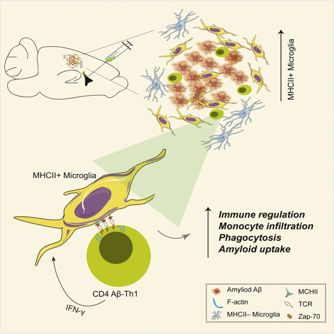
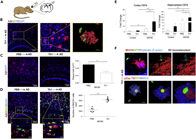
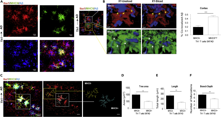
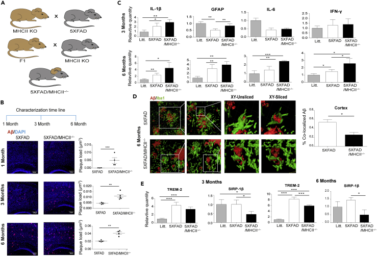
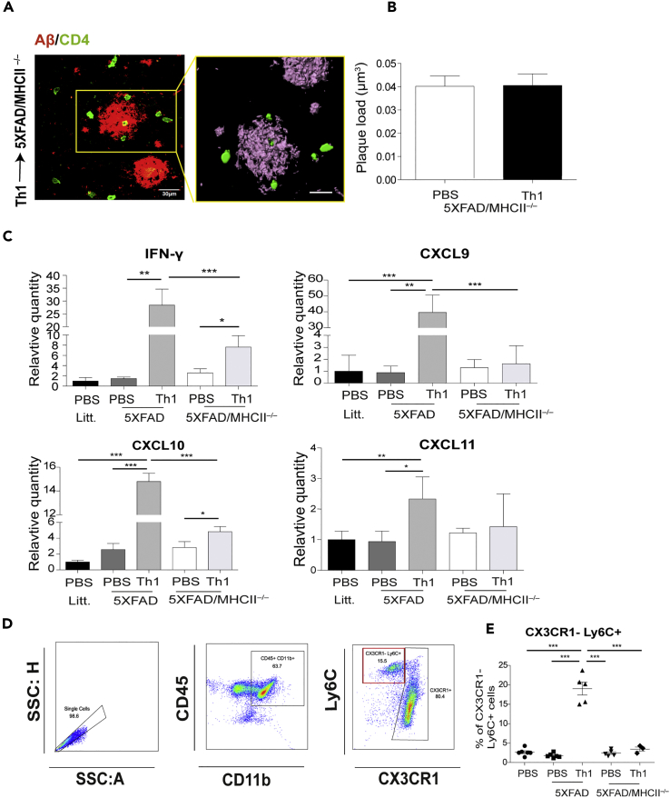
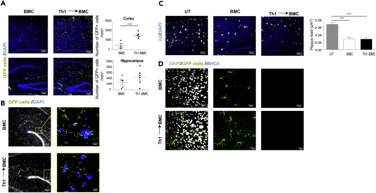
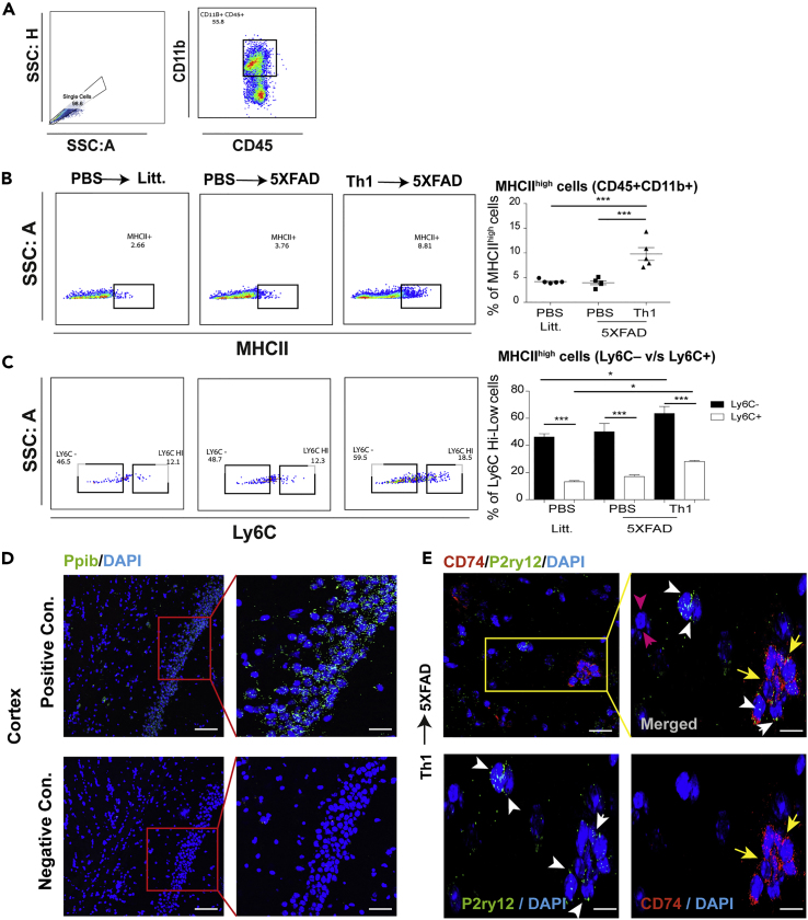
Microglia play a key role in innate immunity in Alzheimer disease (AD), but their role as antigen-presenting cells is as yet unclear. Here we found that amyloid β peptide (Aβ)-specific T helper 1 (Aβ-Th1 cells) T cells polarized to secrete interferon-γ and intracerebroventricularly (ICV) injected to the 5XFAD mouse model of AD induced the differentiation of major histocompatibility complex class II (MHCII)+ microglia with distinct morphology and enhanced plaque clearance capacity than MHCII- microglia. Notably, 5XFAD mice lacking MHCII exhibited an enhanced amyloid pathology in the brain along with exacerbated innate inflammation and reduced phagocytic capacity. Using a bone marrow chimera mouse model, we showed that infiltrating macrophages did not differentiate to MHCII+ cells following ICV injection of Aβ-Th1 cells and did not support T cell-mediated amyloid clearance. Overall, we demonstrate that CD4 T cells induce a P2ry12+ MHCII+ subset of microglia, which play a key role in T cell-mediated effector functions that abrogate AD-like pathology.
Overview
The claims and statistics of this paper haven’t been checked yet.
Run the check — free, no account needed. It reads the open-access text and checks the sources it cites.
Integrity signals
Registry
Cites
Statistics
Figures
Verification
Claims the paper makes
The claims and statistics of this paper haven’t been checked yet.
Run the check — free, no account needed. It reads the open-access text and checks the sources it cites.
Claims other papers make about this one
Each is checked against this paper’s own text: does it say what the citing paper says it does?
The source backs part of the claim, not all of it — or backs it for a different population, condition or comparison.
In the citing paper
“MHCII+ microglia exhibited higher rates of phagocytosis than their MHCII− counterparts in 5xFAD mice (60).”
From Reducing microglial lipid load enhances β amyloid phagocytosis in an Alzheimer’s disease mouse model (2025) · cites this paper as [60]
Experimental — accuracy not yet measuredThe source backs part of the claim, not all of it — or backs it for a different population, condition or comparison.
In the citing paper
“It has previously been shown that the depletion of T cells, B cells, and natural killer (NK) cells contributes to an increase in Aβ deposition [4–6].”
From Control of the temporal development of Alzheimer’s disease pathology by the MR1/MAIT cell axis (2023) · cites this paper as [6]
Experimental — accuracy not yet measuredThe cited source says what the claim attributes to it.
In the citing paper
“a recent study found that 5XFAD mice that lack MHC class II expression exhibit increased amyloid pathology, 21”
From The P522R protective variant of PLCG2 promotes the expression of antigen presentation genes by human microglia in an Alzheimer's disease mouse model (2022) · cites this paper as [21]
The cited source says what the claim attributes to it.
In the citing paper
“Notably, the loss of MHC class II molecules has been shown to increase Aβ plaques [3, 6].”
From Control of the temporal development of Alzheimer’s disease pathology by the MR1/MAIT cell axis (2023) · cites this paper as [6]
The cited source says what the claim attributes to it.
In the citing paper
“Adoptive transfer of T helper 1 (Th1) cells into AD model mice results in a decrease in the amount of Aβ [6].”
From Control of the temporal development of Alzheimer’s disease pathology by the MR1/MAIT cell axis (2023) · cites this paper as [6]
Self-check
Percentages, sums and reported differences re-computed from the paper’s own numbers.
The self-check needs the paper’s full text.
Citations
Cites
Neuroinflammation in Alzheimer's disease
Heneka, M. T., Carson, M. J., Khoury, J. E., Landreth, G. E., Brosseron, F., Feinstein, D. L., Jacobs, A. H., Wyss-Coray, T., Vitorica, J., Ransohoff, R. M., et al. (2015) · The Lancet Neurology · doi:10.1016/s1474-4422(15)70016-5 · Cited by 6,226
The amyloid hypothesis of Alzheimer's disease at 25 years
Selkoe, D. J., & Hardy, J. (2016) · EMBO Molecular Medicine · doi:10.15252/emmm.201606210 · Cited by 6,164
A Unique Microglia Type Associated with Restricting Development of Alzheimer’s Disease
Keren-Shaul, H., Spinrad, A., Weiner, A., Matcovitch-Natan, O., Dvir-Szternfeld, R., Ulland, T. K., David, E., Baruch, K., Lara-Astaiso, D., Toth, B., et al. (2017) · Cell · doi:10.1016/j.cell.2017.05.018 · Cited by 5,493
Microglia-mediated neurotoxicity: uncovering the molecular mechanisms
Block, M. L., Zecca, L., & Hong, J.-S. (2007) · Nature Reviews Neuroscience · doi:10.1038/nrn2038 · Cited by 4,110
Oakley, H., Cole, S. L., Logan, S., Maus, E., Shao, P., Craft, J., Guillozet-Bongaarts, A., Ohno, M., Disterhoft, J., Van Eldik, L., et al. (2006) · The Journal of Neuroscience · doi:10.1523/jneurosci.1202-06.2006 · Cited by 3,530
Identification of a unique TGF-β–dependent molecular and functional signature in microglia
Butovsky, O., Jedrychowski, M. P., Moore, C. S., Cialic, R., Lanser, A. J., Gabriely, G., Koeglsperger, T., Dake, B., Wu, P. M., Doykan, C. E., et al. (2014) · Nature Neuroscience · doi:10.1038/nn.3599 · Cited by 2,543
TREM2 Lipid Sensing Sustains the Microglial Response in an Alzheimer’s Disease Model
Wang, Y., Cella, M., Mallinson, K., Ulrich, J. D., Young, K. L., Robinette, M. L., Gilfillan, S., Krishnan, G. M., Sudhakar, S., Zinselmeyer, B. H., et al. (2015) · Cell · doi:10.1016/j.cell.2015.01.049 · Cited by 1,736
The twilight of immunity: emerging concepts in aging of the immune system
Nikolich-Žugich, J. (2018) · Nature Immunology · doi:10.1038/s41590-017-0006-x · Cited by 1,240
Regulatory T cells are key cerebroprotective immunomodulators in acute experimental stroke
Liesz, A., Suri‐Payer, E., Veltkamp, C., Doerr, H., Sommer, C., Rivest, S., Giese, T., & Veltkamp, R. C. (2009) · Nature Medicine · doi:10.1038/nm.1927 · Cited by 1,137
Tangle and neuron numbers, but not amyloid load, predict cognitive status in Alzheimer’s disease
Giannakopoulos, P., Herrmann, F. R., Bussière, T., Bouras, C., Kövari, E., Perl, D. P., Morrison, J. H., Gold, G., & Hof, P. R. (2003) · Neurology · doi:10.1212/01.wnl.0000063311.58879.01 · Cited by 1,118
Cited by
Sorted by citations
Microglia states and nomenclature: A field at its crossroads
Paolicelli, R. C., Sierra, A., Stevens, B., Tremblay, M.-E., Aguzzi, A., Ajami, B., Amit, I., Audinat, E., Bechmann, I., Bennett, M., et al. (2022) · Neuron · doi:10.1016/j.neuron.2022.10.020 · Cited by 1,973
Neuroinflammation in Alzheimer disease
Heneka, M. T., van der Flier, W. M., Jessen, F., Hoozemanns, J., Thal, D. R., Boche, D., Brosseron, F., Teunissen, C., Zetterberg, H., Jacobs, A. H., et al. (2025) · Nature Reviews Immunology · doi:10.1038/s41577-024-01104-7 · Cited by 638
Self-citationThe neuroimmune axis of Alzheimer’s disease
Jorfi, M., Maaser‐Hecker, A., & Tanzi, R. E. (2023) · Genome Medicine · doi:10.1186/s13073-023-01155-w · Cited by 313
Inflammation context in Alzheimer’s disease, a relationship intricate to define
Novoa, C., Salazar, P., Cisternas, P., Gherardelli, C., Vera-Salazar, R., Zolezzi, J. M., & Inestrosa, N. C. (2022) · Biological Research · doi:10.1186/s40659-022-00404-3 · Cited by 206
Xu, J., Chen, Z., Yu, F., Liu, H., Ma, C., Xie, D., Hu, X., Leak, R. K., Chou, S. H. Y., Stetler, R. A., et al. (2020) · Proceedings of the National Academy of Sciences · doi:10.1073/pnas.2018497117 · Cited by 180
Shao, X., Lu, X., Liao, J., Chen, H., & Fan, X. (2020) · Protein & Cell · doi:10.1007/s13238-020-00727-5 · Cited by 164
CD4+ effector T cells accelerate Alzheimer’s disease in mice
Machhi, J., Yeapuri, P., Lu, Y., Foster, E. G., Chikhale, R. V., Herskovitz, J., Namminga, K. L., Olson, K. E., Abdelmoaty, M. M., Gao, J., et al. (2021) · Journal of Neuroinflammation · doi:10.1186/s12974-021-02308-7 · Cited by 148
Microglial activation states and their implications for Alzheimer's Disease
Valiukas, Z., Tangalakis, K., Apostolopoulos, V., & Feehan, J. (2025) · The Journal of Prevention of Alzheimer s Disease · doi:10.1016/j.tjpad.2024.100013 · Cited by 167
Neuroinflammation in Alzheimer’s disease: insights from peripheral immune cells
Zhang, Q., Yang, G., Luo, Y., Jiang, L., Chi, H., & Tian, G. (2024) · Immunity & Ageing · doi:10.1186/s12979-024-00445-0 · Cited by 138
Insights into T‐cell dysfunction in Alzheimer's disease
Dai, L., & Shen, Y. (2021) · Aging Cell · doi:10.1111/acel.13511 · Cited by 123
Lau, S.-F., Chen, C., Fu, W.-Y., Qu, J. Y., Cheung, T. H., Fu, A. K. Y., & Ip, N. Y. (2020) · Cell Reports · doi:10.1016/j.celrep.2020.107530 · Cited by 114
Elyahu, Y., & Monsonego, A. (2020) · Ageing Research Reviews · doi:10.1016/j.arr.2020.101231 · Cited by 93
Self-citationTau at the interface between neurodegeneration and neuroinflammation
Didonna, A. (2020) · Genes & Immunity · doi:10.1038/s41435-020-00113-5 · Cited by 82
Peripheral and central neuroimmune mechanisms in Alzheimer’s disease pathogenesis
Zhang, S., Gao, Y., Zhao, Y., Huang, T. Y., Zheng, Q., & Wang, X. (2025) · Molecular Neurodegeneration · doi:10.1186/s13024-025-00812-5 · Cited by 78
Common Peripheral Immunity Mechanisms in Multiple Sclerosis and Alzheimer's Disease
Rossi, B., Santos-Lima, B., Terrabuio, E., Zenaro, E., & Constantin, G. (2021) · Frontiers in Immunology · doi:10.3389/fimmu.2021.639369 · Cited by 84
Neuroimmune contributions to Alzheimer’s disease: a focus on human data
Haage, V., & De Jager, P. L. (2022) · Molecular Psychiatry · doi:10.1038/s41380-022-01637-0 · Cited by 74
Amyloid-β specific regulatory T cells attenuate Alzheimer’s disease pathobiology in APP/PS1 mice
Yeapuri, P., Machhi, J., Lu, Y., Abdelmoaty, M. M., Kadry, R., Patel, M., Bhattarai, S., Lu, E., Namminga, K. L., Olson, K. E., et al. (2023) · Molecular Neurodegeneration · doi:10.1186/s13024-023-00692-7 · Cited by 85
The role of the immune system in driving neuroinflammation
Ní Chasaide, C., & Lynch, M. A. (2020) · Brain and Neuroscience Advances · doi:10.1177/2398212819901082 · Cited by 72
Reducing microglial lipid load enhances β amyloid phagocytosis in an Alzheimer’s disease mouse model
Wu, X., Miller, J. A., Lee, B. T. K., Wang, Y., & Ruedl, C. (2025) · Science Advances · doi:10.1126/sciadv.adq6038 · Cited by 67
“MHCII + microglia exhibited higher rates of phagocytosis than their MHCII − counterparts in 5xFAD mice ( 60 ).”
T Cells: A Growing Universe of Roles in Neurodegenerative Diseases
Dressman, D., & Elyaman, W. (2021) · The Neuroscientist · doi:10.1177/10738584211024907 · Cited by 60
Immune and Inflammatory Determinants Underlying Alzheimer’s Disease Pathology
Baulch, J. E., Acharya, M. M., Agrawal, S., Apodaca, L. A., Monteiro, C., & Agrawal, A. (2020) · Journal of Neuroimmune Pharmacology · doi:10.1007/s11481-020-09908-9 · Cited by 56
Claes, C., England, W. E., Danhash, E. P., Kiani Shabestari, S., Jairaman, A., Chadarevian, J. P., Hasselmann, J., Tsai, A. P., Coburn, M. A., Sanchez, J., et al. (2022) · Alzheimer's & Dementia · doi:10.1002/alz.12577 · Cited by 52
Jiang, Y., Zhou, X., Wong, H. Y., Ouyang, L., Ip, F. C. F., Chau, V. M. N., Lau, S., Wu, W., Wong, D. Y. K., Seo, H., et al. (2022) · Nature Aging · doi:10.1038/s43587-022-00241-9 · Cited by 56
Brain clearance of protein aggregates: a close-up on astrocytes
Giusti, V., Kaur, G., Giusto, E., & Civiero, L. (2024) · Molecular Neurodegeneration · doi:10.1186/s13024-024-00703-1 · Cited by 54
Huang, Z., Hamblin, M. R., & Zhang, Q. (2024) · Alzheimer s Research & Therapy · doi:10.1186/s13195-024-01484-x · Cited by 54
Wyatt-Johnson, S. K., & Brutkiewicz, R. R. (2020) · Frontiers in Aging Neuroscience · doi:10.3389/fnagi.2020.592359 · Cited by 50
The aging immune system in Alzheimer’s and Parkinson’s diseases
Heavener, K. S., & Bradshaw, E. M. (2022) · Seminars in Immunopathology · doi:10.1007/s00281-022-00944-6 · Cited by 51
Ghosh, P., Singh, R. B., Ganeshpurkar, A., Pokle, A. V., Singh, R. B., Singh, S. K., & Kumar, A. (2021) · Neurochemistry International · doi:10.1016/j.neuint.2021.105212 · Cited by 44
Zheng, L., He, S., Wang, H., Li, J., Liu, Y., & Liu, S. (2024) · Aging and Disease · doi:10.14336/ad.2024.0206 · Cited by 46
Stone, T. W., & Williams, R. O. (2023) · Journal of Neurochemistry · doi:10.1111/jnc.16015 · Cited by 45
Reduction of the RNA Binding Protein TIA1 Exacerbates Neuroinflammation in Tauopathy
LeBlang, C. J., Medalla, M., Nicoletti, N. W., Hays, E. C., Zhao, J., Shattuck, J. E., Cruz, A. L., Wolozin, B. L., & Luebke, J. I. (2020) · Frontiers in Neuroscience · doi:10.3389/fnins.2020.00285 · Cited by 38
Phagocytic Glial Cells in Brain Homeostasis
Kono, R., Ikegaya, Y., & Koyama, R. (2021) · Cells · doi:10.3390/cells10061348 · Cited by 41
The role of CD56bright NK cells in neurodegenerative disorders
Rodriguez-Mogeda, C., van Ansenwoude, C. M. J., van der Molen, L. H., Strijbis, E. M. M., Mebius, R. E., & de Vries, H. E. (2024) · Journal of Neuroinflammation · doi:10.1186/s12974-024-03040-8 · Cited by 39
Petralla, S., Chirico, F. D., Miti, A., Tartagni, O., Massenzio, F., Poeta, E., Virgili, M., Zuccheri, G., & Monti, B. (2021) · Biomolecules · doi:10.3390/biom11020306 · Cited by 37
T cell aging and Alzheimer’s disease
Guo, L., Li, X., Gould, T. M., Wang, Z., & Cao, W. (2023) · Frontiers in Immunology · doi:10.3389/fimmu.2023.1154699 · Cited by 35
Specificity of Adaptive Immune Responses in Central Nervous System Health, Aging and Diseases
Rickenbach, C., & Gericke, C. (2022) · Frontiers in Neuroscience · doi:10.3389/fnins.2021.806260 · Cited by 33
Tian, Y., Lu, Y., Cao, Y., Dang, C., Wang, N., Tian, K., Luo, Q., Guo, E., Luo, S., Wang, L., et al. (2022) · Frontiers in Aging Neuroscience · doi:10.3389/fnagi.2022.919614 · Cited by 32
Gossypetin ameliorates 5xFAD spatial learning and memory through enhanced phagocytosis against Aβ
Jo, K. W., Lee, D., Cha, D. G., Oh, E.-J., Choi, Y. H., Kim, S., Park, E. S., Kim, J. K., & Kim, K. (2022) · Alzheimer s Research & Therapy · doi:10.1186/s13195-022-01096-3 · Cited by 33
Imaging the Neuroimmune Dynamics Across Space and Time
Carrier, M., Robert, M.-È., Ibáñez, F. G., Desjardins, M., & Tremblay, M. (2020) · Frontiers in Neuroscience · doi:10.3389/fnins.2020.00903 · Cited by 28
Aging reshapes the adaptive immune system from healer to saboteur
Delgado-Pulido, S., Yousefzadeh, M. J., & Mittelbrunn, M. (2025) · Nature Aging · doi:10.1038/s43587-025-00906-1 · Cited by 29
Immune dysfunction in Alzheimer disease
Butovsky, O., Rosenzweig, N., Kleemann, K. L., Jorfi, M., Kuchroo, V. K., Tanzi, R. E., & Weiner, H. L. (2025) · Nature reviews. Neuroscience · doi:10.1038/s41583-025-00997-0 · Cited by 41
Zeng, J., Liu, J., Qu, Q., Zhao, X., & Zhang, J. (2021) · Irish Journal of Medical Science (1971 -) · doi:10.1007/s11845-021-02749-2 · Cited by 28
Jimenez-Ferrer, I., Bäckström, F., Dueñas-Rey, A., Jewett, M., Boza-Serrano, A., Luk, K. C., Deierborg, T., & Swanberg, M. (2021) · Brain, Behavior, and Immunity · doi:10.1016/j.bbi.2020.10.017 · Cited by 26
Glia as antigen-presenting cells in the central nervous system
Sutter, P. A., & Crocker, S. J. (2022) · Current Opinion in Neurobiology · doi:10.1016/j.conb.2022.102646 · Cited by 24
Differential roles of regulatory T cells in Alzheimer's disease
Jafarzadeh, A., Sheikhi, A., Jafarzadeh, Z., & Nemati, M. (2023) · Cellular Immunology · doi:10.1016/j.cellimm.2023.104778 · Cited by 26
Control of the temporal development of Alzheimer’s disease pathology by the MR1/MAIT cell axis
Wyatt-Johnson, S. K., Kersey, H. N., Codocedo, J. F., Newell, K. L., Landreth, G. E., Lamb, B. T., Oblak, A. L., & Brutkiewicz, R. R. (2023) · Journal of Neuroinflammation · doi:10.1186/s12974-023-02761-6 · Cited by 22
An optimized intracerebroventricular injection of CD4+ T cells into mice
Taylor, Z. V., Khand, B., Porgador, A., Monsonego, A., & Eremenko, E. (2021) · STAR Protocols · doi:10.1016/j.xpro.2021.100725 · Cited by 24
Self-citationZaccai, S., Nemirovsky, A., Lerner, L., Alfahel, L., Eremenko, E., Israelson, A., & Monsonego, A. (2024) · Journal of Neuroinflammation · doi:10.1186/s12974-023-03007-1 · Cited by 23
Self-citationLiu, A., Wang, T., Yang, L., & Zhou, Y. (2025) · Brain Sciences · doi:10.3390/brainsci15070675 · Cited by 23
Oligomeric Forms of Human Amyloid-Beta(1–42) Inhibit Antigen Presentation
Gericke, C., Mallone, A., Engelhardt, B., Nitsch, R. M., & Ferretti, M. T. (2020) · Frontiers in Immunology · doi:10.3389/fimmu.2020.01029 · Cited by 20
Alzheimer’s disease and age-related macular degeneration: Shared and distinct immune mechanisms
Butovsky, O., & Rosenzweig, N. (2025) · Immunity · doi:10.1016/j.immuni.2025.04.013 · Cited by 22
King, H., Reiber, M., Philippi, V., Stirling, H., Aulehner, K., Bankstahl, M., Bleich, A., Buchecker, V., Glasenapp, A., Jirkof, P., et al. (2023) · Frontiers in Neuroscience · doi:10.3389/fnins.2023.1143109 · Cited by 19
Inflammation in Posttraumatic Stress Disorder: Dysregulation or Recalibration?
Patas, K., Baker, D. G., Chrousos, G. P., & Agorastos, A. (2023) · Current Neuropharmacology · doi:10.2174/1570159x21666230807152051 · Cited by 21
A glance through the effects of CD4 + T cells, CD8 + T cells, and cytokines on Alzheimer's disease
Afsar, A., Chen, M., Xuan, Z., & Li, Z. (2023) · Computational and Structural Biotechnology Journal · doi:10.1016/j.csbj.2023.10.058 · Cited by 20
Muraleedharan, A., Rotem‐Dai, N., Strominger, I., Anto, N. P., Isakov, N., Monsonego, A., & Livneh, E. (2020) · Glia · doi:10.1002/glia.23921 · Cited by 16
Self-citationImmunological aspects of central neurodegeneration
Niso‐Santano, M., Fuentes, J. M., & Galluzzi, L. (2024) · Cell Discovery · doi:10.1038/s41421-024-00666-z · Cited by 17
Poxleitner, M., Hoffmann, S. H. L., Berezhnoy, G., Ionescu, T. M., González-Menéndez, I., Maier, F. C., Seyfried, D., Ehrlichmann, W., Quintanilla‐Martínez, L., Schmid, A. M., et al. (2024) · Journal of Neuroinflammation · doi:10.1186/s12974-024-03080-0 · Cited by 22
Do microglia metabolize fructose in Alzheimer’s disease?
Sturno, A. M., Hassell, J. E., Lanaspa, M. A., & Bruce, K. D. (2025) · Journal of Neuroinflammation · doi:10.1186/s12974-025-03401-x · Cited by 20
Engineering chimeric antigen receptor CD4 T cells for Alzheimer’s disease
Boskovic, P., Shalita, R., Gao, W., Vernon, H., Deng, Y. L., Colonna, M., Majzner, R. G., Amit, I., & Kipnis, J. (2026) · Proceedings of the National Academy of Sciences · doi:10.1073/pnas.2530977123 · Cited by 16
Wang, Y., Jiang, R., Li, M., Wang, Z., Yang, Y., & Sun, L. (2023) · Journal of Alzheimer s Disease · doi:10.3233/jad-230784 · Cited by 18
Lian, P., Cai, X., Yang, X., Ma, Z., Wang, C., Liu, P., Wu, Y., Cao, X., & Xu, Y. (2024) · APOPTOSIS · doi:10.1007/s10495-024-01943-8 · Cited by 15
Yeapuri, P., Machhi, J., Foster, E. G., Kadry, R., Bhattarai, S., Lu, Y., Sil, S., Sapkota, R., Srivastava, S., Kumar, M., et al. (2025) · Alzheimer s & Dementia · doi:10.1002/alz.70084 · Cited by 15
Fernando, N., Gopalakrishnan, J., Behensky, A. A., Reich, L. N., Liu, C., Bass, V., Bono, M., Montgomery, W., Pace, R. D., Mattapallil, M. J., et al. (2023) · bioRxiv (Cold Spring Harbor Laboratory) · doi:10.1101/2023.03.18.533293 · Cited by 15
Emerging Roles of T Helper Cells in Non-Infectious Neuroinflammation: Savior or Sinner
Liu, W., Fan, M., Lu, W., Zhu, W., Meng, L., & Lu, S. (2022) · Frontiers in Immunology · doi:10.3389/fimmu.2022.872167 · Cited by 16
Lee, E. J., Bae, S., Suh, M., Choi, Y., Choi, Y., Hwang, D. W., & Lee, D. S. (2024) · BMC Genomics · doi:10.1186/s12864-024-10434-8 · Cited by 15
Chen, Y., & Colonna, M. (2022) · Current Opinion in Immunology · doi:10.1016/j.coi.2022.102233 · Cited by 14
Will cellular immunotherapies end neurodegenerative diseases?
Boskovic, P., Gao, W., & Kipnis, J. (2024) · Trends in Immunology · doi:10.1016/j.it.2024.03.006 · Cited by 15
Update on the role of T cells in cognitive impairment
Ruiz‐Fernández, I., Sánchez‐Díaz, R., Ortega‐Sollero, E., & Martı́n, P. (2023) · British Journal of Pharmacology · doi:10.1111/bph.16214 · Cited by 14
Iannucci, J., Dominy, R., Bandopadhyay, S., Arthur, E. M., Noarbe, B. P., Jullienne, A., Krkasharyan, M., Tobin, R. P., Pereverzev, A., Beevers, S., et al. (2024) · Journal of Neuroinflammation · doi:10.1186/s12974-024-03146-z · Cited by 13
Lachish, M., Fainstein, N., Ganz, T., Sofer, L., & Ben‐Hur, T. (2022) · Cells · doi:10.3390/cells11050876 · Cited by 13
Circulatory proteins shape microglia state and boost phagocytosisPreprint
Lu, N., Losada, P. M., Hahn, O., Saksena, A., Tapp, E., Chadarevian, J. P., Dong, W., Shi, S. M., Shuken, S. R., Guldner, I. H., et al. (2024) · bioRxiv (Cold Spring Harbor Laboratory) · doi:10.1101/2024.09.30.615861 · Cited by 12
β-Amyloids and Immune Responses Associated with Alzheimer’s Disease
Kolobova, E. A., Petrushanko, I. Y., Mitkevich, V. A., Makarov, A. А., & Grigorova, I. (2024) · Cells · doi:10.3390/cells13191624 · Cited by 10
Approaches for studying neuroimmune interactions in Alzheimer’s disease
Lin, C., Tian, Y., Tanzi, R. E., & Jorfi, M. (2024) · Trends in Immunology · doi:10.1016/j.it.2024.10.002 · Cited by 11
Afify, R. A. A., Lipsius, K., Wyatt‐Johnson, S. K., & Brutkiewicz, R. R. (2024) · Frontiers in Neuroscience · doi:10.3389/fnins.2024.1488382 · Cited by 11
Immune cells in Alzheimer’s disease: insights into pathogenesis and potential therapeutic targets
Fu, X., Cai, H., Quan, S., Ren, Z., Xu, Y., & Jia, L. (2024) · Medical Review · doi:10.1515/mr-2024-0064 · Cited by 10
Hypercholesterolemia drives microglial dysfunction and weakens response to amyloid plaques
Kaye, S. D., Gold, A., Lin, D., Chen, M., Zhu, J., & Gao, J. (2025) · Experimental Neurology · doi:10.1016/j.expneurol.2025.115272 · Cited by 10
Garg, A., Vo, S., de Souza, I. D., Brant, A. C., Brase, L., Aladyeva, E., Albanus, R. D., Nallapu, A., Fu, H., & Harari, O. (2025) · Alzheimer s Research & Therapy · doi:10.1186/s13195-025-01944-y · Cited by 11
Kadam, R., Gupta, M., Lazarov, O., & Prabhakar, B. S. (2024) · Journal of Leukocyte Biology · doi:10.1093/jleuko/qiae134 · Cited by 8
Recent advances in Alzheimer’s disease pathogenesis and therapeutics from an immune perspective
Nguyen, T.-D., Dang, L. N., Jang, J., & Park, S. (2023) · Journal of Pharmaceutical Investigation · doi:10.1007/s40005-023-00631-0 · Cited by 9
T Cells Trafficking into the Brain in Aging and Alzheimer’s Disease
Ma, Y., Cao, J., Zhang, Y., Su, X.-M., Jing, Y., & Gao, L. (2024) · Journal of Neuroimmune Pharmacology · doi:10.1007/s11481-024-10147-5 · Cited by 11
Immune-microbiome interplay and its implications in neurodegenerative disorders
Uniyal, A., Tiwari, V., Rani, M., & Tiwari, V. (2021) · Metabolic Brain Disease · doi:10.1007/s11011-021-00807-3 · Cited by 9
Dressman, D., Buttrick, T., Cimpean, M., Bennett, D. A., Menon, V., Bradshaw, E. M., Vardarajan, B. N., & Elyaman, W. (2022) · Human Molecular Genetics · doi:10.1093/hmg/ddac126 · Cited by 7
Dissecting the immune response of CD4+ T cells in Alzheimer’s disease
Kostić, M. S., Živković, N., Cvetanović, A., Bašić, J., & Stojanović, I. R. (2024) · Reviews in the Neurosciences · doi:10.1515/revneuro-2024-0090 · Cited by 8
Vijayan, V., Siddique, I. A., Gupta, S., Chopra, E. M., Raj, N., Surolia, A., & Gupta, S. (2025) · Experimental Neurology · doi:10.1016/j.expneurol.2025.115322 · Cited by 6
The association of microbial infection and adaptive immune cell activation in Alzheimer’s disease
Clement, M. (2023) · Discovery Immunology · doi:10.1093/discim/kyad015 · Cited by 6
The therapeutic potential of early exercise in Alzheimer's disease: Focus on the brain-spleen axis
Wang, T., Feng, L., & Tian, Z. (2025) · Ageing Research Reviews · doi:10.1016/j.arr.2025.102980 · Cited by 6
Yang, G., Su, R., Bu, J., Li, Y., Lin, X., Jin, J., Zhang, Y., Zhuang, P., Guo, H., & Yin, Q. (2025) · Metabolic Brain Disease · doi:10.1007/s11011-025-01532-x · Cited by 4
Chihara, N., Tsuji, A., & Matsumoto, R. (2023) · Clinical and Experimental Neuroimmunology · doi:10.1111/cen3.12747 · Cited by 4
Liu, J., Yan, M., Chen, L., Yu, W., & Lü, Y. (2025) · Scientific Reports · doi:10.1038/s41598-025-89980-4 · Cited by 3
Cytokine-induced Neurogenesis for Alzheimer's Disease and Frontotemporal Dementia
Shirasawa, T., & Cobos, L. C. A. (2022) · Personalized Medicine Universe · doi:10.46459/pmu.2022006 · Cited by 3
Yanes, K. J. O., Bui, C. T., Tomasello, J., Morsy, H., Kim, E., Lam, T., Tsourmas, K. I., Ayala, L. A., Green, K. N., Inlay, M. A., et al. (2026) · Journal of Neuroinflammation · doi:10.1186/s12974-025-03666-2 · Cited by 3
Adaptive immunity in the pathogenesis of neurodegeneration
Li, Y., Ulrich, J. D., & Holtzman, D. M. (2026) · Nature Immunology · doi:10.1038/s41590-026-02538-y · Cited by 4
Shi, G., Wang, K., Liu, Q., Wu, C., Zhang, Q., Ma, Y., Ai, Q., & Huang, M. (2025) · Chemico-Biological Interactions · doi:10.1016/j.cbi.2025.111721 · Cited by 2
Jurcău, M. C., Iovanovici, D. C., Jurcău, A., Militaru, M., Udrea, R. B., Comanescu, A. D., & Nunkoo, V. S. (2025) · International Journal of Molecular Sciences · doi:10.3390/ijms26189009 · Cited by 2
Retinal Amyloid Clearance Enhanced by 40-Hz Light Flicker via MHC-II + Microglia Regulation in Mice
Sheng, W., Wang, Q., Huang, Y., Gu, J., Ding, C., Cui, Z., Tang, S., Xia, X., & Chen, J. (2026) · Investigative Ophthalmology & Visual Science · doi:10.1167/iovs.67.1.32 · Cited by 1
Yang, Z., Zhang, M., Zhi, W., Lizhen, Hu, X., Zou, Y., & Wang, L. (2026) · International Journal of Molecular Sciences · doi:10.3390/ijms27031492 · Cited by 3
Wang, R., Feng, Y., Zhou, Z., Jiang, J., Zhang, R., Zou, W., Yang, H., Lv, W., & Shen, Y. (2026) · Frontiers in Aging Neuroscience · doi:10.3389/fnagi.2026.1748418 · Cited by 2
Calderazzo, S. M., Butler, M. L. M. D., Breen, K., Kanner, H., Tripodis, Y., Stein, T. D., Holtzman, D. M., Cherry, J. D., Huber, B. R., & McKee, A. C. (2026) · Acta Neuropathologica Communications · doi:10.1186/s40478-026-02285-z · Cited by 2
Song, C., Lee, Y., & Kim, M. S. (2026) · Frontiers in Immunology · doi:10.3389/fimmu.2026.1813048 · Cited by 1
Lee, E. J., Bae, S., Suh, M., Choi, H., Choi, Y., Hwang, D. W., & Lee, D. S. (2023) · bioRxiv (Cold Spring Harbor Laboratory) · doi:10.1101/2023.01.20.524845 · Cited by 1
Metadata
- Authors
- Mittal, K., Eremenko, E., Berner, O., Elyahu, Y., Strominger, I., Apelblat, D., Nemirovsky, A., Spiegel, I., & Monsonego, A.
- Affiliations
- Ben-Gurion University of the Negev (IL)
- Weizmann Institute of Science (IL)
- Corresponding author
- Monsonego, Alon
- ORCID
- Venue
- iScience
- Publisher
- Elsevier BV
- Published
- 2019-06
- Volume
- 16
- Pages
- 298-311
- ISSN
- 2589-0042 (ISSN-L)
- PMC
- PMC6581663
- OpenAlex
- W2947933080
- Cited by
- 114
- References
- 76
- Citations by year
- 2026: 13 · 2025: 17 · 2024: 28 · 2023: 17 · 2022: 16 · 2021: 10 · 2020: 13
- FWCI
- 4.29
- Type
- journal article
- Language
- en
- Open access
- gold
- doi.org
- Licence
- Locations
- iScience (publishedVersion, cc-by-nc-nd)
- PubMed Central (publishedVersion, cc-by-nc-nd)
- PubMed (publishedVersion)
- DOAJ (DOAJ: Directory of Open Access Journals) (submittedVersion, cc-by-sa)
- Funders
- Israel Science Foundation (684/14)
- Litwin and Gural Foundations
- Topics
- Life Sciences › Neuroscience › Neurology › Neuroinflammation and Neurodegeneration Mechanisms
- Life Sciences › Neuroscience › Cellular and Molecular Neuroscience › Alzheimer's disease research and treatments
- Health Sciences › Medicine › Clinical Neurology › Multiple Sclerosis Research Studies
- Keywords
- Alzheimer's disease, microglia, CD4+ T cells, MHC class II, amyloid-beta, Th1 cells, interferon-gamma, amyloid plaque clearance, P2Y12 receptor, 5xFAD mice, innate immunity, phagocytosis, bone marrow chimeras, infiltrating macrophages
About these checks
Registry, retraction, Cites and statistics results are computed, not judged, with one exception: a printed reference that no index lists is read once by a model before it counts as a citation error. Claims are extracted by a model and kept only when their quote appears verbatim in the text; each cited source is then checked, with a model’s help, for whether it says what the claim relies on. Those verdicts can be wrong — read the claim’s quote and the cited source before relying on one.
Statistics and arithmetic are recomputed, not judged.
Confidence bands are shown only for fields calibrated on our annotated test set. None is calibrated yet, so no band is shown.
What counts as a claim ›How we measure confidence ›How we check this ›
Are you an author, or do you see a problem with this page? Request a review
