

The DOI matches its Crossref record; 76 of the 76 references it cites were found in a registry or an index.
CD4 T Cells Induce A Subset of MHCII-Expressing Microglia that Attenuates Alzheimer Pathology
Open accessAbstract
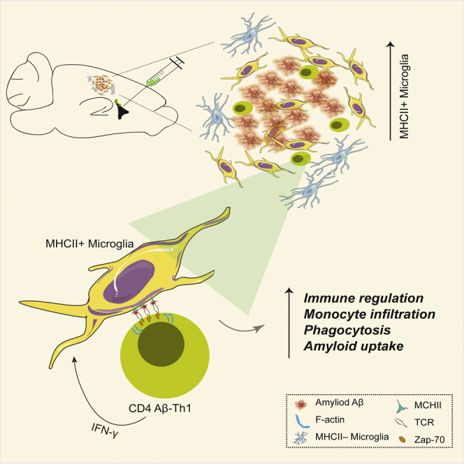
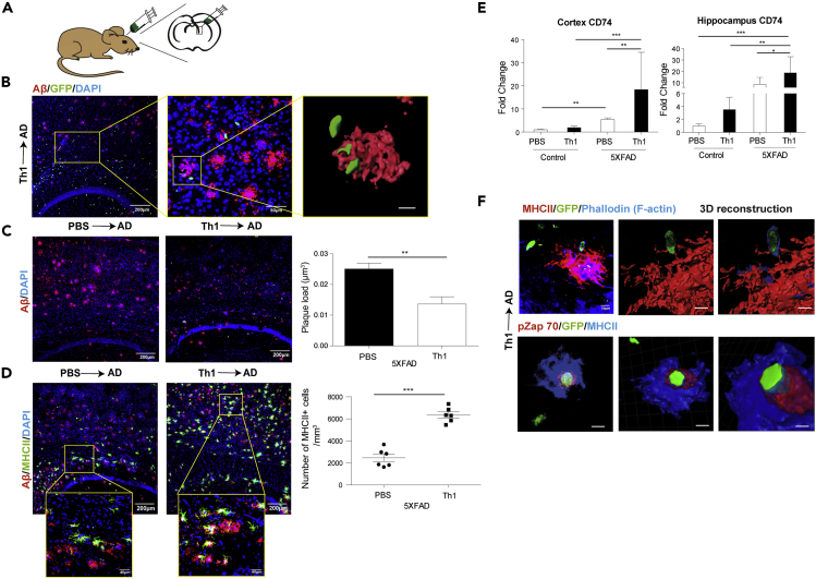
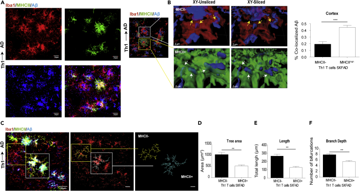
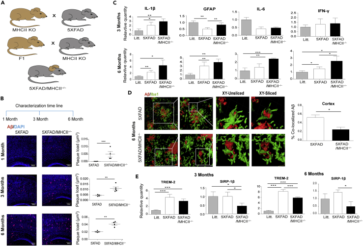
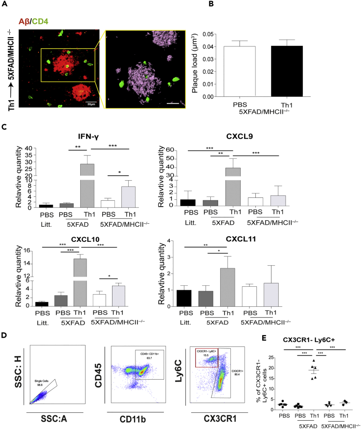
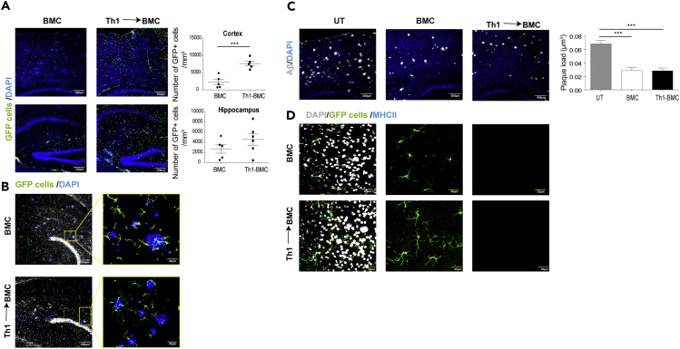
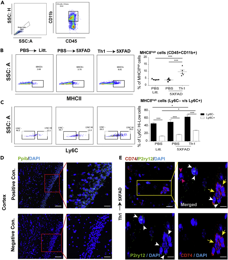
Microglia play a key role in innate immunity in Alzheimer disease (AD), but their role as antigen-presenting cells is as yet unclear. Here we found that amyloid β peptide (Aβ)-specific T helper 1 (Aβ-Th1 cells) T cells polarized to secrete interferon-γ and intracerebroventricularly (ICV) injected to the 5XFAD mouse model of AD induced the differentiation of major histocompatibility complex class II (MHCII)+ microglia with distinct morphology and enhanced plaque clearance capacity than MHCII- microglia. Notably, 5XFAD mice lacking MHCII exhibited an enhanced amyloid pathology in the brain along with exacerbated innate inflammation and reduced phagocytic capacity. Using a bone marrow chimera mouse model, we showed that infiltrating macrophages did not differentiate to MHCII+ cells following ICV injection of Aβ-Th1 cells and did not support T cell-mediated amyloid clearance. Overall, we demonstrate that CD4 T cells induce a P2ry12+ MHCII+ subset of microglia, which play a key role in T cell-mediated effector functions that abrogate AD-like pathology.
Overview
The claims and statistics of this paper haven’t been checked yet.
Run the check — free, no account needed. It reads the open-access text and checks the sources it cites.
Integrity signals
Registry
Cites
Statistics
Figures
Verification
Claims the paper makes
The claims and statistics of this paper haven’t been checked yet.
Run the check — free, no account needed. It reads the open-access text and checks the sources it cites.
Claims other papers make about this one
Each is checked against this paper’s own text: does it say what the citing paper says it does?
The source backs part of the claim, not all of it — or backs it for a different population, condition or comparison.
In the citing paper
“MHCII+ microglia exhibited higher rates of phagocytosis than their MHCII− counterparts in 5xFAD mice (60).”
From Reducing microglial lipid load enhances β amyloid phagocytosis in an Alzheimer’s disease mouse model (2025) · cites this paper as [60]
Experimental — accuracy not yet measuredThe source backs part of the claim, not all of it — or backs it for a different population, condition or comparison.
In the citing paper
“It has previously been shown that the depletion of T cells, B cells, and natural killer (NK) cells contributes to an increase in Aβ deposition [4–6].”
From Control of the temporal development of Alzheimer’s disease pathology by the MR1/MAIT cell axis (2023) · cites this paper as [6]
Experimental — accuracy not yet measuredThe cited source says what the claim attributes to it.
In the citing paper
“a recent study found that 5XFAD mice that lack MHC class II expression exhibit increased amyloid pathology, 21”
From The P522R protective variant of PLCG2 promotes the expression of antigen presentation genes by human microglia in an Alzheimer's disease mouse model (2022) · cites this paper as [21]
The cited source says what the claim attributes to it.
In the citing paper
“Notably, the loss of MHC class II molecules has been shown to increase Aβ plaques [3, 6].”
From Control of the temporal development of Alzheimer’s disease pathology by the MR1/MAIT cell axis (2023) · cites this paper as [6]
The cited source says what the claim attributes to it.
In the citing paper
“Adoptive transfer of T helper 1 (Th1) cells into AD model mice results in a decrease in the amount of Aβ [6].”
From Control of the temporal development of Alzheimer’s disease pathology by the MR1/MAIT cell axis (2023) · cites this paper as [6]
Self-check
Percentages, sums and reported differences re-computed from the paper’s own numbers.
The self-check needs the paper’s full text.
Citations
Cites
Neuroinflammation in Alzheimer's disease
Heneka, M. T., Carson, M. J., Khoury, J. E., Landreth, G. E., Brosseron, F., Feinstein, D. L., Jacobs, A. H., Wyss-Coray, T., Vitorica, J., Ransohoff, R. M., et al. (2015) · The Lancet Neurology · doi:10.1016/s1474-4422(15)70016-5 · Cited by 6,226
The amyloid hypothesis of Alzheimer's disease at 25 years
Selkoe, D. J., & Hardy, J. (2016) · EMBO Molecular Medicine · doi:10.15252/emmm.201606210 · Cited by 6,164
A Unique Microglia Type Associated with Restricting Development of Alzheimer’s Disease
Keren-Shaul, H., Spinrad, A., Weiner, A., Matcovitch-Natan, O., Dvir-Szternfeld, R., Ulland, T. K., David, E., Baruch, K., Lara-Astaiso, D., Toth, B., et al. (2017) · Cell · doi:10.1016/j.cell.2017.05.018 · Cited by 5,493
Microglia-mediated neurotoxicity: uncovering the molecular mechanisms
Block, M. L., Zecca, L., & Hong, J.-S. (2007) · Nature Reviews Neuroscience · doi:10.1038/nrn2038 · Cited by 4,110
Oakley, H., Cole, S. L., Logan, S., Maus, E., Shao, P., Craft, J., Guillozet-Bongaarts, A., Ohno, M., Disterhoft, J., Van Eldik, L., et al. (2006) · The Journal of Neuroscience · doi:10.1523/jneurosci.1202-06.2006 · Cited by 3,530
Identification of a unique TGF-β–dependent molecular and functional signature in microglia
Butovsky, O., Jedrychowski, M. P., Moore, C. S., Cialic, R., Lanser, A. J., Gabriely, G., Koeglsperger, T., Dake, B., Wu, P. M., Doykan, C. E., et al. (2014) · Nature Neuroscience · doi:10.1038/nn.3599 · Cited by 2,543
TREM2 Lipid Sensing Sustains the Microglial Response in an Alzheimer’s Disease Model
Wang, Y., Cella, M., Mallinson, K., Ulrich, J. D., Young, K. L., Robinette, M. L., Gilfillan, S., Krishnan, G. M., Sudhakar, S., Zinselmeyer, B. H., et al. (2015) · Cell · doi:10.1016/j.cell.2015.01.049 · Cited by 1,736
The twilight of immunity: emerging concepts in aging of the immune system
Nikolich-Žugich, J. (2018) · Nature Immunology · doi:10.1038/s41590-017-0006-x · Cited by 1,240
Regulatory T cells are key cerebroprotective immunomodulators in acute experimental stroke
Liesz, A., Suri‐Payer, E., Veltkamp, C., Doerr, H., Sommer, C., Rivest, S., Giese, T., & Veltkamp, R. C. (2009) · Nature Medicine · doi:10.1038/nm.1927 · Cited by 1,137
Tangle and neuron numbers, but not amyloid load, predict cognitive status in Alzheimer’s disease
Giannakopoulos, P., Herrmann, F. R., Bussière, T., Bouras, C., Kövari, E., Perl, D. P., Morrison, J. H., Gold, G., & Hof, P. R. (2003) · Neurology · doi:10.1212/01.wnl.0000063311.58879.01 · Cited by 1,118
Streit, W. J., Walter, S. A., & Pennell, N. A. (1999) · Progress in Neurobiology · doi:10.1016/s0301-0082(98)00069-0 · Cited by 1,096
Inflammation in Alzheimer Disease--A Brief Review of the Basic Science and Clinical Literature
Wyss-Coray, T., & Rogers, J. (2012) · Cold Spring Harbor Perspectives in Medicine · doi:10.1101/cshperspect.a006346 · Cited by 975
Sala Frigerio, C., Wolfs, L., Fattorelli, N., Thrupp, N., Voytyuk, I., Schmidt, I., Mancuso, R., Chen, W.-T., Woodbury, M. E., Srivastava, G., et al. (2019) · Cell Reports · doi:10.1016/j.celrep.2019.03.099 · Cited by 926
T cells from patients with Parkinson’s disease recognize α-synuclein peptides
Sulzer, D., Alcalay, R. N., Garretti, F., Cote, L., Kanter, E., Agin-Liebes, J., Liong, C., McMurtrey, C., Hildebrand, W. H., Mao, X., et al. (2017) · Nature · doi:10.1038/nature22815 · Cited by 866
TREM2-mediated early microglial response limits diffusion and toxicity of amyloid plaques
Wang, Y., Ulland, T. K., Ulrich, J. D., Song, W., Tzaferis, J. A., Hole, J. T., Yuan, P., Mahan, T. E., Shi, Y., Gilfillan, S., et al. (2016) · Journal of Experimental Medicine · doi:10.1084/jem.20151948 · Cited by 840
McGeer, P. L., Itagaki, S., Tago, H., & McGeer, E. G. (1987) · Neuroscience Letters · doi:10.1016/0304-3940(87)90696-3 · Cited by 836
Tau and neurodegenerative disease: the story so far
Iqbal, K., Liu, F., & Gong, C.-X. (2016) · Nature Reviews Neurology · doi:10.1038/nrneurol.2015.225 · Cited by 806
Temporal Tracking of Microglia Activation in Neurodegeneration at Single-Cell Resolution
Mathys, H., Adaikkan, C., Gao, F., Young, J. Z., Manet, E., Hemberg, M., De Jager, P. L., Ransohoff, R. M., Regev, A., & Tsai, L.-H. (2017) · Cell Reports · doi:10.1016/j.celrep.2017.09.039 · Cited by 740
Pathogenic Mechanisms Associated With Different Clinical Courses of Multiple Sclerosis
Lassmann, H. (2019) · Frontiers in Immunology · doi:10.3389/fimmu.2018.03116 · Cited by 711
Hopperton, K. E., Mohammad, D., Trépanier, M. O., Giuliano, V, & Bazinet, R. P. (2018) · Molecular Psychiatry · doi:10.1038/mp.2017.246 · Cited by 567
Walker, D. G., & Lue, L.-F. (2015) · Alzheimer's Research & Therapy · doi:10.1186/s13195-015-0139-9 · Cited by 562
Barkal, A. A., Weiskopf, K., Kao, K. S., Gordon, S. R., Rosental, B., Yiu, Y. Y., George, B. M., Markovic, M., Ring, N. G., Tsai, J. M., et al. (2017) · Nature Immunology · doi:10.1038/s41590-017-0004-z · Cited by 560
Multiple sclerosis: experimental models and reality
Lassmann, H., & Bradl, M. (2017) · Acta Neuropathologica · doi:10.1007/s00401-016-1631-4 · Cited by 525
Cruz Hernández, J. C., Bracko, O., Kersbergen, C. J., Muse, V., Haft-Javaherian, M., Berg, M., Park, L., Vinarcsik, L. K., Ivasyk, I., Rivera, D. A., et al. (2019) · Nature Neuroscience · doi:10.1038/s41593-018-0329-4 · Cited by 508
Baruch, K., Rosenzweig, N., Kertser, A., Deczkowska, A., Sharif, A. M., Spinrad, A., Tsitsou-Kampeli, A., Sarel, A., Cahalon, L., & Schwartz, M. (2015) · Nature Communications · doi:10.1038/ncomms8967 · Cited by 491
Reactive Glia not only Associates with Plaques but also Parallels Tangles in Alzheimer’s Disease
Serrano-Pozo, A., Mielke, M. L., Gómez-Isla, T., Betensky, R. A., Growdon, J. H., Frosch, M. P., & Hyman, B. T. (2011) · The American Journal of Pathology · doi:10.1016/j.ajpath.2011.05.047 · Cited by 488
Occurrence of T cells in the brain of Alzheimer's disease and other neurological diseases
Togo, T., Akiyama, H., Iseki, E., Kondo, H., Ikeda, K., Kato, M., Oda, T., Tsuchiya, K., & Kosaka, K. (2002) · Journal of Neuroimmunology · doi:10.1016/s0165-5728(01)00496-9 · Cited by 481
Baruch, K., Deczkowska, A., Rosenzweig, N., Tsitsou-Kampeli, A., Sharif, A. M., Matcovitch-Natan, O., Kertser, A., David, E., Amit, I., & Schwartz, M. (2016) · Nature Medicine · doi:10.1038/nm.4022 · Cited by 425
Marsh, S. E., Abud, E. M., Lakatos, A., Karimzadeh, A., Yeung, S. T., Davtyan, H., Fote, G. M., Lau, L., Weinger, J. G., Lane, T. E., et al. (2016) · Proceedings of the National Academy of Sciences · doi:10.1073/pnas.1525466113 · Cited by 411
Neuroinflammation: Microglia and T Cells Get Ready to Tango
Schetters, S. T. T., Gómez‐Nicola, D., García-Vallejo, J. J., & van Kooyk, Y. (2018) · Frontiers in Immunology · doi:10.3389/fimmu.2017.01905 · Cited by 400
T lymphocytes potentiate endogenous neuroprotective inflammation in a mouse model of ALS
Chiu, I. M., Chen, A., Zheng, Y., Kosaras, B., Tsiftsoglou, S. A., Vartanian, T. K., Brown, R. H., & Carroll, M. C. (2008) · Proceedings of the National Academy of Sciences · doi:10.1073/pnas.0804610105 · Cited by 380
Regulatory T cells delay disease progression in Alzheimer-like pathology
Dansokho, C., Ahmed, D. A., Aïd, S., Toly‐Ndour, C., Chaigneau, T., Calle, V., Cagnard, N., Holzenberger, M., Piaggio, E., Aucouturier, P., et al. (2016) · Brain · doi:10.1093/brain/awv408 · Cited by 375
Increased T cell reactivity to amyloid β protein in older humans and patients with Alzheimer disease
Monsonego, A., Zota, V., Karni, A., Krieger, J. I., Bar-Or, A., Bitan, G., Budson, A. E., Sperling, R., Selkoe, D. J., & Weiner, H. L. (2003) · Journal of Clinical Investigation · doi:10.1172/jci200318104 · Cited by 367
Self-citationVanRyzin, J. W., Marquardt, A. E., Argue, K. J., Vecchiarelli, H. A., Ashton, S. E., Arambula, S. E., Hill, M. N., & McCarthy, M. M. (2019) · Neuron · doi:10.1016/j.neuron.2019.02.006 · Cited by 272
Aging Microglia—Phenotypes, Functions and Implications for Age-Related Neurodegenerative Diseases
Spittau, B. (2017) · Frontiers in Aging Neuroscience · doi:10.3389/fnagi.2017.00194 · Cited by 271
Inflammation in Alzheimer's disease: relevance to pathogenesis and therapy
Zotova, E., Nicoll, J. A. R., Kalaria, R. N., Holmes, C., & Boche, D. (2010) · Alzheimer s Research & Therapy · doi:10.1186/alzrt24 · Cited by 269
Davies, D. S., Ma, J., Jegathees, T., & Goldsbury, C. (2017) · Brain Pathology · doi:10.1111/bpa.12456 · Cited by 251
A Brief Overview of Tauopathy: Causes, Consequences, and Therapeutic Strategies
Orr, M. E., Sullivan, A. C., & Frost, B. E. (2017) · Trends in Pharmacological Sciences · doi:10.1016/j.tips.2017.03.011 · Cited by 242
The roles of inflammation and immune mechanisms in Alzheimer's disease
Eldik, L. J. V., Carrillo, M. C., Cole, P. E., Feuerbach, D., Greenberg, B., Hendrix, J. A., Kennedy, M. E., Kozauer, N., Margolin, R. A., Molinuevo, J. L., et al. (2016) · Alzheimer s & Dementia Translational Research & Clinical Interventions · doi:10.1016/j.trci.2016.05.001 · Cited by 238
TREM2 and the neuroimmunology of Alzheimer's disease
Hickman, S. E., & El Khoury, J. (2014) · Biochemical Pharmacology · doi:10.1016/j.bcp.2013.11.021 · Cited by 221
Immunotherapeutic Approaches to Alzheimer's Disease
Monsonego, A., & Weiner, H. L. (2003) · Science · doi:10.1126/science.1088469 · Cited by 217
Self-citationWhen aging reaches CD4+ T-cells: phenotypic and functional changes
Moro‐García, M. A. (2013) · Frontiers in Immunology · doi:10.3389/fimmu.2013.00107 · Cited by 216
Kamphuis, W., Kooijman, L., Schetters, S., Orre, M., & Hol, E. M. (2016) · Biochimica et Biophysica Acta (BBA) - Molecular Basis of Disease · doi:10.1016/j.bbadis.2016.07.007 · Cited by 209
Inflammatory changes parallel the early stages of Alzheimer disease
Parachikova, A., Agadjanyan, M. G., Cribbs, D. H., Blurton-Jones, M., Perreau, V., Rogers, J., Beach, T. G., & Cotman, C. W. (2007) · Neurobiology of Aging · doi:10.1016/j.neurobiolaging.2006.08.014 · Cited by 203
Immune Surveillance of the CNS following Infection and Injury
Russo, M. V., & McGavern, D. B. (2015) · Trends in Immunology · doi:10.1016/j.it.2015.08.002 · Cited by 191
Sex differences in microglial phagocytosis in the neonatal hippocampus
Nelson, L. H., Warden, S., & Lenz, K. M. (2017) · Brain, Behavior, and Immunity · doi:10.1016/j.bbi.2017.03.010 · Cited by 189
Tooyama, I., Kimura, H., Akiyama, H., & McGeer, P. L. (1990) · Brain Research · doi:10.1016/0006-8993(90)91496-4 · Cited by 180
Landel, V., Baranger, K., Virard, I., Loriod, B., Khrestchatisky, M., Rivera, S., Benech, P., & Féron, F. (2014) · Molecular Neurodegeneration · doi:10.1186/1750-1326-9-33 · Cited by 176
Neuroprotective effects of CD4+CD25+Foxp3+ regulatory T cells in a 3xTg-AD Alzheimer's disease model
Baek, H., Ye, M., Kang, G., Lee, C., Lee, G., Choi, D. B., Jung, J., Kim, H., Lee, S., Kim, J. S., et al. (2016) · Oncotarget · doi:10.18632/oncotarget.12469 · Cited by 174
Monsonego, A., Imitola, J., Petrovic, S., Zota, V. E., Nemirovsky, A., Baron, R., Fisher, Y., Owens, T., & Weiner, H. L. (2006) · Proceedings of the National Academy of Sciences · doi:10.1073/pnas.0506209103 · Cited by 165
Self-citationDual effect of CD4 + CD25 + regulatory T cells in neurodegeneration: A dialogue with microglia
Kipnis, J., Avidan, H., Caspi, R. R., & Schwartz, M. (2004) · Proceedings of the National Academy of Sciences · doi:10.1073/pnas.0404842101 · Cited by 163
Targeting innate immunity for neurodegenerative disorders of the central nervous system
Andreasson, K. I., Bachstetter, A. D., Colonna, M., Ginhoux, F., Holmes, C., Lamb, B., Landreth, G., Lee, D. C., Low, D., Lynch, M. A., et al. (2016) · Journal of Neurochemistry · doi:10.1111/jnc.13667 · Cited by 126
Self-citationMayo, L., da Cunha, A. P., Madi, A., Beynon, V., Yang, Z., Alvarez, J. I., Prat, A., Sobel, R. A., Kobzik, L., Lassmann, H., et al. (2016) · Brain · doi:10.1093/brain/aww113 · Cited by 120
Re, F., Belyanskaya, S. L., Riese, R. J., Cipriani, B., Fischer, F. R., Granucci, F., Ricciardi-Castagnoli, P., Brosnan, C., Stern, L. J., Strominger, J. L., et al. (2002) · The Journal of Immunology · doi:10.4049/jimmunol.169.5.2264 · Cited by 109
Mastrangelo, M. A., Sudol, K. L., Narrow, W. C., & Bowers, W. J. (2009) · The American Journal of Pathology · doi:10.2353/ajpath.2009.090059 · Cited by 100
Cytotoxic CD4+ T Cells Drive Multiple Sclerosis Progression
Peeters, L. M., Vanheusden, M., Somers, V. A., Wijmeersch, B. V., Stinissen, P., Broux, B., & Hellings, N. (2017) · Frontiers in Immunology · doi:10.3389/fimmu.2017.01160 · Cited by 97
Gottfried‐Blackmore, A., Kaunzner, U. W., Idoyaga, J., Felger, J. C., McEwen, B. S., & Bulloch, K. (2009) · Proceedings of the National Academy of Sciences · doi:10.1073/pnas.0911509106 · Cited by 93
Wlodarczyk, A., Cédile, O., Jensen, K. N., Jasson, A., Mony, J. T., Khorooshi, R., & Owens, T. (2015) · Frontiers in Immunology · doi:10.3389/fimmu.2015.00463 · Cited by 88
Gaikwad, S. M., Larionov, S., Wang, Y., Dannenberg, H., Matozaki, T., Monsonego, A., Thal, D. R., & Neumann, H. (2009) · American Journal Of Pathology · doi:10.2353/ajpath.2009.090147 · Cited by 86
Self-citationDual destructive and protective roles of adaptive immunity in neurodegenerative disorders
Anderson, K. M., Olson, K. E., Estes, K. A., Flanagan, K., Gendelman, H. E., & Mosley, R. L. (2014) · Translational Neurodegeneration · doi:10.1186/2047-9158-3-25 · Cited by 83
Zhu, C., Kros, J. M., van der Weiden, M., Zheng, P., Cheng, C., & Mustafa, D. A. M. (2017) · Acta Neuropathologica Communications · doi:10.1186/s40478-016-0405-5 · Cited by 81
Microglial Activation Milieu Controls Regulatory T Cell Responses
Ebner, F., Brandt, C., Thiele, P., Richter, D., Schliesser, U., Siffrin, V., Schueler, J., Stubbe, T., Ellinghaus, A., Meisel, C., et al. (2013) · The Journal of Immunology · doi:10.4049/jimmunol.1203331 · Cited by 78
Neuroprotection by IFN-γviaastrocyte-secreted IL-6 in acute neuroinflammation
Sun, L., Li, Y., Jia, X., Wang, Q., Li, Y., Hu, M., Tian, L., Yang, J., Xing, W., Zhang, W., et al. (2017) · Oncotarget · doi:10.18632/oncotarget.16990 · Cited by 71
Ishii, H., Tanabe, S., Ueno, M., Kubo, T., Kayama, H., Serada, S., Fujimoto, M., Takeda, K., Naka, T., & Yamashita, T. (2013) · Cell Death and Disease · doi:10.1038/cddis.2013.234 · Cited by 69
SIRP/CD47 signaling in neurological disorders
Zhang, H., Li, F., Yang, Y., Chen, J., & Hu, X. (2015) · Brain Research · doi:10.1016/j.brainres.2015.03.012 · Cited by 67
Th1 Polarization of T Cells Injected into the Cerebrospinal Fluid Induces Brain Immunosurveillance
Fisher, Y., Strominger, I., Biton, S., Nemirovsky, A., Baron, R., & Monsonego, A. (2013) · The Journal of Immunology · doi:10.4049/jimmunol.1301707 · Cited by 67
Self-citationNew candidates for CD4 T cell pathogenicity in experimental neuroinflammation and multiple sclerosis
Hoppmann, N., Graetz, C., Paterka, M., Poisa-Beiro, L., Larochelle, C., Hasan, M., Lill, C. M., Zipp, F., & Siffrin, V. (2015) · Brain · doi:10.1093/brain/awu408 · Cited by 66
HLA-DR Alleles in Amyloid β-Peptide Autoimmunity: A Highly Immunogenic Role for the DRB1*1501 Allele
Zota, V. E., Nemirovsky, A., Baron, R., Fisher, Y., Selkoe, D. J., Altmann, D. M., Weiner, H. L., & Monsonego, A. (2009) · The Journal of Immunology · doi:10.4049/jimmunol.0900620 · Cited by 60
Self-citationInducing and Administering Tregs to Treat Human Disease
Perdigoto, A. L., Chatenoud, L., Bluestone, J. A., & Herold, K. C. (2016) · Frontiers in Immunology · doi:10.3389/fimmu.2015.00654 · Cited by 51
T cells in the central nervous system: messengers of destruction or purveyors of protection?
Walsh, J. T., Watson, N., & Kipnis, J. (2014) · Immunology · doi:10.1111/imm.12187 · Cited by 51
TREM2 analysis and increased risk of Alzheimer's disease
Finelli, D., Rollinson, S. J., Harris, J., Jones, M. S., Richardson, A. M. T., Gerhard, A., Snowden, J. S., Mann, D. M. A., & Pickering-Brown, S. M. (2014) · Neurobiology of Aging · doi:10.1016/j.neurobiolaging.2014.08.001 · Cited by 45
New immunosuppressive approaches: Oral administration of CD3-specific antibody to treat autoimmunity
Ochi, H., Abraham, M., Ishikawa, H., Frenkel, D., Yang, K., Basso, A. S., Wu, H. D., Chen, M., Gandhi, R., Miller, A., et al. (2008) · Journal of the Neurological Sciences · doi:10.1016/j.jns.2008.07.027 · Cited by 40
TREM2 enables amyloid β clearance by microglia.
Rivest, S. (2015) · Cell Research · doi:10.1038/cr.2015.37 · Cited by 34
Linking T cells to Alzheimer's disease: from neurodegeneration to neurorepair
Bryson, K. J., & Lynch, M. A. (2015) · Current Opinion in Pharmacology · doi:10.1016/j.coph.2015.10.003 · Cited by 33
T cells and Parkinson's disease
Mosley, R. L., & Gendelman, H. E. (2017) · The Lancet Neurology · doi:10.1016/s1474-4422(17)30276-4 · Cited by 26
Harpaz, I., Bhattacharya, U., Elyahu, Y., Strominger, I., & Monsonego, A. (2017) · Frontiers in Immunology · doi:10.3389/fimmu.2017.00283 · Cited by 22
Self-citation
Cited by
Sorted by citations
Microglia states and nomenclature: A field at its crossroads
Paolicelli, R. C., Sierra, A., Stevens, B., Tremblay, M.-E., Aguzzi, A., Ajami, B., Amit, I., Audinat, E., Bechmann, I., Bennett, M., et al. (2022) · Neuron · doi:10.1016/j.neuron.2022.10.020 · Cited by 1,973
Neuroinflammation in Alzheimer disease
Heneka, M. T., van der Flier, W. M., Jessen, F., Hoozemanns, J., Thal, D. R., Boche, D., Brosseron, F., Teunissen, C., Zetterberg, H., Jacobs, A. H., et al. (2025) · Nature Reviews Immunology · doi:10.1038/s41577-024-01104-7 · Cited by 638
Self-citationThe neuroimmune axis of Alzheimer’s disease
Jorfi, M., Maaser‐Hecker, A., & Tanzi, R. E. (2023) · Genome Medicine · doi:10.1186/s13073-023-01155-w · Cited by 313
Inflammation context in Alzheimer’s disease, a relationship intricate to define
Novoa, C., Salazar, P., Cisternas, P., Gherardelli, C., Vera-Salazar, R., Zolezzi, J. M., & Inestrosa, N. C. (2022) · Biological Research · doi:10.1186/s40659-022-00404-3 · Cited by 206
Xu, J., Chen, Z., Yu, F., Liu, H., Ma, C., Xie, D., Hu, X., Leak, R. K., Chou, S. H. Y., Stetler, R. A., et al. (2020) · Proceedings of the National Academy of Sciences · doi:10.1073/pnas.2018497117 · Cited by 180
Metadata
- Authors
- Mittal, K., Eremenko, E., Berner, O., Elyahu, Y., Strominger, I., Apelblat, D., Nemirovsky, A., Spiegel, I., & Monsonego, A.
- Affiliations
- Ben-Gurion University of the Negev (IL)
- Weizmann Institute of Science (IL)
- Corresponding author
- Monsonego, Alon
- ORCID
- Venue
- iScience
- Publisher
- Elsevier BV
- Published
- 2019-06
- Volume
- 16
- Pages
- 298-311
- ISSN
- 2589-0042 (ISSN-L)
- PMC
- PMC6581663
- OpenAlex
- W2947933080
- Cited by
- 114
- References
- 76
- Citations by year
- 2026: 13 · 2025: 17 · 2024: 28 · 2023: 17 · 2022: 16 · 2021: 10 · 2020: 13
- FWCI
- 4.29
- Type
- journal article
- Language
- en
- Open access
- gold
- doi.org
- Licence
- Locations
- iScience (publishedVersion, cc-by-nc-nd)
- PubMed Central (publishedVersion, cc-by-nc-nd)
- PubMed (publishedVersion)
- DOAJ (DOAJ: Directory of Open Access Journals) (submittedVersion, cc-by-sa)
- Funders
- Israel Science Foundation (684/14)
- Litwin and Gural Foundations
- Topics
- Life Sciences › Neuroscience › Neurology › Neuroinflammation and Neurodegeneration Mechanisms
- Life Sciences › Neuroscience › Cellular and Molecular Neuroscience › Alzheimer's disease research and treatments
- Health Sciences › Medicine › Clinical Neurology › Multiple Sclerosis Research Studies
- Keywords
- Alzheimer's disease, microglia, CD4+ T cells, MHC class II, amyloid-beta, Th1 cells, interferon-gamma, amyloid plaque clearance, P2Y12 receptor, 5xFAD mice, innate immunity, phagocytosis, bone marrow chimeras, infiltrating macrophages
About these checks
Registry, retraction, Cites and statistics results are computed, not judged, with one exception: a printed reference that no index lists is read once by a model before it counts as a citation error. Claims are extracted by a model and kept only when their quote appears verbatim in the text; each cited source is then checked, with a model’s help, for whether it says what the claim relies on. Those verdicts can be wrong — read the claim’s quote and the cited source before relying on one.
Statistics and arithmetic are recomputed, not judged.
Confidence bands are shown only for fields calibrated on our annotated test set. None is calibrated yet, so no band is shown.
What counts as a claim ›How we measure confidence ›How we check this ›
Are you an author, or do you see a problem with this page? Request a review
