

The DOI matches its Crossref record; 2 of the 2 references it cites were found in a registry or an index.
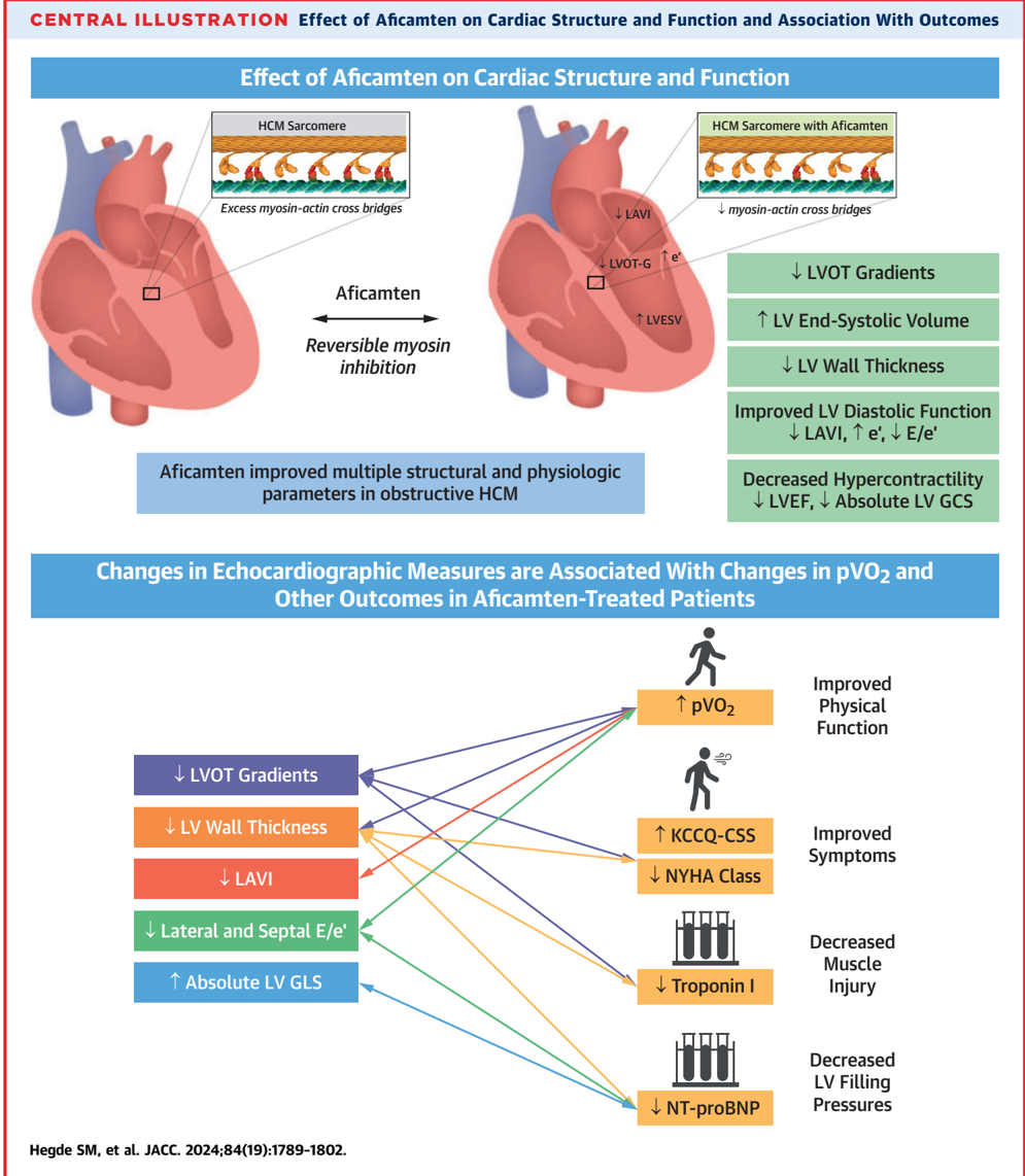
Impact of Aficamten on Echocardiographic Cardiac Structure and Function in Symptomatic Obstructive Hypertrophic Cardiomyopathy
Overview
The claims and statistics of this paper haven’t been checked yet.
Run the check — free, no account needed. It reads the open-access text and checks the sources it cites.
Integrity signals
Registry
Cites
Statistics
Figures
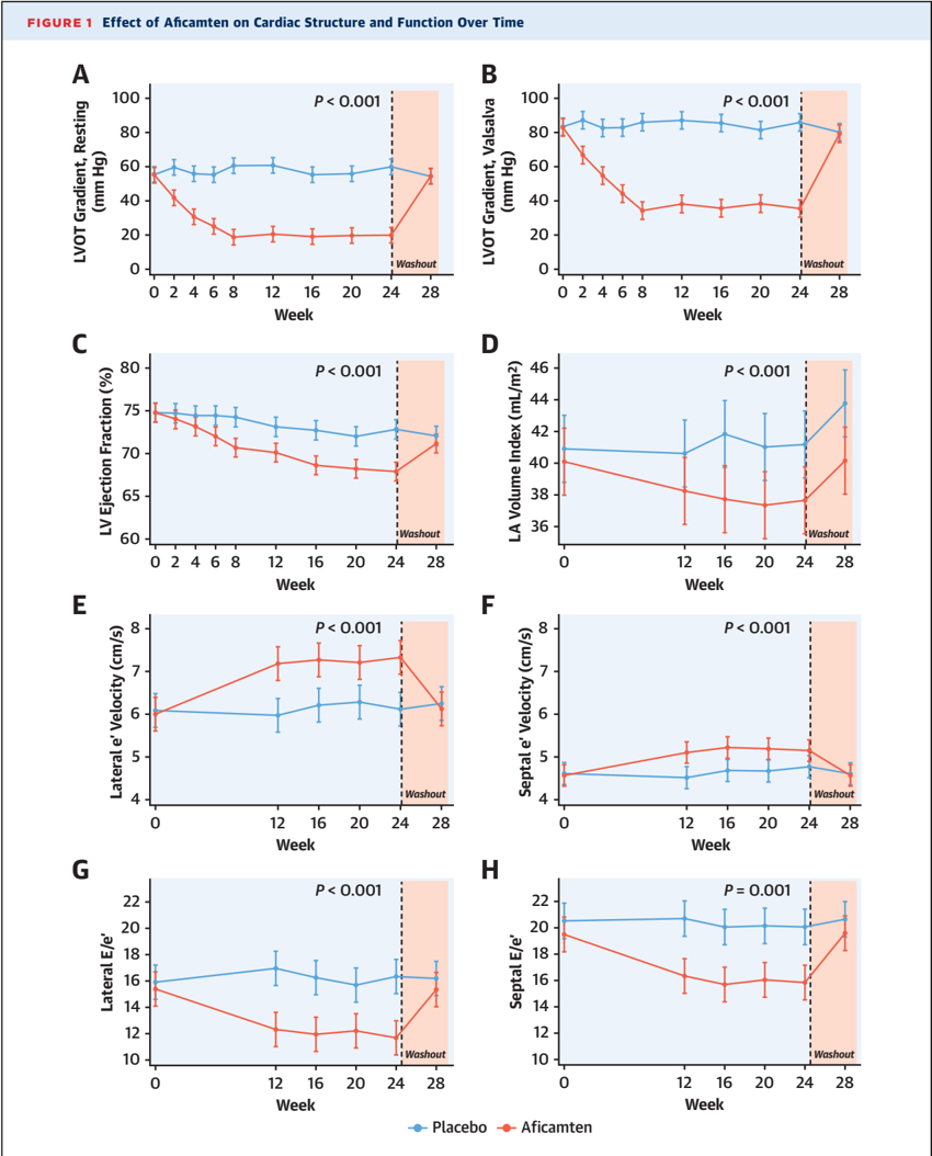
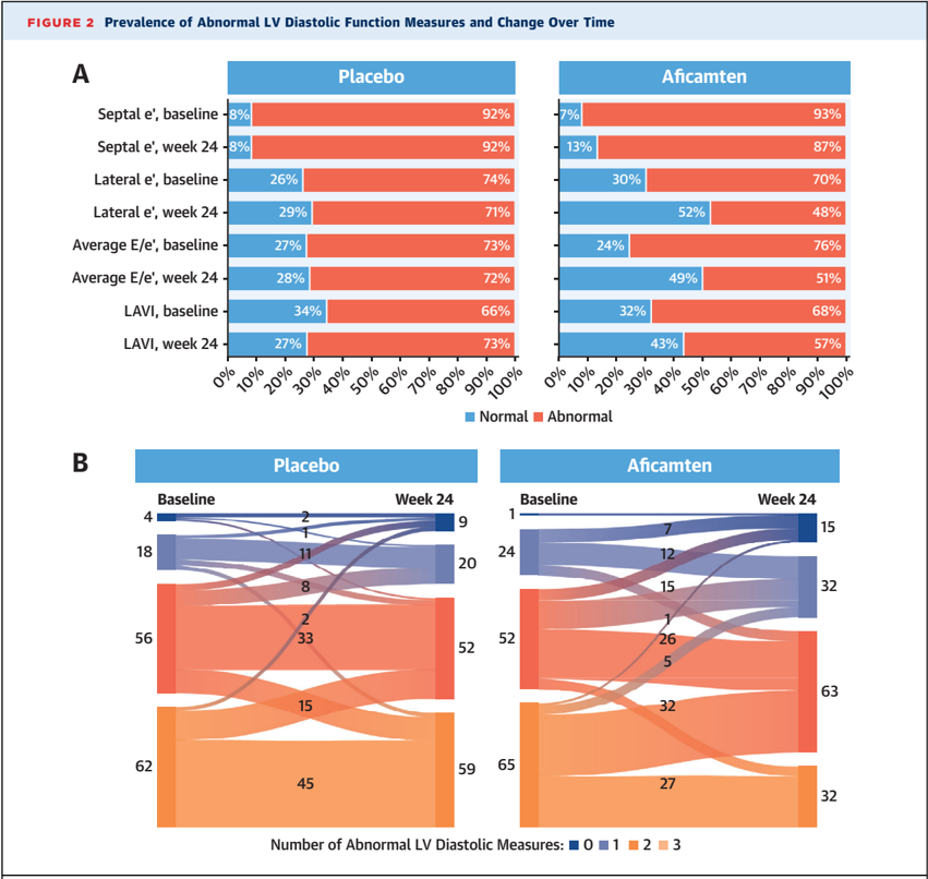
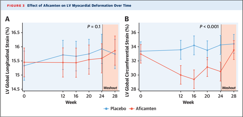
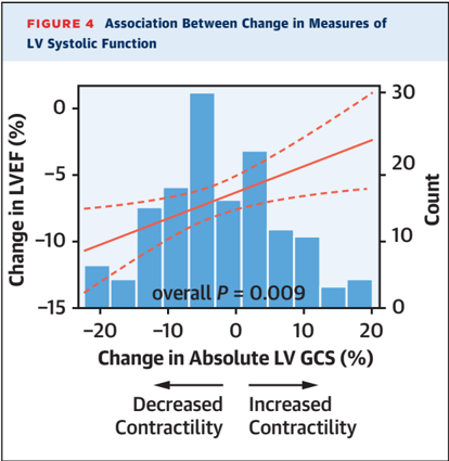
Verification
Claims the paper makes
The claims and statistics of this paper haven’t been checked yet.
Run the check — free, no account needed. It reads the open-access text and checks the sources it cites.
Claims other papers make about this one
Each is checked against this paper’s own text: does it say what the citing paper says it does?
The cited source says what the claim attributes to it.
In the citing paper
“Similarly, data from SEQUOIA-HCM show that patients taking aficamten had improvements in echocardiographic diastolic parameters compared with placebo.”
From Current management of hypertrophic cardiomyopathy (2025) · cites this paper as [152]
Self-check
Percentages, sums and reported differences re-computed from the paper’s own numbers.
The self-check needs the paper’s full text.
Citations
Cites
Sherrid, M. V., Barac, I., McKenna, W. J., Elliott, P. M., Dickie, S., Chojnowska, L., Casey, S., & Maron, B. J. (2005) · Journal of the American College of Cardiology · doi:10.1016/j.jacc.2005.01.012 · Cited by 410
Sherrid, M. V., Shetty, A., Winson, G., Kim, B., Musat, D., Alviar, C. L., Homel, P., Balaram, S. K., & Swistel, D. G. (2013) · Circulation Heart Failure · doi:10.1161/circheartfailure.112.000122 · Cited by 178
Research works printed but not linked: 51
- Ommen, 2024, 2024 AHA/ACC/AMSSM/HRS/PACES/SCMR guideline for the management of hypertrophic cardiomyopathy: a report of the American Heart Association/American College of Cardiology Joint Committee on Clinical Practice Guidelines, J Am Coll Cardiol, 83, 2324, 10.1016/j.jacc.2024.02.014 · DOI
- Geske, 2018, Hypertrophic cardiomyopathy: clinical update, JACC Heart Fail, 6, 364, 10.1016/j.jchf.2018.02.010 · DOI
- Maron, 2003, Effect of left ventricular outflow tract obstruction on clinical outcome in hypertrophic cardiomyopathy, N Engl J Med, 348, 295, 10.1056/NEJMoa021332 · DOI · Paper page
- Ommen, 2005, Long-term effects of surgical septal myectomy on survival in patients with obstructive hypertrophic cardiomyopathy, J Am Coll Cardiol, 46, 470, 10.1016/j.jacc.2005.02.090 · DOI
- Nagueh, 2022, J Am Soc Echocardiogr, 35, 533, 10.1016/j.echo.2022.03.012 · DOI
- Cardim, 2015, Role of multimodality cardiac imaging in the management of patients with hypertrophic cardiomyopathy: an expert consensus of the European Association of Cardiovascular Imaging Endorsed by the Saudi Heart Association, Eur Heart J Cardiovasc Imaging, 16, 280, 10.1093/ehjci/jeu291 · DOI
- Olivotto, 2020, Mavacamten for treatment of symptomatic obstructive hypertrophic cardiomyopathy (EXPLORER-HCM): a randomised, double-blind, placebo-controlled, phase 3 trial, Lancet, 396, 759, 10.1016/S0140-6736(20)31792-X · DOI · Paper page
- Desai, 2023, Mavacamten in patients with hypertrophic cardiomyopathy referred for septal reduction: week 56 results from the VALOR-HCM randomized clinical trial, JAMA Cardiol, 8, 968, 10.1001/jamacardio.2023.3342 · DOI
- Hegde, 2021, Effect of mavacamten on echocardiographic features in symptomatic patients with obstructive hypertrophic cardiomyopathy, J Am Coll Cardiol, 78, 2518, 10.1016/j.jacc.2021.09.1381 · DOI
- Heydari, 2023, 3-Dimensional strain analysis of hypertrophic cardiomyopathy: insights from the NHLBI International HCM Registry, JACC Cardiovasc Imaging, 16, 478, 10.1016/j.jcmg.2022.10.005 · DOI
- Chuang, 2021, Discovery of aficamten (CK-274), a next-generation cardiac myosin inhibitor for the treatment of hypertrophic cardiomyopathy, J Med Chem, 64, 14142, 10.1021/acs.jmedchem.1c01290 · DOI
- Malik, 2022, A phase 1 dose-escalation study of the cardiac myosin inhibitor aficamten in healthy participants, JACC Basic Transl Sci, 7, 763, 10.1016/j.jacbts.2022.04.008 · DOI
- Maron, 2023, Phase 2 study of aficamten in patients with obstructive hypertrophic cardiomyopathy, J Am Coll Cardiol, 81, 34, 10.1016/j.jacc.2022.10.020 · DOI
- Maron, 2024, Aficamten for symptomatic obstructive hypertrophic cardiomyopathy, N Engl J Med, 390, 1849, 10.1056/NEJMoa2401424 · DOI · Paper page
- Coats, 2024, Exercise capacity in patients with obstructive hypertrophic cardiomyopathy: SEQUOIA-HCM baseline characteristics and study design, JACC Heart Fail, 12, 199, 10.1016/j.jchf.2023.10.004 · DOI
- Lang, 2015, Recommendations for cardiac chamber quantification by echocardiography in adults: an update from the American Society of Echocardiography and the European Association of Cardiovascular Imaging, J Am Soc Echocardiogr, 28, 1, 10.1016/j.echo.2014.10.003 · DOI · Paper page
- Shah, 2014, Rationale and design of a multicenter echocardiographic study to assess the relationship between cardiac structure and function and heart failure risk in a biracial cohort of community-dwelling elderly persons: the Atherosclerosis Risk in Communities study, Circ Cardiovasc Imaging, 7, 173, 10.1161/CIRCIMAGING.113.000736 · DOI
- Nagueh, 2016, Recommendations for the evaluation of left ventricular diastolic function by echocardiography: an update from the American Society of Echocardiography and the European Association of Cardiovascular Imaging, J Am Soc Echocardiogr, 29, 277, 10.1016/j.echo.2016.01.011 · DOI · Paper page
- Shah, 2017, Contemporary assessment of left ventricular diastolic function in older adults: the Atherosclerosis Risk in Communities study, Circulation, 135, 426, 10.1161/CIRCULATIONAHA.116.024825 · DOI
- Corona-Villalobos, 2016, Left ventricular wall thickness in patients with hypertrophic cardiomyopathy: a comparison between cardiac magnetic resonance imaging and echocardiography, Int J Cardiovasc Imaging, 32, 945, 10.1007/s10554-016-0858-4 · DOI
- Hindieh, 2017, Discrepant measurements of maximal left ventricular wall thickness between cardiac magnetic resonance imaging and echocardiography in patients with hypertrophic cardiomyopathy, Circ Cardiovasc Imaging, 10, 10.1161/CIRCIMAGING.117.006309 · DOI
- Mazur, 2001, Regression of left ventricular hypertrophy after nonsurgical septal reduction therapy for hypertrophic obstructive cardiomyopathy, Circulation, 103, 1492, 10.1161/01.CIR.103.11.1492 · DOI
- Finocchiaro, 2016, Impact of septal reduction on left atrial size and diastole in hypertrophic cardiomyopathy, Echocardiography, 33, 686, 10.1111/echo.13158 · DOI
- Moravsky, 2013, Myocardial mechanical remodeling after septal myectomy for severe obstructive hypertrophic cardiomyopathy, J Am Soc Echocardiogr, 26, 893, 10.1016/j.echo.2013.05.012 · DOI
- Dabrowski, 2019, Reduction of left ventricular mass, left atrial size, and N-terminal pro-B-type natriuretic peptide level following alcohol septal ablation in patients with hypertrophic obstructive cardiomyopathy, Kardiol Pol, 77, 181, 10.5603/KP.a2018.0242 · DOI
- Ho, 2009, Echocardiographic strain imaging to assess early and late consequences of sarcomere mutations in hypertrophic cardiomyopathy, Circ Cardiovasc Genet, 2, 314, 10.1161/CIRCGENETICS.109.862128 · DOI
- Villemain, 2019, Myocardial stiffness evaluation using noninvasive shear wave imaging in healthy and hypertrophic cardiomyopathic adults, JACC Cardiovasc Imaging, 12, 1135, 10.1016/j.jcmg.2018.02.002 · DOI
- Weissler-Snir, 2019, Left atrial remodeling postseptal myectomy for severe obstructive hypertrophic cardiomyopathy: Analysis by two-dimensional speckle-tracking echocardiography, Echocardiography, 36, 276, 10.1111/echo.14226 · DOI
- Nagueh, 1999, Changes in left ventricular diastolic function 6 months after nonsurgical septal reduction therapy for hypertrophic obstructive cardiomyopathy, Circulation, 99, 344, 10.1161/01.CIR.99.3.344 · DOI
- Collier, 2017, A test in context: myocardial strain measured by speckle-tracking echocardiography, J Am Coll Cardiol, 69, 1043, 10.1016/j.jacc.2016.12.012 · DOI
- Afonso, 2012, Two-dimensional strain profiles in patients with physiological and pathological hypertrophy and preserved left ventricular systolic function: a comparative analyses, BMJ Open, 2, 10.1136/bmjopen-2012-001390 · DOI
- Tower-Rader, 2019, Prognostic value of global longitudinal strain in hypertrophic cardiomyopathy: a systematic review of existing literature, JACC Cardiovasc Imaging, 12, 1930, 10.1016/j.jcmg.2018.07.016 · DOI
- Kobayashi, 2013, Association between septal strain rate and histopathology in symptomatic hypertrophic cardiomyopathy patients undergoing septal myectomy, Am Heart J, 166, 10.1016/j.ahj.2013.06.011 · DOI
- Yingchoncharoen, 2013, Normal ranges of left ventricular strain: a meta-analysis, J Am Soc Echocardiogr, 26, 185, 10.1016/j.echo.2012.10.008 · DOI
- Cui, 2023, Preoperative left ventricular longitudinal strain predicts outcome of septal myectomy for obstructive hypertrophic cardiomyopathy, J Thorac Cardiovasc Surg, 166, 492, 10.1016/j.jtcvs.2021.09.058 · DOI
- Calin, 2020, Role of advanced left ventricular imaging in adults with aortic stenosis, Heart, 106, 962, 10.1136/heartjnl-2019-315211 · DOI
- Carasso, 2011, Relation of myocardial mechanics in severe aortic stenosis to left ventricular ejection fraction and response to aortic valve replacement, Am J Cardiol, 107, 1052, 10.1016/j.amjcard.2010.11.032 · DOI
- Dahl, 2019, Assessment of subclinical left ventricular dysfunction in aortic stenosis, JACC Cardiovasc Imaging, 12, 163, 10.1016/j.jcmg.2018.08.040 · DOI
- Streeter, 1969, Fiber orientation in the canine left ventricle during diastole and systole, Circ Res, 24, 339, 10.1161/01.RES.24.3.339 · DOI
- Sengupta, 2006, Left ventricular structure and function: basic science for cardiac imaging, J Am Coll Cardiol, 48, 1988, 10.1016/j.jacc.2006.08.030 · DOI
- Wessly, 2023, Early observations on effects of mavacamten on left atrial function in obstructive hypertrophic cardiomyopathy patients, JACC Cardiovasc Imaging, 16, 1633, 10.1016/j.jcmg.2023.06.008 · DOI
- Sulaiman, 2021, The combined role of NT-proBNP and LV-GLS in the detection of early subtle chemotherapy-induced cardiotoxicity in breast cancer female patients, Egypt Heart J, 73, 20, 10.1186/s43044-021-00142-z · DOI
- Dong, 2022, Early predictive value of NT-proBNP combined with echocardiography in anthracyclines induced cardiotoxicity, Front Surg, 9, 10.3389/fsurg.2022.898172 · DOI
- Erez, 2024, Exercise limitation in hypertrophic cardiomyopathy: combined stress echocardiography and cardiopulmonary exercise test, ESC Heart Fail, 11, 2287, 10.1002/ehf2.14776 · DOI
- Saijo, 2022, Impact of left atrial strain mechanics on exercise intolerance and need for septal reduction therapy in hypertrophic cardiomyopathy, Eur Heart J Cardiovasc Imaging, 23, 238, 10.1093/ehjci/jeab001 · DOI
- Zegkos, 2023, Left atrial myopathy is associated with exercise incapacity and ventilatory inefficiency in hypertrophic cardiomyopathy, Heart Lung Circ, 32, 215, 10.1016/j.hlc.2022.10.002 · DOI
- Sachdev, 2005, Left atrial volumetric remodeling is predictive of functional capacity in nonobstructive hypertrophic cardiomyopathy, Am Heart J, 149, 730, 10.1016/j.ahj.2004.07.017 · DOI
- Yang, 2009, Left atrial volume index: a predictor of adverse outcome in patients with hypertrophic cardiomyopathy, J Am Soc Echocardiogr, 22, 1338, 10.1016/j.echo.2009.09.016 · DOI
- Zhu, 2023, Medical therapies to improve left ventricular outflow obstruction and diastolic function in hypertrophic cardiomyopathy, JACC Adv, 2, 10.1016/j.jacadv.2023.100622 · DOI
- Dybro, 2021, Randomized trial of metoprolol in patients with obstructive hypertrophic cardiomyopathy, J Am Coll Cardiol, 78, 2505, 10.1016/j.jacc.2021.07.065 · DOI
- Dybro, 2023, Metoprolol improves left ventricular longitudinal strain at rest and during exercise in obstructive hypertrophic cardiomyopathy, J Am Soc Echocardiogr, 36, 196, 10.1016/j.echo.2022.09.008 · DOI
Cited by
Sorted by citations
Current management of hypertrophic cardiomyopathy
Sikand, N., Stendahl, J., Sen, S., Lampert, R., & Day, S. (2025) · BMJ · doi:10.1136/bmj-2023-077274 · Cited by 26
Metadata
- Authors
- Hegde, S. M., Claggett, B. L., Wang, X., Jering, K., Prasad, N., Roshanali, F., Masri, A., Nassif, M. E., Barriales-Villa, R., Abraham, T. P., Cardim, N., Coats, C. J., Kramer, C. M., Maron, M. S., Michels, M., Olivotto, I., Saberi, S., Jacoby, D. L., Heitner, S. B., Kupfer, S., Meng, L., Wohltman, A., Malik, F. I., & Solomon, S. D.
- Venue
- Journal of the American College of Cardiology
- Publisher
- Elsevier BV
- Published
- 2024-11
- Volume
- 84
- Issue
- 19
- Pages
- 1789-1802
- ISSN
- 0735-1097
- Cited by
- 47
- References
- 2
- Language
- en
- Licence
- Funders
- Cytokinetics Inc
About these checks
Registry, retraction, Cites and statistics results are computed, not judged, with one exception: a printed reference that no index lists is read once by a model before it counts as a citation error. Claims are extracted by a model and kept only when their quote appears verbatim in the text; each cited source is then checked, with a model’s help, for whether it says what the claim relies on. Those verdicts can be wrong — read the claim’s quote and the cited source before relying on one.
Statistics and arithmetic are recomputed, not judged.
Confidence bands are shown only for fields calibrated on our annotated test set. None is calibrated yet, so no band is shown.
What counts as a claim ›How we measure confidence ›How we check this ›
Are you an author, or do you see a problem with this page? Request a review
