

The DOI matches its Crossref record; 51 of the 51 references it cites were found in a registry or an index.
The aged rhesus macaque manifests Braak stage III/IV Alzheimer's‐like pathology
Open accessAbstract
Introduction: An animal model of late‐onset Alzheimer's disease is needed to research what causes degeneration in the absence of dominant genetic insults and why the association cortex is particularly vulnerable to degeneration. Methods: We studied the progression of tau and amyloid cortical pathology in the aging rhesus macaque using immunoelectron microscopy and biochemical assays. Results: Aging macaques exhibited the same qualitative pattern and sequence of tau and amyloid cortical pathology as humans, reaching Braak stage III/IV. Pathology began in the young‐adult entorhinal cortex with protein kinase A‐phosphorylation of tau, progressing to fibrillation with paired helical filaments and mature tangles in oldest animals. Tau pathology in the dorsolateral prefrontal cortex paralleled but lagged behind the entorhinal cortex, not afflicting the primary visual cortex. Discussion: The aging rhesus macaque provides the long‐sought animal model for exploring the etiology of late‐onset Alzheimer's disease and for testing preventive strategies.
Overview
The claims and statistics of this paper haven’t been checked yet.
Run the check — free, no account needed. It reads the open-access text and checks the sources it cites.
Integrity signals
Registry
Cites
Statistics
Figures
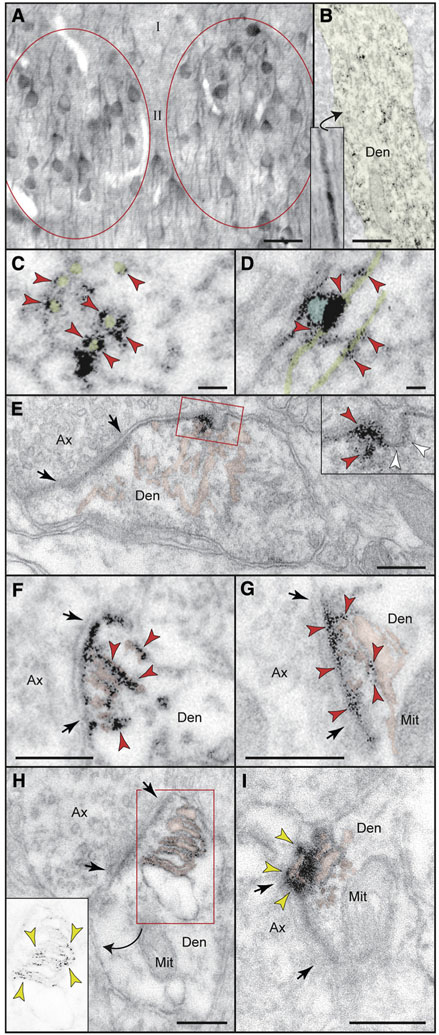
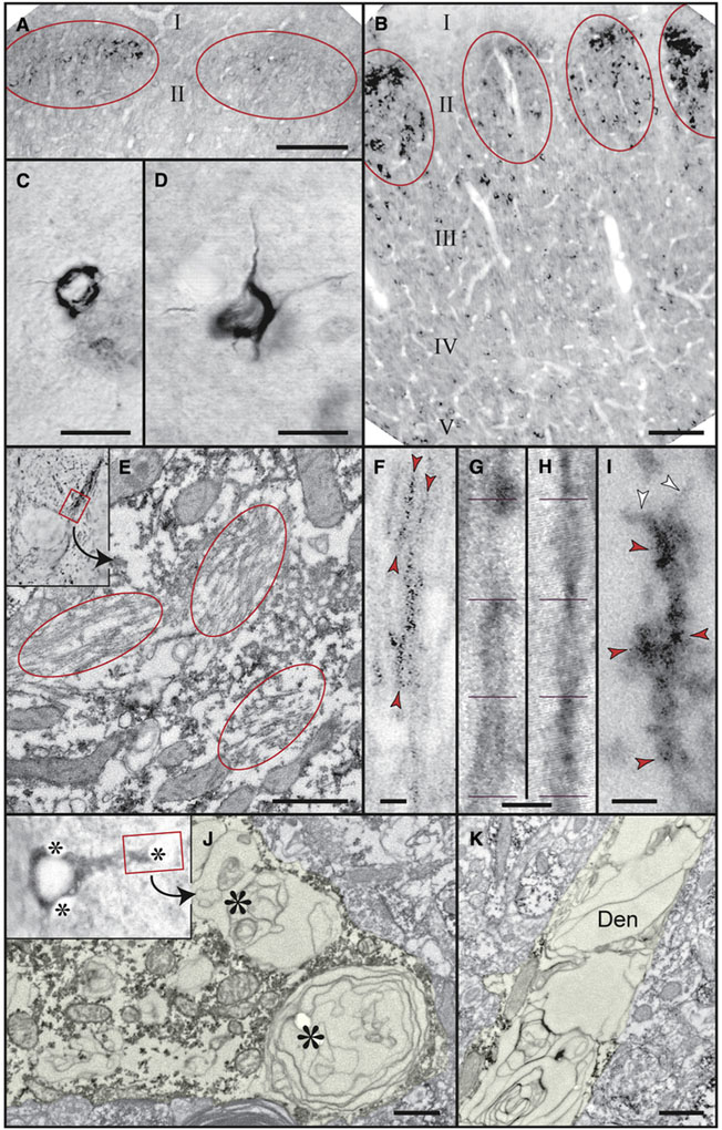
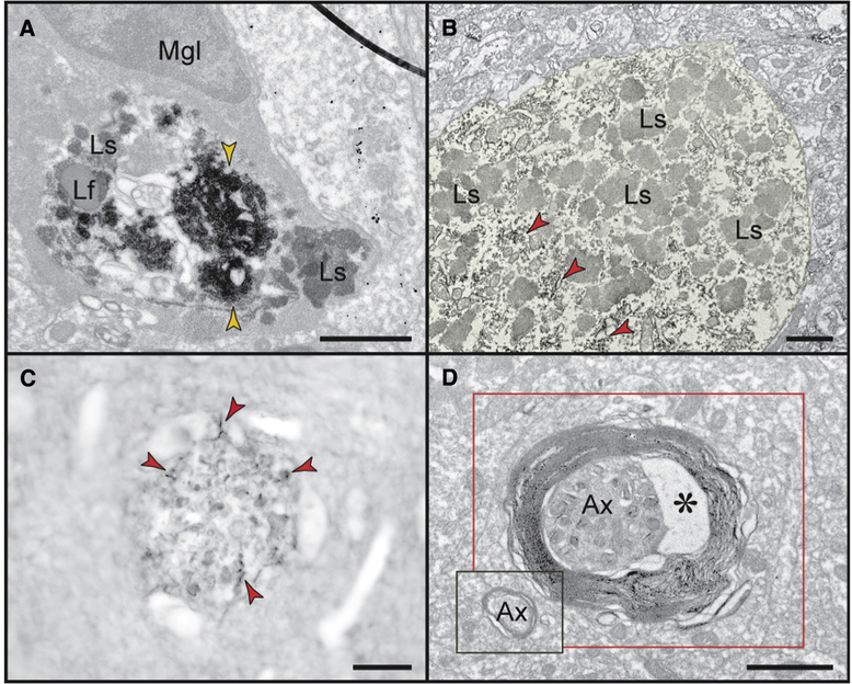
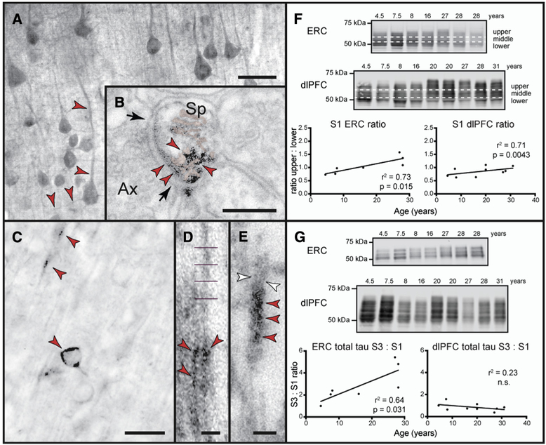
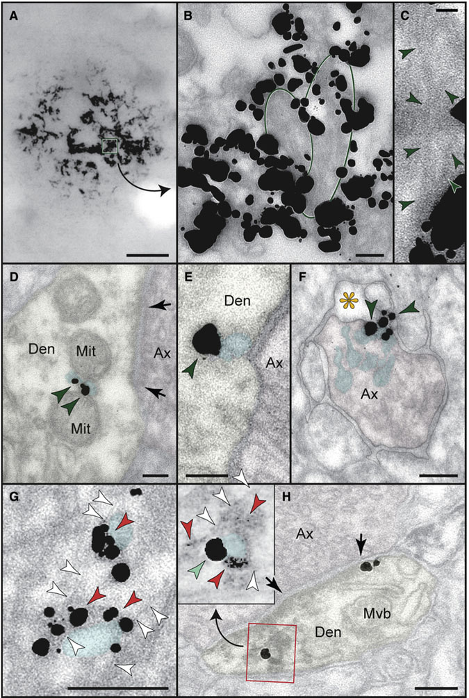
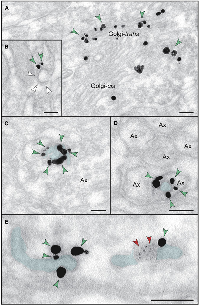
Verification
Claims the paper makes
The claims and statistics of this paper haven’t been checked yet.
Run the check — free, no account needed. It reads the open-access text and checks the sources it cites.
Claims other papers make about this one
Each is checked against this paper’s own text: does it say what the citing paper says it does?
In the citing paper
“Recent studies have indicated that adult rhesus monkeys can effectively model AD-related neurodegeneration in primate brain, and may represent a more appropriate model for AD [636, 637].”
From Molecular and cellular mechanisms underlying the pathogenesis of Alzheimer’s disease (2020) · cites this paper as [637]
Self-check
Percentages, sums and reported differences re-computed from the paper’s own numbers.
The self-check needs the paper’s full text.
Citations
Cites
Amyloid oligomers exacerbate tau pathology in a mouse model of tauopathy
Selenica, M.-L. B., Brownlow, M., Jimenez, J. P., Lee, D. C., Pena, G., Dickey, C. A., Gordon, M. N., & Morgan, D. (2013) · Neurodegenerative Diseases · doi:10.1159/000337230 · Cited by 10,359
Braak, H., Alafuzoff, I., Arzberger, T., Kretzschmar, H., & Del Tredici, K. (2006) · Acta Neuropathologica · doi:10.1007/s00401-006-0127-z · Cited by 3,172
Tau in physiology and pathology
Wang, Y., & Mandelkow, E. (2016) · Nature Reviews Neuroscience · doi:10.1038/nrn.2015.1 · Cited by 2,251
Stages of the Pathologic Process in Alzheimer Disease: Age Categories From 1 to 100 Years
Braak, H., Thal, D. R., Ghebremedhin, E., & Del Tredici, K. (2011) · Journal of Neuropathology & Experimental Neurology · doi:10.1097/nen.0b013e318232a379 · Cited by 2,194
Amyloid Precursor Protein Processing and Alzheimer's Disease
O'Brien, R. J., & Wong, P. C. (2011) · Annual Review of Neuroscience · doi:10.1146/annurev-neuro-061010-113613 · Cited by 2,061
Nelson, P. T., Alafuzoff, I., Bigio, E. H., Bouras, C., Braak, H., Cairns, N. J., Castellani, R. J., Crain, B. J., Davies, P., Tredici, K. D., et al. (2012) · Journal of Neuropathology & Experimental Neurology · doi:10.1097/nen.0b013e31825018f7 · Cited by 2,050 · cited 5×
“NFTs accumulate within degenerating neurons and correlate with cognitive deficits [4,5].”
BackgroundResultInfluentialIntracellular amyloid-β in Alzheimer's disease
LaFerla, F. M., Green, K. N., & Oddo, S. (2007) · Nature Reviews Neuroscience · doi:10.1038/nrn2168 · Cited by 1,942
Gene regulation and DNA damage in the ageing human brain
Lu, T., Pan, Y., Kao, S.-Y., Li, C., Kohane, I., Chan, J., & Yankner, B. A. (2004) · Nature · doi:10.1038/nature02661 · Cited by 1,874
Alzheimer's Disease and the Amyloid-β Peptide
Murphy, M. P., & LeVine, H. (2010) · Journal of Alzheimer's Disease · doi:10.3233/jad-2010-1221 · Cited by 1,750
Formation of Neurofibrillary Tangles in P301L Tau Transgenic Mice Induced by Aβ42 Fibrils
Götz, J., Chen, F., van Dorpe, J., & Nitsch, R. M. (2001) · Science · doi:10.1126/science.1062097 · Cited by 1,526
Cited by
Sorted by citations
Molecular and cellular mechanisms underlying the pathogenesis of Alzheimer’s disease
Guo, T., Zhang, D., Zeng, Y., Huang, T. Y., Xu, H., & Zhao, Y. (2020) · Molecular Neurodegeneration · doi:10.1186/s13024-020-00391-7 · Cited by 994
Synaptic degeneration in Alzheimer disease
Tzioras, M., McGeachan, R. I., Durrant, C. S., & Spires-Jones, T. L. (2023) · Nature Reviews Neurology · doi:10.1038/s41582-022-00749-z · Cited by 504
Hypothesis: Tau pathology is an initiating factor in sporadic Alzheimer's disease
Arnsten, A. F. T., Datta, D., Del Tredici, K., & Braak, H. (2021) · Alzheimer's & Dementia · doi:10.1002/alz.12192 · Cited by 361
Self-citationFranjic, D., Skarica, M., Ma, S., Arellano, J. I., Tebbenkamp, A. T. N., Choi, J., Xu, C., Li, Q., Morozov, Y. M., Andrijevic, D., et al. (2022) · Neuron · doi:10.1016/j.neuron.2021.10.036 · Cited by 308
Self-citationFernández-Calle, R., Konings, S. C., Frontiñán-Rubio, J., García-Revilla, J., Camprubí-Ferrer, L., Svensson, M., Martinson, I., Boza-Serrano, A., Venero, J. L., Nielsen, H. M., et al. (2022) · Molecular Neurodegeneration · doi:10.1186/s13024-022-00566-4 · Cited by 278
Cools, R., & Arnsten, A. F. T. (2021) · Neuropsychopharmacology · doi:10.1038/s41386-021-01100-8 · Cited by 216
Self-citationMicroglia in Aging and Alzheimer’s Disease: A Comparative Species Review
Edler, M. K., Mhatre-Winters, I., & Richardson, J. R. (2021) · Cells · doi:10.3390/cells10051138 · Cited by 126
Oligomeric Aβ in the monkey brain impacts synaptic integrity and induces accelerated cortical aging
Beckman, D., Ott, S., Donis-Cox, K., Janssen, W. G., Bliss-Moreau, E., Rudebeck, P. H., Baxter, M. G., & Morrison, J. H. (2019) · Proceedings of the National Academy of Sciences · doi:10.1073/pnas.1902301116 · Cited by 111
A novel tau‐based rhesus monkey model of Alzheimer's pathogenesis
Beckman, D., Chakrabarty, P., Ott, S. P., Dao, A., Zhou, E., Janssen, W. G. M., Donis‐Cox, K., Muller, S., Kordower, J. H., & Morrison, J. H. (2021) · Alzheimer s & Dementia · doi:10.1002/alz.12318 · Cited by 112
Mullane, K., & Williams, M. J. (2020) · Biochemical Pharmacology · doi:10.1016/j.bcp.2020.113945 · Cited by 109
O’Dell, R. S., Mecca, A. P., Chen, M.-K., Naganawa, M., Toyonaga, T., Lu, Y., Godek, T. A., Harris, J. E., Bartlett, H. H., Banks, E. R., et al. (2021) · Alzheimer s Research & Therapy · doi:10.1186/s13195-020-00742-y · Cited by 107
Self-citationDatta, D., Leslie, S. N., Wang, M., Morozov, Y. M., Yang, S., Mentone, S., Zeiss, C. J., Duque, A., RAKIC, P. T., Horváth, T. L., et al. (2021) · Alzheimer s & Dementia · doi:10.1002/alz.12325 · Cited by 102
Self-citationNeuroprosthetics: from sensorimotor to cognitive disorders
Gupta, A., Vardalakis, N., & Wagner, F. B. (2023) · Communications Biology · doi:10.1038/s42003-022-04390-w · Cited by 113
Alzheimer’s disease: A clinical perspective and future nonhuman primate research opportunities
Haque, R. U., & Levey, A. I. (2019) · Proceedings of the National Academy of Sciences · doi:10.1073/pnas.1912954116 · Cited by 99
Latimer, C. S., Shively, C. A., Keene, C. D., Jorgensen, M. J., Andrews, R. N., Register, T. C., Montine, T. J., Wilson, A. M., Neth, B. J., Mintz, A., et al. (2019) · Alzheimer's & Dementia · doi:10.1016/j.jalz.2018.06.3057 · Cited by 98
Galvin, V. C., Yang, S. T., Paspalas, C. D., Yang, Y., Jin, L. E., Datta, D., Morozov, Y. M., Lightbourne, T. C., Lowet, A. S., Rakic, P., et al. (2020) · Neuron · doi:10.1016/j.neuron.2020.02.030 · Cited by 79
Self-citationThe genie in the bottle-magnified calcium signaling in dorsolateral prefrontal cortex
Arnsten, A. F. T., Datta, D., & Wang, M. (2020) · Molecular Psychiatry · doi:10.1038/s41380-020-00973-3 · Cited by 79
Self-citationDatta, D., & Arnsten, A. (2019) · Brain Sciences · doi:10.3390/brainsci9050113 · Cited by 78
Self-citationDatta, D., Leslie, S. N., Morozov, Y. M., Duque, A., Rakic, P., van Dyck, C. H., Nairn, A. C., & Arnsten, A. F. T. (2020) · Journal of Neuroinflammation · doi:10.1186/s12974-019-1683-1 · Cited by 76
Self-citationArnsten, A. F. T., Datta, D., Leslie, S., Yang, S.-T., Wang, M., & Nairn, A. C. (2019) · Proceedings of the National Academy of Sciences · doi:10.1073/pnas.1903671116 · Cited by 74
Self-citationMckean, N. E., Handley, R. R., & Snell, R. G. (2021) · International Journal of Molecular Sciences · doi:10.3390/ijms222313168 · Cited by 81
Gilvesy, A., Husén, E., Maglóczky, Z., Mihály, O. S., Hortobágyi, T., Kanatani, S., Heinsen, H., Renier, N., Hökfelt, T. G. M., Mulder, J., et al. (2022) · Acta Neuropathologica · doi:10.1007/s00401-022-02477-6 · Cited by 77
Tredici, K. D., & Braak, H. (2019) · Current Opinion in Neurobiology · doi:10.1016/j.conb.2019.11.008 · Cited by 68
Amyloid-β: A double agent in Alzheimer's disease?
Yu, H., & Wu, J. (2021) · Biomedicine & Pharmacotherapy · doi:10.1016/j.biopha.2021.111575 · Cited by 62
Arnsten, A. F. T., Datta, D., & Preuss, T. M. (2021) · American Journal of Primatology · doi:10.1002/ajp.23254 · Cited by 66
Self-citationAge-related decline in cognitive flexibility in female chimpanzees
Agnès Lacreuse, A., Parr, L. A., Chennareddi, L. S., & Herndon, J. G. (2018) · Neurobiology of Aging · doi:10.1016/j.neurobiolaging.2018.08.018 · Cited by 57
Cognitive aging is associated with redistribution of synaptic weights in the hippocampus
Buss, E. W., Corbett, N. J., Roberts, J. G., Ybarra, N., Musial, T. F., Simkin, D., Molina-Campos, E., Oh, K.-J., Nielsen, L. L., Ayala, G. D., et al. (2021) · Proceedings of the National Academy of Sciences · doi:10.1073/pnas.1921481118 · Cited by 52
Prefrontal cortex and cognitive aging in macaque monkeys
Upright, N. A., & Baxter, M. G. (2021) · American Journal of Primatology · doi:10.1002/ajp.23250 · Cited by 44
Bimber, B. N., Yan, M. Y., Peterson, S. M., & Ferguson, B. M. (2019) · BMC Genomics · doi:10.1186/s12864-019-5559-7 · Cited by 46
Langerscheidt, F., Wied, T., Kabbani, M. A. A., van Eimeren, T., Wunderlich, G., & Zempel, H. (2024) · Journal of Neurology · doi:10.1007/s00415-024-12314-3 · Cited by 52
Comparative neuropathology in aging primates: A perspective
Freire‐Cobo, C., Edler, M. K., Varghese, M., Munger, E. L., Laffey, J., Raia, S., In, S. S., Wicinski, B. A., Medalla, M., Perez, S. E., et al. (2021) · American Journal of Primatology · doi:10.1002/ajp.23299 · Cited by 46
The common marmoset as a model of neurodegeneration
Perez‐Cruz, C., & Rodríguez-Callejas, J. D. (2023) · Trends in Neurosciences · doi:10.1016/j.tins.2023.02.002 · Cited by 39
Arnsten, A. F. T., Tredici, K. D., Barthélemy, N. R., Gabitto, M. I., van Dyck, C. H., Lein, E. S., Braak, H., & Datta, D. (2025) · Alzheimer s & Dementia · doi:10.1002/alz.70404 · Cited by 39
Self-citationThe Rhesus Macaque as a Translational Model for Neurodegeneration and Alzheimer’s Disease
Stonebarger, G. A., Bimonte‐Nelson, H. A., & Urbanski, H. F. (2021) · Frontiers in Aging Neuroscience · doi:10.3389/fnagi.2021.734173 · Cited by 38
Genetically modified non-human primate models for research on neurodegenerative diseases
Pan, M.-T., 暨南大学粤港澳中枢神经再生研究院, 广., Zhang, H., Li, X., & Guo, X. (2024) · 动物学研究 · doi:10.24272/j.issn.2095-8137.2023.197 · Cited by 36
Shastri, D., Raj, V., & Lee, S.-K. (2024) · Ageing Research Reviews · doi:10.1016/j.arr.2024.102379 · Cited by 34
Li, Z.-H., 650223,中国, 中. 昆., He, X., Li, H., He, R., Hu, X., 650204,中国, 中. 昆., 671000,中国, 大. 大., 200031,中国, 中., 650223,中国, 中. 昆., et al. (2020) · 动物学研究 · doi:10.24272/j.issn.2095-8137.2020.088 · Cited by 32
Modelling behaviors relevant to brain disorders in the nonhuman primate: Are we there yet?
Scott, J. T., & Bourne, J. A. (2021) · Progress in Neurobiology · doi:10.1016/j.pneurobio.2021.102183 · Cited by 31
de Sousa, A. A., Dames, B. A. R., Graff, E. C., Mohamedelhassan, R., Vassilopoulos, T., & Charvet, C. J. (2023) · Communications Biology · doi:10.1038/s42003-023-05034-3 · Cited by 36
Jiang, Z., Wang, J., Qin, Y., Liu, S., Luo, B., Bai, F., Wei, H., Zhang, S., Wei, J., Ding, G., et al. (2024) · Alzheimer s Research & Therapy · doi:10.1186/s13195-024-01392-0 · Cited by 32
Clinical relevance of animal models in aging-related dementia research
Padmanabhan, P., & Götz, J. (2023) · Nature Aging · doi:10.1038/s43587-023-00402-4 · Cited by 30
Experimental modeling of Alzheimer's disease: Translational lessons from cross‐taxon analyses
Yenkoyan, K. B., Kotova, M. M., Apukhtin, K. V., Galstyan, D. S., Amstislavskaya, T. G., Strekalova, T. V., de Abreu, M. S., Chavushyan, V. A., Lim, L. W., Yang, L., et al. (2025) · Alzheimer s & Dementia · doi:10.1002/alz.70273 · Cited by 30
Stonebarger, G. A., Urbanski, H. F., Woltjer, R. L., Vaughan, K. L., Ingram, D., Schultz, P., Calderazzo, S. M., Siedeman, J., Mattison, J., Rosene, D. L., et al. (2020) · GeroScience · doi:10.1007/s11357-020-00259-0 · Cited by 27
Self-citationChiu, M., Yang, S., Chen, T., Lin, C., Yang, F., Chen, W., Zetterberg, H., & Blennow, K. (2021) · ACS Chemical Neuroscience · doi:10.1021/acschemneuro.1c00010 · Cited by 29
PET Imaging in Animal Models of Alzheimer’s Disease
Chen, B., Marquez-Nostra, B. V., Belitzky, E., Toyonaga, T., Tong, J., Huang, Y. H., & Cai, Z. (2022) · Frontiers in Neuroscience · doi:10.3389/fnins.2022.872509 · Cited by 27
Darricau, M., Dou, C., Kinet, R., Zhu, T., Zhou, L., Li, X., Bedel, A., Claverol, S., Tokarski, C., Katsinelos, T., et al. (2023) · Alzheimer s & Dementia · doi:10.1002/alz.13604 · Cited by 30
Datta, D., Perone, I., Wijegunawardana, D., Liang, F., Morozov, Y. M., Arellano, J. I., Duque, A., Xie, Z., van Dyck, C. H., Joyce, M. K. P., et al. (2024) · Alzheimer s & Dementia · doi:10.1002/alz.13737 · Cited by 28
Self-citationAmyloid β and tau pathology in brains of aged pinniped species (sea lion, seal, and walrus)
Takaichi, Y., CHAMBERS, J. K., Takahashi, K., Soeda, Y., Koike, R., Katsumata, E., Kita, C., Matsuda, F., HARITANI, M., Takashima, A., et al. (2021) · Acta Neuropathologica Communications · doi:10.1186/s40478-020-01104-3 · Cited by 26
Rhesus monkeys as a translational model for late‐onset Alzheimer's disease
Souder, D. C., Dreischmeier, I. A., Smith, A., Wright, S., Martin, S. A., Sagar, M. A. K., Eliceiri, K. W., Salamat, S. M., Bendlin, B. B., Colman, R. J., et al. (2021) · Aging Cell · doi:10.1111/acel.13374 · Cited by 25
Maggiore, A., Latina, V., D’Erme, M., Amadoro, G., & Coccurello, R. (2024) · Ageing Research Reviews · doi:10.1016/j.arr.2024.102578 · Cited by 25
Rodríguez-Callejas, J. D., Fuchs, E., & Perez‐Cruz, C. (2023) · Neurobiology of Aging · doi:10.1016/j.neurobiolaging.2023.04.010 · Cited by 23
Zinc in Regulating Protein Kinases and Phosphatases in Neurodegenerative Diseases
Zhang, H.-L., Wang, X., & Liu, R. (2022) · Biomolecules · doi:10.3390/biom12060785 · Cited by 20
Cognitive Deficits and Alzheimer’s Disease-Like Pathologies in the Aged Chinese Tree Shrew
Li, H., Xiang, B.-L., Li, X., Li, C., Li, Y., Miao, Y., Ma, G., Ma, Y., Chen, J., Zhang, Q., et al. (2023) · Molecular Neurobiology · doi:10.1007/s12035-023-03663-7 · Cited by 21
Diet, psychosocial stress, and Alzheimer's disease–related neuroanatomy in female nonhuman primates
Frye, B. M., Craft, S., Register, T. C., Andrews, R. N., Appt, S. E., Vitolins, M. Z., Uberseder, B., Silverstein‐Metzler, M. G., Chen, H., Whitlow, C. T., et al. (2020) · Alzheimer s & Dementia · doi:10.1002/alz.12232 · Cited by 19
Neuropsychology of cognitive aging in rhesus monkeys
Baxter, M. G., Roberts, M. T., Roberts, J. A., & Rapp, P. R. (2023) · Neurobiology of Aging · doi:10.1016/j.neurobiolaging.2023.06.011 · Cited by 18
Jin, Z., Lu, Y., Tang, H., & Cui, H. (2025) · Alzheimer s & Dementia · doi:10.1002/alz.70472 · Cited by 24
Wang, J., Wang, M., Moshiri, A., Harris, R. A., Raveendran, M., Nguyen, T., Kim, S., Young, L. J., Wang, K., Wiseman, R., et al. (2024) · Nature Communications · doi:10.1038/s41467-024-49922-6 · Cited by 18
Towards developing a rhesus monkey model of early Alzheimer's disease focusing on women's health
Beckman, D., & Morrison, J. H. (2021) · American Journal of Primatology · doi:10.1002/ajp.23289 · Cited by 17
Molecular Diversity Among Adult Human Hippocampal and Entorhinal CellsPreprint
Franjic, D., Choi, J., Škarica, M., Xu, C., Li, Q., Ma, S., Tebbenkamp, A. T. N., Santpere, G., Arellano, J. I., Gudelj, I., et al. (2020) · bioRxiv (Cold Spring Harbor Laboratory) · doi:10.1101/2019.12.31.889139 · Cited by 16
Philippens, I. H. C. H. M., & Langermans, J. A. M. (2021) · Vaccines · doi:10.3390/vaccines9040388 · Cited by 16
Animal models of Alzheimer’s disease embrace diversity
Neff, E. P. (2019) · Lab Animal · doi:10.1038/s41684-019-0377-8 · Cited by 17
Jester, H. M., Gosrani, S. P., Ding, H., Zhou, X., Ko, M., & Ma, T. (2022) · Journal of Alzheimer s Disease · doi:10.3233/jad-215303 · Cited by 15
Wakeman, D. R., Weed, M. R., Perez, S. E., Cline, E. N., Viola, K. L., Wilcox, K. C., Moddrelle, D. S., Nisbett, E. Z., Kurian, A., Bell, A., et al. (2022) · Neuropathology and Applied Neurobiology · doi:10.1111/nan.12800 · Cited by 18
Datta, D., Perone, I., Morozov, Y. M., Arellano, J. I., Duque, A., RAKIC, P. T., van Dyck, C. H., & Arnsten, A. F. T. (2023) · Cerebral Cortex · doi:10.1093/cercor/bhad382 · Cited by 16
Self-citationUtility of spontaneous animal models of Alzheimer’s disease in preclinical efficacy studies
Zeiss, C. J. (2020) · Cell and Tissue Research · doi:10.1007/s00441-020-03198-6 · Cited by 16
Leslie, S. N., Kanyo, J. E., Datta, D., Wilson, R. S., Zeiss, C. J., Duque, A., Lam, T. T., Arnsten, A. F. T., & Nairn, A. C. (2021) · Frontiers in Aging Neuroscience · doi:10.3389/fnagi.2021.767322 · Cited by 14
Self-citationTimofeeva, A. M., Aulova, K. S., & Nevinsky, G. A. (2025) · Brain Sciences · doi:10.3390/brainsci15050486 · Cited by 17
Huhe, H., Shapley, S. M., Duong, D. M., Wu, F., Ha, S.-K., Choi, S., Kofler, J., Mou, Y., Guimarães, T. R., Thathiah, A., et al. (2024) · Alzheimer s & Dementia · doi:10.1002/alz.14366 · Cited by 12
Datta, D., & Arnsten, A. F. T. (2025) · Alzheimer s & Dementia · doi:10.1002/alz.14477 · Cited by 11
Self-citationBathla, S., Datta, D., Liang, F., Barthélemy, N. R., Wiseman, R., Slusher, B. S., Asher, J. L., Zeiss, C. J., Ekanayake‐Alper, D., Holden, D., et al. (2023) · Alzheimer s & Dementia Translational Research & Clinical Interventions · doi:10.1002/trc2.12431 · Cited by 11
Self-citationBrain aging and Alzheimer’s disease, a perspective from non-human primates
Ferrer, I. (2024) · Aging · doi:10.18632/aging.206143 · Cited by 10
Merino‐Serrais, P., Soria, J. M., Arrabal, C. A., Ortigado-López, A., Esparza, M. Á. G., Muñoz, A., Hernández, F., Ávila, J., DeFelipe, J., & León‐Espinosa, G. (2024) · Experimental Neurology · doi:10.1016/j.expneurol.2024.115049 · Cited by 10
Aged rhesus monkeys: Cognitive performance categorizations and preclinical drug testing
Plagenhoef, M. R., Callahan, P. M., Beck, W. D., Blake, D. T., & Terry, A. V. (2021) · Neuropharmacology · doi:10.1016/j.neuropharm.2021.108489 · Cited by 10
Modeling neurodegenerative diseases using non-human primates: advances and challenges
Li, B., He, D., Li, X., & Guo, X. (2022) · Ageing and Neurodegenerative Diseases · doi:10.20517/and.2022.14 · Cited by 11
Bearded capuchin monkeys as a model for Alzheimer’s disease
Rodriguez, R. D., Tavares, M. C. H., Brucki, S. M. D., Takada, L. T., Otaduy, M. C. G., Martin, M. D. G. M., Suemoto, C. K., Grinberg, L. T., Leite, C. C., Tomaz, C. A. B., et al. (2024) · Scientific Reports · doi:10.1038/s41598-024-56791-y · Cited by 10
Singhaarachchi, P. H., Antal, P., Calon, F., Culmsee, C., Delpech, J.-C., Feldotto, M., Geertsema, J., Hoeksema, E. E., Kőrösi, A., Layé, S., et al. (2025) · Progress in Neurobiology · doi:10.1016/j.pneurobio.2025.102821 · Cited by 13
Cerebral Aβ deposition in an Aβ-precursor protein-transgenic rhesus monkey
Chan, A. W. S., Cho, I. K., Li, C., Zhang, X., Patel, S., Rusnak, R., Raper, J., Bachevalier, J., Moran, S. P., Chi, T., et al. (2022) · Aging Brain · doi:10.1016/j.nbas.2022.100044 · Cited by 10
The NAAG’ing Concerns of Modeling Human Alzheimer’s Disease in Mice
Hollinger, K. R., Alt, J., Rais, R., Kaplin, A. I., & Slusher, B. S. (2019) · Journal of Alzheimer s Disease · doi:10.3233/jad-181251 · Cited by 9
Non-human Primate Models of Alzheimer’s Disease
Li, Y., & Gu, B. J. (2023) · Journal of Exploratory Research in Pharmacology · doi:10.14218/jerp.2023.00006 · Cited by 8
Expression of tau and phosphorylated tau in the brain of normal and hemiparkinsonian rhesus macaques
Gambardella, J. C., Schoephoerster, W., Bondarenko, V., Yandell, B. S., & Emborg, M. E. (2023) · The Journal of Comparative Neurology · doi:10.1002/cne.25490 · Cited by 8
Domain-Specific Cognitive Impairment Reflects Prefrontal Dysfunction in Aged Common Marmosets
Glavis‐Bloom, C., Vanderlip, C. R., Reynolds, J. H., & Reynolds, J. H. (2023) · eNeuro · doi:10.1523/eneuro.0187-23.2023 · Cited by 8
Chami, M., & Checler, F. (2020) · Current Alzheimer Research · doi:10.2174/1567205017666200225102941 · Cited by 8
Senile plaques and phosphorylated tau deposition in a super-aged rhesus monkey (Macaca mulatta)
Iwaide, S., Nakayama, Y., CHAMBERS, J. K., Uchida, K., Nakagawa, D., Yamanashi, Y., Bando, H., & MURAKAMI, T. (2023) · Journal of Veterinary Medical Science · doi:10.1292/jvms.23-0313 · Cited by 8
Barnes, C. A., Permenter, M. R., Vogt, J. A., Chen, K., & Beach, T. G. (2024) · The Journal of Comparative Neurology · doi:10.1002/cne.25670 · Cited by 8
Arnsten, A. F. T., Perone, I., Wang, M., Yang, S., Uchendu, S., Bolat, D., & Datta, D. (2025) · Frontiers in Aging Neuroscience · doi:10.3389/fnagi.2025.1610350 · Cited by 7
Self-citationThe aged female rhesus macaque as a translational model for human menopause and hormone therapy
Kohama, S. G., & Urbanski, H. F. (2024) · Hormones and Behavior · doi:10.1016/j.yhbeh.2024.105658 · Cited by 8
Metzler, L. A. P., Metzger, J. M., Gerred, K., Emborg, M. E., & Kapoor, A. (2025) · Experimental Gerontology · doi:10.1016/j.exger.2025.112736 · Cited by 7
Tau and tauopathies across primate species: implications for modeling neurodegenerative disorders
Colwell, J. C., & Emborg, M. E. (2025) · Frontiers in Aging Neuroscience · doi:10.3389/fnagi.2025.1598245 · Cited by 10
Targeting Calpain-2 for Alzheimer’s Disease Treatment
Arnsten, A. F. T., & Baudry, M. (2023) · Medical Research Archives · doi:10.18103/mra.v11i2.3487 · Cited by 6
Self-citationCrimins, J. L., Puri, R., Calakos, K. C., Yuk, F., Janssen, W. G. M., Hara, Y., Rapp, P. R., & Morrison, J. H. (2018) · The Journal of Comparative Neurology · doi:10.1002/cne.24576 · Cited by 6
Self-citationZeiss, C. J., Huttner, A. J., Nairn, A. C., Arnsten, A. F. T., Datta, D., Strittmatter, S. M., Wyk, B. V., & Duque, A. (2025) · Journal of Alzheimer s Disease · doi:10.1177/13872877251323787 · Cited by 5
Self-citationBathla, S., Datta, D., Bolat, D., Woo, E., Duque, A., Arellano, J. I., Arnsten, A. F. T., & Nairn, A. C. (2025) · Frontiers in Aging Neuroscience · doi:10.3389/fnagi.2025.1549770 · Cited by 5
Self-citationAmyloid-β Pathology Is the Common Nominator Proteinopathy of the Primate Brain Aging
Ferrer, I. (2024) · Journal of Alzheimer s Disease · doi:10.3233/jad-240389 · Cited by 7
Pathological Markers of Alzheimer’s Disease and Related Dementia in the Rhesus Macaque Amygdala
Thomas, J. L., Nilaver, B. I., Lomniczi, A., Brown, D. I., Appleman, M., Kohama, S. G., & Urbanski, H. F. (2024) · Journal of Alzheimer s Disease Reports · doi:10.3233/adr-230184 · Cited by 5
Nonhuman primates as models for aging and Alzheimer’s disease
Edler, M. K., Munger, E. L., Groetz, H., & Raghanti, M. A. (2021) · Elsevier eBooks · doi:10.1016/b978-0-12-818000-6.00047-0 · Cited by 6
A comparison of behavioral paradigms assessing spatial memory in tree shrews
Li, C.-J., Hui, Y.-Q., Zhang, R., Zhou, H.-Y., Cai, X., & Lu, L. (2023) · Cerebral Cortex · doi:10.1093/cercor/bhad283 · Cited by 5
Wang, W., Huang, R., Lv, L., Ma, X., Li, Z., Zhang, Y., Wu, J., Wu, S., Xu, J., Hu, Y., et al. (2026) · Proceedings of the National Academy of Sciences · doi:10.1073/pnas.2529565123 · Cited by 4
Inhibition of brain glutamate carboxypeptidase II (GCPII) to enhance cognitive function
Wiseman, R., Bigos, K. L., Arnsten, A. F. T., & Slusher, B. S. (2024) · Advances in pharmacology · doi:10.1016/bs.apha.2024.10.018 · Cited by 4
Self-citationFuture directions in animal models of Alzheimer’s disease
Beckman, D., Baxter, M. G., & Morrison, J. H. (2018) · Journal of Neuroscience Research · doi:10.1002/jnr.24328 · Cited by 4
Naturally occurring Alzheimer’s disease in rhesus monkeysPreprint
Li, Z., He, X., Wu, S., Huang, R., Li, H., Wang, Z., Wang, L., Qin, D., Kong, Y., Guo, Y., et al. (2022) · bioRxiv (Cold Spring Harbor Laboratory) · doi:10.1101/2022.10.20.513120 · Cited by 5
Metadata
- Authors
- Paspalas, C. D., Carlyle, B. C., Leslie, S., Preuss, T. M., Crimins, J. L., Huttner, A. J., van Dyck, C. H., Rosene, D. L., Nairn, A. C., & Arnsten, A. F. T.
- Affiliations
- Yale University (US)
- Emory University (US)
- Department of Neuroscience Yale School of Medicine New Haven CT USA
- Boston University (US)
- Corresponding author
- Paspalas, Constantinos D.
- Arnsten, Amy F.T.
- ORCID
- Venue
- Alzheimer's & Dementia
- Publisher
- Wiley
- Published
- 2017-12-11
- Volume
- 14
- Issue
- 5
- Pages
- 680-691
- ISSN
- 1552-5260 (ISSN-L)
- 1552-5279
- PMC
- PMC6178089
- OpenAlex
- W2772269246
- Cited by
- 140
- References
- 51
- Citations by year
- 2026: 14 · 2025: 22 · 2024: 21 · 2023: 20 · 2022: 11 · 2021: 27 · 2020: 13 · 2019: 8 · 2018: 4
- FWCI
- 5.06
- Type
- journal article
- Language
- en
- Open access
- hybrid
- doi.org
- Licence
- Locations
- Alzheimer s & Dementia (publishedVersion, cc-by-nc-nd)
- PubMed (publishedVersion)
- PubMed Central (acceptedVersion, cc-by-nc-nd)
- Funders
- National Institute on Aging
- National Institutes of Health (DP1AG047744, P50‐AG047270, RO1 AG043640)
- Yerkes National Primate Research Center, Emory University (OD P51OD11132)
- Topics
- Life Sciences › Neuroscience › Cellular and Molecular Neuroscience › Alzheimer's disease research and treatments
- Health Sciences › Medicine › Psychiatry and Mental health › Dementia and Cognitive Impairment Research
- Life Sciences › Neuroscience › Neurology › Neuroinflammation and Neurodegeneration Mechanisms
- Keywords
- Alzheimer's disease, rhesus monkeys
- MeSH terms
- Aging, Alzheimer Disease, Amyloid, Animals, Brain, Disease Models, Animal, Macaca mulatta, Phosphorylation, Microscopy, Immunoelectron, Neurofibrillary Tangles, tau Proteins, Prefrontal Cortex, Disease Progression, Entorhinal Cortex, Plaque, Amyloid
About these checks
Registry, retraction, Cites and statistics results are computed, not judged, with one exception: a printed reference that no index lists is read once by a model before it counts as a citation error. Claims are extracted by a model and kept only when their quote appears verbatim in the text; each cited source is then checked, with a model’s help, for whether it says what the claim relies on. Those verdicts can be wrong — read the claim’s quote and the cited source before relying on one.
Statistics and arithmetic are recomputed, not judged.
Confidence bands are shown only for fields calibrated on our annotated test set. None is calibrated yet, so no band is shown.
What counts as a claim ›How we measure confidence ›How we check this ›
Are you an author, or do you see a problem with this page? Request a review
