

The DOI matches its Crossref record; 1 of the 1 references it cites were found in a registry or an index.
Outer surface lipoproteins from the Lyme disease spirochete exploit the molecular switch mechanism of the complement protease C1s
Open accessPublished version of a preprint
Abstract
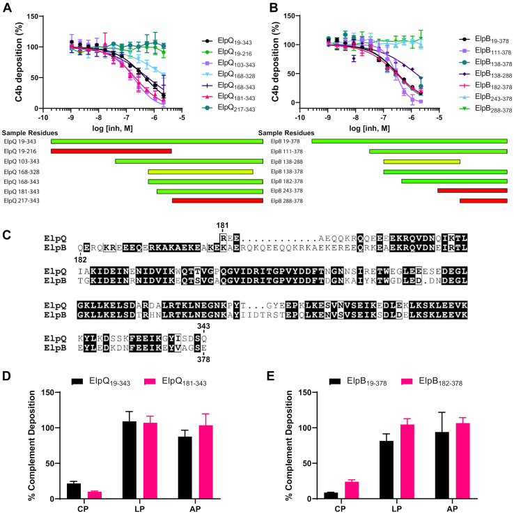
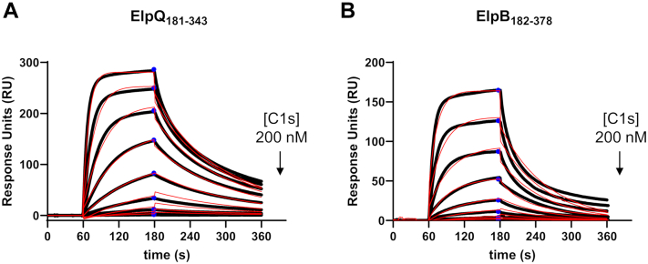
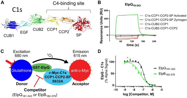
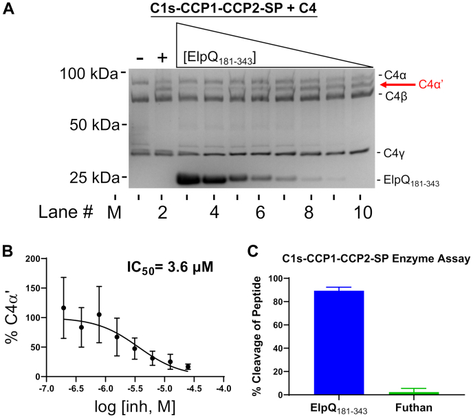
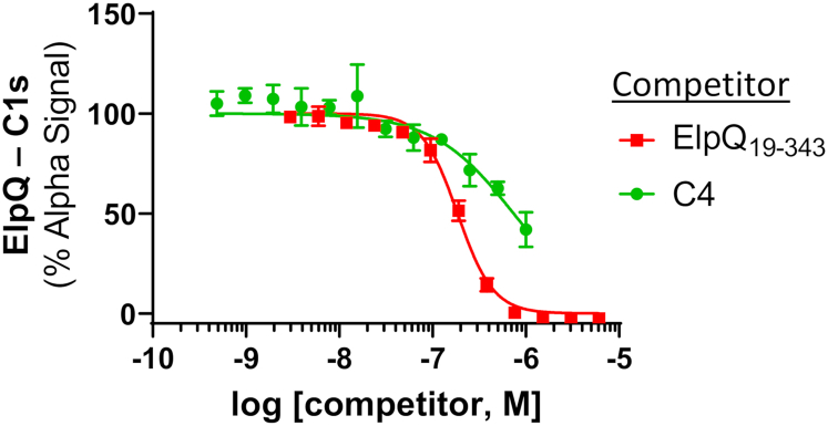
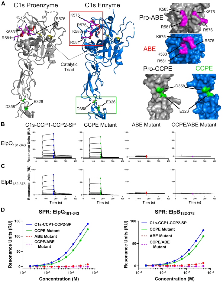
Proteolytic cascades comprise several important physiological systems, including a primary arm of innate immunity called the complement cascade. To safeguard against complement-mediated attack, the etiologic agent of Lyme disease, Borreliella burgdorferi, produces numerous outer surface-localized lipoproteins that contribute to successful complement evasion. Recently, we discovered a pair of B. burgdorferi surface lipoproteins of the OspEF-related protein family-termed ElpB and ElpQ-that inhibit antibody-mediated complement activation. In this study, we investigate the molecular mechanism of ElpB and ElpQ complement inhibition using an array of biochemical and biophysical approaches. In vitro assays of complement activation show that an independently folded homologous C-terminal domain of each Elp protein maintains full complement inhibitory activity and selectively inhibits the classical pathway. Using binding assays and complement component C1s enzyme assays, we show that binding of Elp proteins to activated C1s blocks complement component C4 cleavage by competing with C1s-C4 binding without occluding the active site. C1s-mediated C4 cleavage is dependent on activation-induced binding sites, termed exosites. To test whether these exosites are involved in Elp-C1s binding, we performed site-directed mutagenesis, which showed that ElpB and ElpQ binding require C1s residues in the anion-binding exosite located on the serine protease domain of C1s. Based on these results, we propose a model whereby ElpB and ElpQ exploit activation-induced conformational changes that are normally important for C1s-mediated C4 cleavage. Our study expands the known complement evasion mechanisms of microbial pathogens and reveals a novel molecular mechanism for selective C1s inhibition by Lyme disease spirochetes.
Overview
The claims and statistics of this paper haven’t been checked yet.
Run the check — free, no account needed. It reads the open-access text and checks the sources it cites.
Integrity signals
Registry
Cites
Statistics
Figures
Verification
Claims the paper makes
The claims and statistics of this paper haven’t been checked yet.
Run the check — free, no account needed. It reads the open-access text and checks the sources it cites.
Self-check
Percentages, sums and reported differences re-computed from the paper’s own numbers.
The self-check needs the paper’s full text.
Citations
Cites
Highly accurate protein structure prediction with AlphaFold
Jumper, J., Evans, R., Pritzel, A., Green, T., Figurnov, M., Ronneberger, O., Tunyasuvunakool, K., Bates, R., Žídek, A., Potapenko, A., et al. (2021) · Nature · doi:10.1038/s41586-021-03819-2 · Cited by 43,764
Research works printed but not linked: 65
- Merle, 2015, Complement system part I - molecular mechanisms of activation and regulation, Front. Immunol., 6, 262, 10.3389/fimmu.2015.00262 · DOI · Paper page
- Merle, 2015, Complement system part II: role in immunity, Front. Immunol., 6, 257, 10.3389/fimmu.2015.00257 · DOI
- Walport, 2001, Complement. First of two parts, N. Engl. J. Med., 344, 1058, 10.1056/NEJM200104053441406 · DOI
- Walport, 2001, Complement. Second of two parts, N. Engl. J. Med., 344, 1140, 10.1056/NEJM200104123441506 · DOI
- Schechter, 1967, On the size of the active site in proteases. I. Papain, Biochem. Biophys. Res. Commun., 27, 157, 10.1016/S0006-291X(67)80055-X · DOI
- Duncan, 2012, Identification of a catalytic exosite for complement component C4 on the serine protease domain of C1s, J. Immunol., 189, 2365, 10.4049/jimmunol.1201085 · DOI
- Perry, 2013, A molecular switch governs the interaction between the human complement protease C1s and its substrate, complement C4, J. Biol. Chem., 288, 15821, 10.1074/jbc.M113.464545 · DOI
- Gaboriaud, 2000, Crystal structure of the catalytic domain of human complement c1s: a serine protease with a handle, EMBO J., 19, 1755, 10.1093/emboj/19.8.1755 · DOI
- Kidmose, 2012, Structural basis for activation of the complement system by component C4 cleavage, Proc. Natl. Acad. Sci. U. S. A., 109, 15425, 10.1073/pnas.1208031109 · DOI
- Bock, 2007, Exosites in the substrate specificity of blood coagulation reactions, J. Thromb. Haemost., 5 Suppl 1, 81, 10.1111/j.1538-7836.2007.02496.x · DOI
- Schmidt, 2016, Protection of host cells by complement regulators, Immunol. Rev., 274, 152, 10.1111/imr.12475 · DOI
- Ricklin, 2013, Complement in immune and inflammatory disorders: therapeutic interventions, J. Immunol., 190, 3839, 10.4049/jimmunol.1203200 · DOI
- Lambris, 2008, Complement evasion by human pathogens, Nat. Rev. Microbiol., 6, 132, 10.1038/nrmicro1824 · DOI
- Garcia, 2016, Novel evasion mechanisms of the classical complement pathway, J. Immunol., 197, 2051, 10.4049/jimmunol.1600863 · DOI
- Kugeler, 2021, Estimating the frequency of Lyme disease diagnoses, United States, 2010-2018, Emerg. Infect. Dis., 27, 616, 10.3201/eid2702.202731 · DOI
- Skare, 2020, Complement evasion by Lyme disease spirochetes, Trends Microbiol., 28, 889, 10.1016/j.tim.2020.05.004 · DOI
- Coburn, 2021, Lyme disease pathogenesis, Curr. Issues Mol. Biol., 42, 473
- Kraiczy, 2013, Complement regulator-acquiring surface proteins of Borrelia burgdorferi: structure, function and regulation of gene expression, Ticks Tick Borne Dis., 4, 26, 10.1016/j.ttbdis.2012.10.039 · DOI
- Lin, 2020, New insights into CRASP-mediated complement evasion in the Lyme disease enzootic cycle, Front. Cell. Infect. Microbiol.
- Kraiczy, 2001, Mechanism of complement resistance of pathogenic Borrelia burgdorferi isolates, Int. Immunopharmacol., 1, 393, 10.1016/S1567-5769(00)00041-2 · DOI
- Hartmann, 2006, Functional characterization of BbCRASP-2, a distinct outer membrane protein of Borrelia burgdorferi that binds host complement regulators factor H and FHL-1, Mol. Microbiol., 61, 1220, 10.1111/j.1365-2958.2006.05318.x · DOI
- McDowell, 2006, Evidence that the BBA68 protein (BbCRASP-1) of the Lyme disease spirochetes does not contribute to factor H-mediated immune evasion in humans and other animals, Infect. Immun., 74, 3030, 10.1128/IAI.74.5.3030-3034.2006 · DOI
- McDowell, 2003, Comprehensive analysis of the factor H binding capabilities of Borrelia species associated with Lyme disease: delineation of two distinct classes of factor H binding proteins, Infect. Immun., 71, 3597, 10.1128/IAI.71.6.3597-3602.2003 · DOI
- Bykowski, 2008, Borrelia burgdorferi complement regulator-acquiring surface proteins (BbCRASPs): expression patterns during the mammal-tick infection cycle, Int. J. Med. Microbiol., 298, 249, 10.1016/j.ijmm.2007.10.002 · DOI
- Kenedy, 2009, CspA-mediated binding of human factor H inhibits complement deposition and confers serum resistance in Borrelia burgdorferi, Infect. Immun., 77, 2773, 10.1128/IAI.00318-09 · DOI
- Rogers, 2009, Comparative analysis of the properties and ligand binding characteristics of CspZ, a factor H binding protein, derived from Borrelia burgdorferi isolates of human origin, Infect. Immun., 77, 4396, 10.1128/IAI.00393-09 · DOI
- Hallström, 2013, CspA from Borrelia burgdorferi inhibits the terminal complement pathway, mBio, 4, 10.1128/mBio.00481-13 · DOI
- Garcia, 2016, Borrelia burgdorferi BBK32 inhibits the classical pathway by blocking activation of the C1 complement complex, PLoS Pathog., 12, 10.1371/journal.ppat.1005404 · DOI
- Xie, 2019, Structural determination of the complement inhibitory domain of Borrelia burgdorferi BBK32 provides insight into classical pathway complement evasion by Lyme disease spirochetes, PLoS Pathog., 15, 10.1371/journal.ppat.1007659 · DOI
- Garrigues, 2021, A structural basis for inhibition of the complement initiator protease C1r by Lyme disease spirochetes, J. Immunol., 207, 2856, 10.4049/jimmunol.2100815 · DOI
- Pereira, 2022, Lipoproteome screening of the Lyme disease agent identifies inhibitors of antibody-mediated complement killing, Proc. Natl. Acad. Sci. U. S. A., 119, 10.1073/pnas.2117770119 · DOI
- Dowdell, 2017, Comprehensive spatial analysis of the Borrelia burgdorferi lipoproteome reveals a compartmentalization bias toward the bacterial surface, J. Bacteriol., 199, 10.1128/JB.00658-16 · DOI
- Stevenson, 1996, A family of genes located on four separate 32-kilobase circular plasmids in Borrelia burgdorferi B31, J. Bacteriol., 178, 3508, 10.1128/jb.178.12.3508-3516.1996 · DOI
- Akins, 1999, Molecular and evolutionary analysis of Borrelia burgdorferi 297 circular plasmid-encoded lipoproteins with OspE- and OspF-like leader peptides, Infect. Immun., 67, 1526, 10.1128/IAI.67.3.1526-1532.1999 · DOI
- Marconi, 1996, Molecular and evolutionary analyses of a variable series of genes in Borrelia burgdorferi that are related to ospE and ospF, constitute a gene family, and share a common upstream homology box, J. Bacteriol., 178, 5615, 10.1128/jb.178.19.5615-5626.1996 · DOI
- Brissette, 2008, Lyme borreliosis spirochete Erp proteins, their known host ligands, and potential roles in mammalian infection, Int. J. Med. Microbiol., 298 Suppl 1, 257, 10.1016/j.ijmm.2007.09.004 · DOI
- Stevenson, 2022, The consistent tick-vertebrate infectious cycle of the lyme disease spirochete enables Borrelia burgdorferi to control protein expression by monitoring its physiological status, J. Bacteriol., 204, 10.1128/jb.00606-21 · DOI
- Brissette, 2009, The Borrelia burgdorferi outer-surface protein ErpX binds mammalian laminin, Microbiology (Reading), 155, 863, 10.1099/mic.0.024604-0 · DOI
- Stevenson, 1998, Borrelia burgdorferi Erp proteins are immunogenic in mammals infected by tick bite, and their synthesis is inducible in cultured bacteria, Infect. Immun., 66, 2648, 10.1128/IAI.66.6.2648-2654.1998 · DOI
- Bielefeld-Sevigny, 2009, AlphaLISA immunoassay platform- the “no-wash” high-throughput alternative to ELISA, Assay Drug Dev. Technol., 7, 90, 10.1089/adt.2009.9996 · DOI
- Hart, 2021, Host tropism determination by convergent evolution of immunological evasion in the Lyme disease system, PLoS Pathog., 17, 10.1371/journal.ppat.1009801 · DOI
- Bhattacharjee, 2013, Structural basis for complement evasion by Lyme disease pathogen Borrelia burgdorferi, J. Biol. Chem., 288, 18685, 10.1074/jbc.M113.459040 · DOI
- Brangulis, 2015, Crystal structures of the Erp protein family members ErpP and ErpC from Borrelia burgdorferi reveal the reason for different affinities for complement regulator factor H, Biochim. Biophys. Acta, 1854, 349, 10.1016/j.bbapap.2014.12.025 · DOI
- Zückert, 2014, Secretion of bacterial lipoproteins: through the cytoplasmic membrane, the periplasm and beyond, Biochim. Biophys. Acta, 1843, 1509, 10.1016/j.bbamcr.2014.04.022 · DOI
- Zückert, 2019, Protein secretion in spirochetes, Microbiol. Spectr., 10.1128/microbiolspec.PSIB-0026-2019 · DOI
- Booth, 2022, Borrelia miyamotoi FbpA and FbpB are immunomodulatory outer surface lipoproteins with distinct structures and functions, Front. Immunol., 13, 886733, 10.3389/fimmu.2022.886733 · DOI
- Clark, 2011, A structural basis for Staphylococcal complement subversion: X-ray structure of the complement-binding domain of Staphylococcus aureus protein Sbi in complex with ligand C3d, Mol. Immunol., 48, 452, 10.1016/j.molimm.2010.09.017 · DOI
- Hammel, 2007, Characterization of Ehp, a secreted complement inhibitory protein from Staphylococcus aureus, J. Biol. Chem., 282, 30051, 10.1074/jbc.M704247200 · DOI
- Hammel, 2007, A structural basis for complement inhibition by Staphylococcus aureus, Nat. Immunol., 8, 430, 10.1038/ni1450 · DOI
- Ricklin, 2008, Cutting edge: members of the Staphylococcus aureus extracellular fibrinogen-binding protein family inhibit the interaction of C3d with complement receptor 2, J. Immunol., 181, 7463, 10.4049/jimmunol.181.11.7463 · DOI
- Amdahl, 2013, Staphylococcal Ecb protein and host complement regulator factor H enhance functions of each other in bacterial immune evasion, J. Immunol., 191, 1775, 10.4049/jimmunol.1300638 · DOI
- Rooijakkers, 2009, Structural and functional implications of the alternative complement pathway C3 convertase stabilized by a staphylococcal inhibitor, Nat. Immunol., 10, 721, 10.1038/ni.1756 · DOI
- Takacs, 2020, A CRISPR interference platform for selective downregulation of gene expression in Borrelia burgdorferi, Appl. Environ. Microbiol., 87, e02519
- Gaboriaud, 2013, The serine protease domain of MASP-3: enzymatic properties and crystal structure in complex with ecotin, PLoS One, 8, 10.1371/journal.pone.0067962 · DOI
- Nagy, 2022, Synergy of protease-binding sites within the ecotin homodimer is crucial for inhibition of MASP enzymes and for blocking lectin pathway activation, J. Biol. Chem., 298, 101985, 10.1016/j.jbc.2022.101985 · DOI
- Farady, 2010, Mechanisms of macromolecular protease inhibitors, Chembiochem, 11, 2341, 10.1002/cbic.201000442 · DOI
- Troisi, 2021, Exosite binding in thrombin: a global structural/dynamic overview of complexes with aptamers and other ligands, Int. J. Mol. Sci., 22, 10803, 10.3390/ijms221910803 · DOI
- Fuentes-Prior, 1997, Structure of the thrombin complex with triabin, a lipocalin-like exosite-binding inhibitor derived from a triatomine bug, Proc. Natl. Acad. Sci. U. S. A., 94, 11845, 10.1073/pnas.94.22.11845 · DOI
- Skrzypczak-Jankun, 1991, Structure of the hirugen and hirulog 1 complexes of α-thrombin, J. Mol. Biol., 221, 1379, 10.1016/0022-2836(91)90939-4 · DOI
- Geisbrecht, 2006, An optimized system for expression and purification of secreted bacterial proteins, Protein Expr. Purif., 46, 23, 10.1016/j.pep.2005.09.003 · DOI
- Rushing, 2020, Targeting the initiator protease of the classical pathway of complement using fragment-based drug discovery, Molecules, 25, 4016, 10.3390/molecules25174016 · DOI · Paper page
- Rossi, 1998, Baculovirus-mediated expression of truncated modular fragments from the catalytic region of human complement serine protease C1s: evidence for the involvement of both complement control protein modules in the recognition of the C4 protein substrate, J. Biol. Chem., 273, 1232, 10.1074/jbc.273.2.1232 · DOI
- Yung-Chi, 1973, Relationship between the inhibition constant (K1) and the concentration of inhibitor which causes 50 per cent inhibition (I50) of an enzymatic reaction, Biochem. Pharmacol., 22, 3099, 10.1016/0006-2952(73)90196-2 · DOI · Paper page
- Madeira, 2022, Search and sequence analysis tools services from EMBL-EBI in 2022, Nucleic Acids Res., 50, W276, 10.1093/nar/gkac240 · DOI
- Robert, 2014, Deciphering key features in protein structures with the new ENDscript server, Nucleic Acids Res., 42, W320, 10.1093/nar/gku316 · DOI · Paper page
Metadata
- Authors
- Garrigues, R. J., Thomas, S., Leong, J. M., & Garcia, B. L.
- Affiliations
- East Carolina University (US)
- Tufts University (US)
- Corresponding author
- Garcia, Brandon L.
- Venue
- Journal of Biological Chemistry
- Publisher
- Elsevier BV
- Published
- 2022-11
- Volume
- 298
- Issue
- 11
- Pages
- 102557
- ISSN
- 0021-9258 (ISSN-L)
- PMC
- PMC9637899
- OpenAlex
- W4298009040
- Cited by
- 11
- References
- 1
- Citations by year
- 2026: 1 · 2025: 5 · 2024: 4 · 2023: 1
- FWCI
- 0.95
- Type
- journal article
- Language
- en
- Open access
- gold
- doi.org
- Licence
- Locations
- Journal of Biological Chemistry (publishedVersion, cc-by)
- bioRxiv (Cold Spring Harbor Laboratory) (acceptedVersion, cc-by-nc-nd)
- PubMed Central (publishedVersion, cc-by)
- PubMed (publishedVersion)
- The Scholarship East Carolina University's Institutional Repository (East Carolina University) (submittedVersion)
- Funders
- U.S. Public Health Service (R01-AI146930)
- National Institutes of Health
- National Institute of Allergy and Infectious Diseases (R01-AI121401)
- Topics
- Life Sciences › Immunology and Microbiology › Immunology › Complement system in diseases
- Life Sciences › Immunology and Microbiology › Parasitology › Vector-borne infectious diseases
- Life Sciences › Immunology and Microbiology › Microbiology › Bacterial Infections and Vaccines
- Keywords
- Borrelia burgdorferi, Lyme disease, complement evasion, complement C1, surface lipoproteins, exosite, anion-binding exosite, serine protease, classical complement pathway, antibody-mediated complement activation, complement inhibition
- MeSH terms
- Complement System Proteins, Complement C1s, Complement C4, Humans, Lipoproteins, Lyme Disease, Endopeptidases, Borrelia burgdorferi, Serine Proteases
About these checks
Registry, retraction, Cites and statistics results are computed, not judged, with one exception: a printed reference that no index lists is read once by a model before it counts as a citation error. Claims are extracted by a model and kept only when their quote appears verbatim in the text; each cited source is then checked, with a model’s help, for whether it says what the claim relies on. Those verdicts can be wrong — read the claim’s quote and the cited source before relying on one.
Statistics and arithmetic are recomputed, not judged.
Confidence bands are shown only for fields calibrated on our annotated test set. None is calibrated yet, so no band is shown.
What counts as a claim ›How we measure confidence ›How we check this ›
Are you an author, or do you see a problem with this page? Request a review
How to cite
Cite CoChat for the checks on this page, and the paper for its own findings.
- This page
- CoChat. (2026). Citation check: Outer surface lipoproteins from the Lyme disease spirochete exploit the molecular switch mechanism of the complement protease C1s. https://app.cochat.ai/papers/10.1016/j.jbc.2022.102557
- The paper
- Garrigues, R. J., Thomas, S., Leong, J. M., & Garcia, B. L. (2022). Outer surface lipoproteins from the Lyme disease spirochete exploit the molecular switch mechanism of the complement protease C1s. Journal of Biological Chemistry. https://doi.org/10.1016/j.jbc.2022.102557
