

The DOI matches its Crossref record; 49 of the 49 references it cites were found in a registry or an index.
The 2.3 Å Structure of A21, a Protein Component of the Conserved Poxvirus Entry-Fusion Complex
Open accessPublished version of a preprint
Abstract
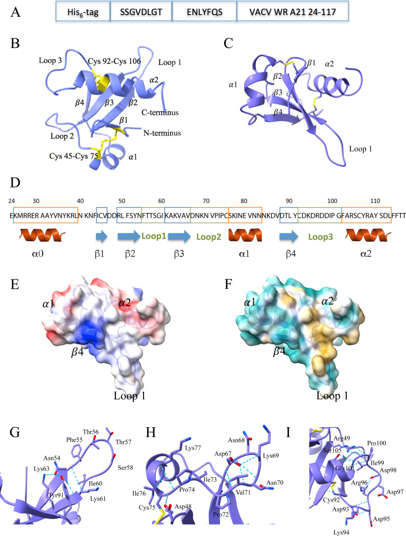
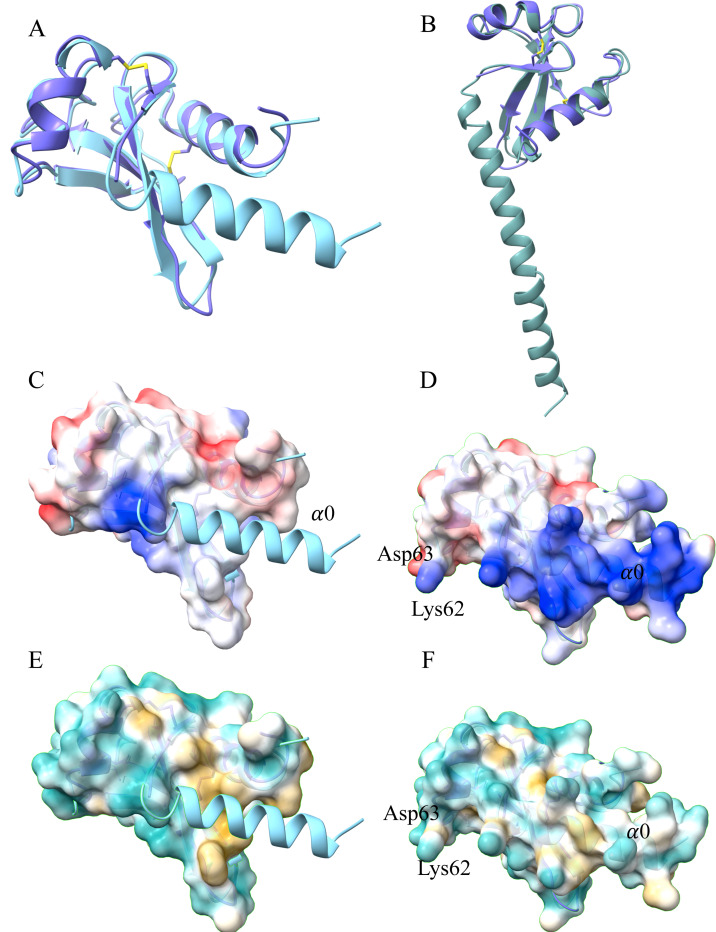
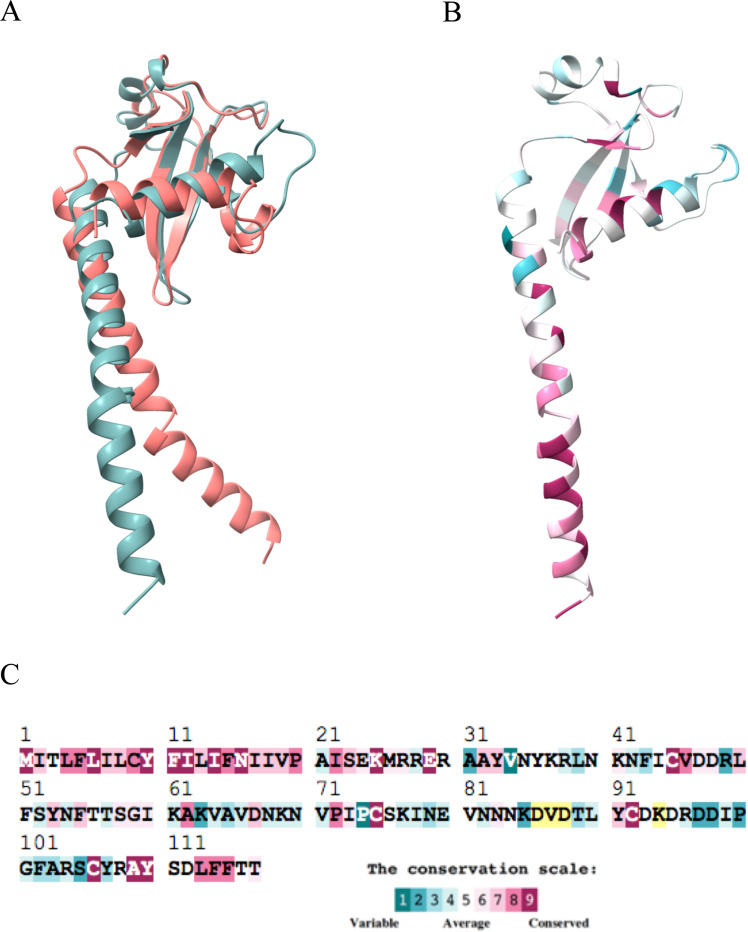
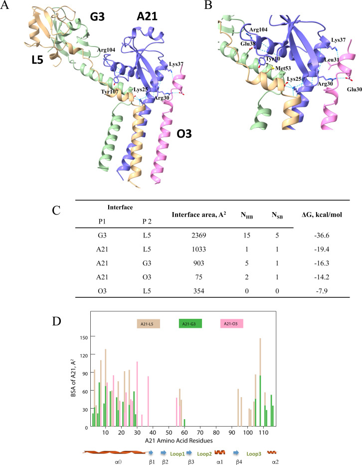
Poxviruses are exceptional in having an entry-fusion complex (EFC) consisting of eleven conserved proteins embedded in the membrane of mature virions. With the goal of understanding the function of the EFC, extensive efforts have been made to determine the structures and roles of its components, and to date, structures have been determined for nine of the eleven proteins. Here, we report the crystal structure of A21, the 10th EFC protein, comprising two α-helices clasping a twisted antiparallel β-sheet stabilized by two conserved disulfide bonds. The stability of each of the three A21 loops is provided by hydrogen bonds between main-chain atoms and several highly conserved residues, making the overall fold of A21 and its orthologs resilient to evolutionary change. Based on AlphaFold modeling and phylogenetic analysis of A21, we suggest that its highly conserved N-terminal transmembrane domain and C-terminal α-helix enable A21 integration into EFC, where it primarily interacts with the G3/L5 subcomplex and the smallest of EFC components, the O3 protein.
Overview
The claims and statistics of this paper haven’t been checked yet.
Run the check — free, no account needed. It reads the open-access text and checks the sources it cites.
Integrity signals
Registry
Cites
Statistics
Figures
Verification
Claims the paper makes
The claims and statistics of this paper haven’t been checked yet.
Run the check — free, no account needed. It reads the open-access text and checks the sources it cites.
Self-check
Percentages, sums and reported differences re-computed from the paper’s own numbers.
The self-check needs the paper’s full text.
Citations
Cites
Highly accurate protein structure prediction with AlphaFold
Jumper, J., Evans, R., Pritzel, A., Green, T., Figurnov, M., Ronneberger, O., Tunyasuvunakool, K., Bates, R., Žídek, A., Potapenko, A., et al. (2021) · Nature · doi:10.1038/s41586-021-03819-2 · Cited by 43,764
Features and development of Coot
Emsley, P., Lohkamp, B., Scott, W. G., & Cowtan, K. (2010) · Acta Crystallographica Section D Biological Crystallography · doi:10.1107/s0907444910007493 · Cited by 29,915
IQ-TREE: A Fast and Effective Stochastic Algorithm for Estimating Maximum-Likelihood Phylogenies
Nguyen, L.-T., Schmidt, H. A., von Haeseler, A., & Minh, B. Q. (2015) · Molecular Biology and Evolution · doi:10.1093/molbev/msu300 · Cited by 26,046
PHENIX : a comprehensive Python-based system for macromolecular structure solution
Adams, P. D., Afonine, P. V., Bunkóczi, G., Chen, V. B., Davis, I. W., Echols, N., Headd, J. J., Hung, L.-W., Kapral, G. J., Grosse-Kunstleve, R. W., et al. (2010) · Acta Crystallographica Section D Biological Crystallography · doi:10.1107/s0907444909052925 · Cited by 23,716
MAFFT: a novel method for rapid multiple sequence alignment based on fast Fourier transform
Katoh, K. (2002) · Nucleic Acids Research · doi:10.1093/nar/gkf436 · Cited by 17,027
Kabsch, W. (2010) · Acta Crystallographica Section D Biological Crystallography · doi:10.1107/s0907444909047337 · Cited by 16,463
Jones, T. A., Zou, J. Y., Cowan, S. W., & Kjeldgaard, M. (1991) · Acta Crystallographica Section A Foundations of Crystallography · doi:10.1107/s0108767390010224 · Cited by 12,648
Inference of Macromolecular Assemblies from Crystalline State
Krissinel, E., & Henrick, K. (2007) · Journal of Molecular Biology · doi:10.1016/j.jmb.2007.05.022 · Cited by 9,984
Solvent content of protein crystals
Matthews, B. W. (1968) · Journal of Molecular Biology · doi:10.1016/0022-2836(68)90205-2 · Cited by 7,961
ConSurf 2005: the projection of evolutionary conservation scores of residues on protein structures
Landau, M., Mayrose, I., Rosenberg, Y., Glaser, F., Martz, E., Pupko, T., & Ben-Tal, N. (2005) · Nucleic Acids Research · doi:10.1093/nar/gki370 · Cited by 1,413
Version 1.2 of the Crystallography and NMR system
Brunger, A. T. (2007) · Nature Protocols · doi:10.1038/nprot.2007.406 · Cited by 1,336
Kantardjieff, K. A., & Rupp, B. (2003) · Protein Science · doi:10.1110/ps.0350503 · Cited by 758
Using evolutionary data to make sense of macromolecules with a “face‐lifted” ConSurf
Yariv, B., Yariv, E., Kessel, A., Masrati, G., Chorin, A. B., Martz, E., Mayrose, I., Pupko, T., & Ben‐Tal, N. (2023) · Protein Science · doi:10.1002/pro.4582 · Cited by 428
Expression and Purification of Soluble His6-Tagged TEV Protease
Tropea, J. E., Cherry, S., & Waugh, D. S. (2009) · Methods in Molecular Biology · doi:10.1007/978-1-59745-196-3_19 · Cited by 371
Crystal contacts as nature's docking solutions
Krissinel, E. (2010) · Journal of Computational Chemistry · doi:10.1002/jcc.21303 · Cited by 365
Poxvirus Cell Entry: How Many Proteins Does it Take?
Moss, B. (2012) · Viruses · doi:10.3390/v4050688 · Cited by 264
Self-citationComplete pathway for protein disulfide bond formation encoded by poxviruses
Senkevich, T. G., White, C. L., Koonin, E. V., & Moss, B. (2002) · Proceedings of the National Academy of Sciences · doi:10.1073/pnas.062163799 · Cited by 182
Self-citationPoxvirus multiprotein entry–fusion complex
Senkevich, T. G., Ojeda, S., Townsley, A. C., Nelson, G. E., & Moss, B. (2005) · Proceedings of the National Academy of Sciences · doi:10.1073/pnas.0509239102 · Cited by 172
Self-citationMembrane fusion during poxvirus entry
Moss, B. (2016) · Seminars in Cell and Developmental Biology · doi:10.1016/j.semcdb.2016.07.015 · Cited by 170
Self-citationLaliberte, J. P., Weisberg, A. S., & Moss, B. (2011) · PLoS Pathogens · doi:10.1371/journal.ppat.1002446 · Cited by 122
Self-citationVaccinia Virus L1 Protein Is Required for Cell Entry and Membrane Fusion
Bisht, H., Weisberg, A. S., & Moss, B. (2008) · Journal of Virology · doi:10.1128/jvi.00852-08 · Cited by 119
Self-citationVaccinia Virus Entry into Cells Is Dependent on a Virion Surface Protein Encoded by the A28L Gene
Senkevich, T. G., Ward, B. M., & Moss, B. A. (2004) · Journal of Virology · doi:10.1128/jvi.78.5.2357-2366.2004 · Cited by 108
Self-citationSenkevich, T. G., & Moss, B. A. (2005) · Journal of Virology · doi:10.1128/jvi.79.8.4744-4754.2005 · Cited by 103
Self-citationVaccinia Virus A21 Virion Membrane Protein Is Required for Cell Entry and Fusion
Townsley, A. C., Senkevich, T. G., & Moss, B. (2005) · Journal of Virology · doi:10.1128/jvi.79.15.9458-9469.2005 · Cited by 103
Self-citationTownsley, A. C., Senkevich, T. G., & Moss, B. (2005) · Journal of Virology · doi:10.1128/jvi.79.17.10988-10998.2005 · Cited by 96
Self-citationOjeda, S., Senkevich, T. G., & Moss, B. (2005) · Journal of Virology · doi:10.1128/jvi.80.1.51-61.2006 · Cited by 96
Self-citationThe 1.51-Å structure of the poxvirus L1 protein, a target of potent neutralizing antibodies
Su, H.-P., Garman, S. C., Allison, T. J., Fogg, C. N., Moss, B., & Garboczi, D. N. (2005) · Proceedings of the National Academy of Sciences · doi:10.1073/pnas.0501103102 · Cited by 87
Self-citationBrown, E. L., Senkevich, T. G., & Moss, B. (2006) · Journal of Virology · doi:10.1128/jvi.01149-06 · Cited by 85
Self-citationUnderstanding the biology of monkeypox virus to prevent future outbreaks
Moss, B. A. (2024) · Nature Microbiology · doi:10.1038/s41564-024-01690-1 · Cited by 83
Self-citationWeichenberger, C. X., & Rupp, B. (2014) · Acta Crystallographica Section D Biological Crystallography · doi:10.1107/s1399004714005550 · Cited by 77
Matamoros, T., Alejo, A., Rodríguez, J. M., Hernáez, B., Guerra, M., Fraile‐Ramos, A., & Andrés, G. (2020) · mBio · doi:10.1128/mbio.00789-20 · Cited by 77
Structure of M11L: A myxoma virus structural homolog of the apoptosis inhibitor, Bcl‐2
Douglas, A. E., Corbett, K. D., Berger, J. M., McFadden, G., & Handel, T. M. (2007) · Protein Science · doi:10.1110/ps.062720107 · Cited by 76
Vaccinia Virus G9 Protein Is an Essential Component of the Poxvirus Entry-Fusion Complex
Ojeda, S., Domi, A., & Moss, B. (2006) · Journal of Virology · doi:10.1128/jvi.00987-06 · Cited by 74
Self-citationSatheshkumar, P. S., & Moss, B. (2009) · Journal of Virology · doi:10.1128/jvi.01744-09 · Cited by 60
Self-citationWagenaar, T. R., Ojeda, S., & Moss, B. (2008) · Journal of Virology · doi:10.1128/jvi.00162-08 · Cited by 58
Self-citationDauter, Z., & Dauter, M. (2001) · Structure · doi:10.1016/s0969-2126(01)00565-2 · Cited by 42
Nelson, G. E., Wagenaar, T. R., & Moss, B. (2008) · Journal of Virology · doi:10.1128/jvi.00434-08 · Cited by 40
Self-citationInteraction between the G3 and L5 proteins of the vaccinia virus entry–fusion complex
Wolfe, C. L., & Moss, B. (2011) · Virology · doi:10.1016/j.virol.2011.01.014 · Cited by 31
Self-citationDiesterbeck, U. S., Gittis, A. G., Garboczi, D. N., & Moss, B. A. (2018) · Scientific Reports · doi:10.1038/s41598-018-34244-7 · Cited by 28
Self-citationSchin, A. M., Diesterbeck, U. S., & Moss, B. A. (2021) · Journal of Virology · doi:10.1128/jvi.00852-21 · Cited by 25
Self-citationWidespread Distribution and Evolution of Poxviral Entry-Fusion Complex Proteins in Giant Viruses
Kao, S., Kao, C.-F., Chang, W., & Ku, C. (2023) · Microbiology Spectrum · doi:10.1128/spectrum.04944-22 · Cited by 24
Phasing on Rapidly Soaked Ions
Nagem, R. A. P., Polikarpov, I., & Dauter, Z. (2003) · Methods in enzymology on CD-ROM/Methods in enzymology · doi:10.1016/s0076-6879(03)74005-1 · Cited by 23
Su, H.-P., Lin, D. Y.-W., & Garboczi, D. N. (2006) · Journal of Virology · doi:10.1128/jvi.00521-06 · Cited by 20
Self-citationStructural basis of poxvirus A16/G9 binding for sub-complex formation
Yang, F., Lin, S., Chen, Z., Yue, D., Yang, M., He, B., Cao, Y., Dong, H., Li, J., Zhao, Q., et al. (2023) · Emerging Microbes & Infections · doi:10.1080/22221751.2023.2179351 · Cited by 14
Lin, S., Yue, D., Yang, F., Chen, Z., He, B., Cao, Y., Dong, H., Li, J., Zhao, Q., & Lu, G. (2022) · Emerging Microbes & Infections · doi:10.1080/22221751.2022.2160661 · Cited by 13
Kao, C.-F., Tsai, M.-H., Carillo, K. J., Tzou, D.-L., & Chang, W. (2023) · PLOS Pathogens · doi:10.1371/journal.ppat.1011500 · Cited by 13
Structural and functional analyses of viral H2 protein of the vaccinia virus entry fusion complex
Kao, C.-F., Liu, C.-Y., Hsieh, C.-L., Carillo, K. J. D., Tzou, D.-L. M., Wang, H.-C., & Chang, W. (2023) · Journal of Virology · doi:10.1128/jvi.01343-23 · Cited by 12
Moss, B. A., & Smith, G. L. (2021) · PLoS Pathogens · doi:10.1371/journal.ppat.1009911 · Cited by 6
Self-citationCarillo, K. J., & Tzou, D. L. (2023) · Worldwide Protein Data Bank · doi:10.2210/pdb8wt5/pdb · Cited by 1
Metadata
- Authors
- Diesterbeck, U. S., Muslinkina, L. A., Gittis, A. G., Singh, K., Moss, B., & Garboczi, D. N.
- Affiliations
- National Institutes of Health (US)
- Corresponding author
- Moss, Bernard
- Garboczi, David N.
- ORCID
- Venue
- Journal of Molecular Biology
- Publisher
- Elsevier BV
- Published
- 2025-06
- Volume
- 437
- Issue
- 12
- Pages
- 169097
- ISSN
- 0022-2836 (ISSN-L)
- PMC
- PMC12190314
- OpenAlex
- W4408596215
- Cited by
- 9
- References
- 49
- Citations by year
- 2026: 5 · 2025: 4
- FWCI
- 4.52
- Type
- journal article
- Language
- en
- Open access
- green
- doi.org
- Licence
- public-domain
- www.elsevier.com/tdm/userlicense/1.0/ (tdm)
- www.elsevier.com/legal/tdmrep-license (tdm)
- Locations
- Journal of Molecular Biology (publishedVersion)
- bioRxiv (Cold Spring Harbor Laboratory) (acceptedVersion, public-domain)
- PubMed (publishedVersion)
- PubMed Central (publishedVersion)
- PubMed Central (submittedVersion, public-domain)
- Funders
- Division of Intramural Research
- National Institute of Allergy and Infectious Diseases
- Topics
- Life Sciences › Immunology and Microbiology › Virology › Poxvirus research and outbreaks
- Life Sciences › Biochemistry, Genetics and Molecular Biology › Structural Biology › Enzyme Structure and Function
- Life Sciences › Biochemistry, Genetics and Molecular Biology › Molecular Biology › Protein Structure and Dynamics
- Keywords
- protein crystal structure, transmembrane domain, protein evolution, phylogenetic analysis
- MeSH terms
- Amino Acid Sequence, Models, Molecular, Phylogeny, Poxviridae, Protein Conformation, Viral Proteins, Crystallography, X-Ray, Virus Internalization
About these checks
Registry, retraction, Cites and statistics results are computed, not judged, with one exception: a printed reference that no index lists is read once by a model before it counts as a citation error. Claims are extracted by a model and kept only when their quote appears verbatim in the text; each cited source is then checked, with a model’s help, for whether it says what the claim relies on. Those verdicts can be wrong — read the claim’s quote and the cited source before relying on one.
Statistics and arithmetic are recomputed, not judged.
Confidence bands are shown only for fields calibrated on our annotated test set. None is calibrated yet, so no band is shown.
What counts as a claim ›How we measure confidence ›How we check this ›
Are you an author, or do you see a problem with this page? Request a review
Data sources
Bibliographic data: Semantic Scholar, OpenAlex, Crossref.
Contains information from the Semantic Scholar Academic Graph Datasets, made available under the ODC Attribution License (ODC-BY 1.0).
Semantic Scholar’s data is described in: Kinney, R., et al. (2023). The Semantic Scholar Open Data Platform. arXiv:2301.10140.
