

The DOI matches its Crossref record; 4 of the 4 references it cites were found in a registry or an index.
Vulnerability to simple faints is predicted by regional differences in brain anatomy
Open accessAbstract
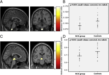
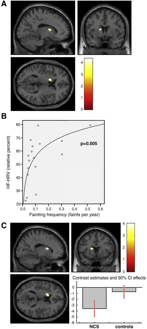
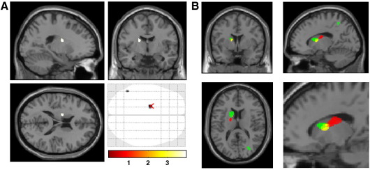
Neurocardiogenic syncope (NCS, simple fainting) is a common and typically benign familial condition, which rarely may result in traumatic injury or hypoxic convulsions. NCS is associated with emotional triggers, anxiety states and stress. However, the etiology of NCS, as a psychophysiological process, is poorly understood. We therefore investigated the relationship between NCS and brain anatomy. We studied a non-clinical sample of eighteen individuals with histories characteristic of NCS, and nineteen matched controls who had never fainted. We recorded fainting frequency, resting heart rate variability measures and anxiety levels. Structural T1-weighted magnetic resonance images (MRI) were acquired at 1.5 T. Associations between brain morphometry (regional gray and white matter volumes) and NCS, resting physiology and anxiety were tested using voxel-based morphometry (VBM). Compared to controls, NCS participants had lower regional brain volume within medulla and midbrain (a priori regions of interest). Moreover, across NCS individuals, lower gray matter volume in contiguous regions of left caudate nucleus predicted enhanced parasympathetic cardiac tone, fainting frequency and anxiety levels. Our findings provide preliminary evidence for a hierarchical anatomical basis to NCS. First, differences in the volume of brainstem centers supporting cardiovascular homeostasis may relate to constitutional predisposition to NCS. Second, differences in the structural organization of the caudate nucleus in NCS individuals may relate to fainting frequency via interactions between emotional state and parasympathetic control of the heart. These observations highlight the application of VBM to the identification of neurovisceral mechanisms relevant to psychosomatic medicine and the neuroscience of emotion.
Overview
The claims and statistics of this paper haven’t been checked yet.
Run the check — free, no account needed. It reads the open-access text and checks the sources it cites.
Integrity signals
Registry
Cites
Statistics
Figures
Verification
Claims the paper makes
The claims and statistics of this paper haven’t been checked yet.
Run the check — free, no account needed. It reads the open-access text and checks the sources it cites.
Claims other papers make about this one
Each is checked against this paper’s own text: does it say what the citing paper says it does?
The cited source says what the claim attributes to it.
In the citing paper
“Critchley and co-workers reported evidence for structural variations in midbrain and medulla regions involved in BP and baroreflex regulation in patients with neurocardiogenic syncope (Beacher et al., 2009), which could serve as dispositional factors contributing to vasomotor abnormalities and thus fainting risk.”
From The psychophysiology of blood-injection-injury phobia: Looking beyond the diphasic response paradigm (2010) · cites this paper as Beacher et al., 2009
Self-check
Percentages, sums and reported differences re-computed from the paper’s own numbers.
The self-check needs the paper’s full text.
Citations
Cites
Vagal and sympathetic mechanisms in patients with orthostatic vasovagal syncope
Morillo, C. A., Eckberg, D. L., Ellenbogen, K. A., Beightol, L. A., Hoag, J. B., Tahvanainen, K. U. O., Kuusela, T. A., & Diedrich, A. M. (1997) · Circulation · doi:10.1161/01.cir.96.8.2509 · Cited by 280
Vasovagal syncope in medical students and their first-degree relatives
Serletis, A., Rose, S., Sheldon, A. G., & Sheldon, R. S. (2006) · European Heart Journal · doi:10.1093/eurheartj/ehl147 · Cited by 229
Saxena, S., Brody, A. L., Ho, M. L., Alborzian, S., Ho, M. K., Maidment, K. M., Huang, S.-C., Wu, H.-M., Au, S. C., & Baxter, L. R. (2001) · Biological Psychiatry · doi:10.1016/s0006-3223(01)01123-4 · Cited by 209
Physiological responses in blood phobics
Öst, L.-G., Sterner, U., & Lindahl, I.-L. (1984) · Behaviour Research and Therapy · doi:10.1016/0005-7967(84)90099-8 · Cited by 177
Research works printed but not linked: 59
- Ashburner, 2007, A fast diffeomorphic image registration algorithm, Neuroimage, 38, 95, 10.1016/j.neuroimage.2007.07.007 · DOI · Paper page
- Ashburner, 2000, Voxel-based morphometry—the methods, Neuroimage, 11, 805, 10.1006/nimg.2000.0582 · DOI · Paper page
- Bergouignan, 2009, Can voxel based morphometry, manual segmentation and automated segmentation equally detect hippocampal volume differences in acute depression?, Neuroimage, 45, 29, 10.1016/j.neuroimage.2008.11.006 · DOI
- Brignole, 2004, Guidelines on management (diagnosis and treatment) of syncope-update 2004. Executive summary, task force on Syncope, European Society of Cardiology, Eur. Heart J., 25, 2054, 10.1016/j.ehj.2004.09.004 · DOI
- Cavun, 2001, Evidence that hemorrhagic hypotension is mediated by the ventrolateral periaqueductal gray region, Am. J. Physiol., 281, R747
- Cohen, 2000, An association between anxiety and neurocardiogenic syncope during head-up tilt table testing, Pacing Clin. Electrophysiol., 23, 837, 10.1111/j.1540-8159.2000.tb00852.x · DOI
- Della Nave, 2008, Brain structural damage in Friedreich's ataxia, J. Neurol. Neurosurg. Psychiatry, 79, 82, 10.1136/jnnp.2007.124297 · DOI
- Donadio, 2007, Arousal elicits exaggerated inhibition of sympathetic nerve activity in phobic syncope patients, Brain, 130, 1653, 10.1093/brain/awm037 · DOI
- Eickhoff, 2005, A new SPM toolbox for combining probabilistic cytoarchitectonic maps and functional imaging data, NeuroImage, 25, 1325, 10.1016/j.neuroimage.2004.12.034 · DOI · Paper page
- Ellenbogen, 1997, Neural monitoring of vasovagal syncope, Pacing Clin. Electrophysiol., 20, 788, 10.1111/j.1540-8159.1997.tb03905.x · DOI
- Friedman, 1993, Autonomic characteristics of nonclinical panic and blood phobia, Biol. Psychiatry, 34, 298, 10.1016/0006-3223(93)90087-T · DOI
- Furlan, 1998, Cardiac autonomic patterns preceding occasional vasovagal reactions in healthy humans, Circulation, 98, 1756, 10.1161/01.CIR.98.17.1756 · DOI
- Ginestroni, 2008, Brain structural damage in spinocerebellar ataxia type 1: a VBM study, J. Neurol., 255, 1153, 10.1007/s00415-008-0860-4 · DOI
- Golanov, 2001, Neurons of nucleus of the solitary tract synchronize the EEG and elevate cerebral blood flow via a novel medullary area, Brain Res., 892, 1, 10.1016/S0006-8993(00)02949-8 · DOI
- Good, 2001, A voxel-based morphometric study of ageing in 465 normal adult human brains, NeuroImage, 14, 21, 10.1006/nimg.2001.0786 · DOI · Paper page
- Gracie, 2006, The role of psychological factors in response to treatment in neurocardiogenic (vasovagal) syncope, Europace, 8, 636, 10.1093/europace/eul073 · DOI
- Gray, 2009, Following one's heart: cardiac rhythms gate central initiation of sympathetic reflexes, J. Neurosci., 29, 1817, 10.1523/JNEUROSCI.3363-08.2009 · DOI
- Grubb, 2005, Clinical practice. Neurocardiogenic syncope, N. Engl. J. Med., 352, 1004, 10.1056/NEJMcp042601 · DOI
- Grubb, 1991, Cerebral vasoconstriction during head-upright tilt-induced vasovagal syncope. A paradoxic and unexpected response, Circulation, 84, 1157, 10.1161/01.CIR.84.3.1157 · DOI
- Hainsworth, 2004, Pathophysiology of syncope, Clin. Auton. Res., 14, 18, 10.1007/s10286-004-1004-2 · DOI
- Hauser, 2006, Visualization and quantification of disease progression in multiple system atrophy, Mov. Disord., 21, 1674, 10.1002/mds.21032 · DOI
- Henderson, 2000, Caudal midline medulla mediates behaviourally-coupled but not baroreceptor-mediated vasodepression, Neuroscience, 98, 779, 10.1016/S0306-4522(00)00117-2 · DOI
- Heslop, 2002, Haemorrhage-evoked compensation and decompensation are mediated by distinct caudal midline medullary regions in the urethane-anaesthetised rat, Neuroscience, 113, 555, 10.1016/S0306-4522(02)00161-6 · DOI
- Jardine, 1998, Autonomic control of vasovagal syncope, Am. J. Physiol., 274, H2110
- Kapoor, 2000, Syncope, N. Engl. J. Med., 343, 1856, 10.1056/NEJM200012213432507 · DOI
- Kochiadakis, 1997, Autonomic nervous system activity during tilt testing in syncopal patients, estimated by power spectral analysis of heart rate variability, Pacing Clin. Electrophysiol., 20, 1332, 10.1111/j.1540-8159.1997.tb06788.x · DOI
- Kochiadakis, 1998, Spectral analysis of heart rate variability during tilt-table testing in patients with vasovagal syncope, Int. J. Cardiol., 64, 185, 10.1016/S0167-5273(98)00039-4 · DOI
- Kouakam, 1999, Inadequate sympathovagal balance in response to orthostatism in patients with unexplained syncope and a positive head up tilt test, Heart, 82, 312, 10.1136/hrt.82.3.312 · DOI
- Kouakam, 2002, Prevalence and prognostic significance of psychiatric disorders in patients evaluated for recurrent unexplained syncope, Am. J. Cardiol., 89, 530, 10.1016/S0002-9149(01)02292-5 · DOI
- Lee, 1996, Excessive myocardial contraction in vasovagal syncope demonstrated by echocardiography during head-up tilt, Clin. Cardiol., 19, 137, 10.1002/clc.4960190212 · DOI
- Lipsitz, 1990, Spectral characteristics of heart rate variability before and during postural tilt: relations to aging and risk of syncope, Circulation, 81, 1803, 10.1161/01.CIR.81.6.1803 · DOI
- Maldjian, 2003, An automated method for neuroanatomic and cytoarchitectonic Atlas-based Interrogation of fMRI data sets, NeuroImage, 19, 1233, 10.1016/S1053-8119(03)00169-1 · DOI
- Mark, 1983, The Bezold–Jarisch reflex revisited: clinical implications of inhibitory reflexes originating in the heart, J. Am. Coll. Cardiol., 1, 90, 10.1016/S0735-1097(83)80014-X · DOI
- Mathias, 1998, Frequency of family history in vasovagal syncope, Lancet, 352, 33, 10.1016/S0140-6736(05)79513-1 · DOI
- Mathias, 2001, Observations on recurrent syncope and presyncope in 641 patients, Lancet, 357, 348, 10.1016/S0140-6736(00)03642-4 · DOI
- Minnerop, 2007, Voxel-based morphometry and voxel-based relaxometry in multiple system atrophy—a comparison between clinical subtypes and correlations with clinical parameters, Neuroimage, 36, 1086, 10.1016/j.neuroimage.2007.04.028 · DOI
- Morillo, 1994, Time and frequency domain analysis of heart rate variability during orthostatic stress in patients with neurally mediated syncope, Am. J. Cardiol., 74, 1258, 10.1016/0002-9149(94)90559-2 · DOI
- Morino, 1994, Effects of microinjection of N-methyl-d-aspartic acid into the nucleus tractus solitarii on cerebral blood flow in anesthetized rats, J. Auton. Nerv. Syst., 49, 159, 10.1016/0165-1838(94)90135-X · DOI
- Mosqueda-Garcia, 1997, Sympathetic and baroreceptor reflex function in neurally mediated syncope evoked by tilt, J. Clin. Invest., 99, 2736, 10.1172/JCI119463 · DOI · Paper page
- Oberg, 1972, Increased activity in left ventricular receptors during occlusion of caval veins in the cat. A possible cause of the vaso-vagal reaction, Acta Physiol. Scand., 85, 164, 10.1111/j.1748-1716.1972.tb05247.x · DOI
- Piccirillo, 2004, Heart rate and blood pressure variability in subjects with vasovagal syncope, Clin. Sci., 107, 55, 10.1042/CS20030327 · DOI
- Robertson, 1999, Neurally mediated syncope: pathophysiology and implications for treatment, Am. J. Med. Sci., 317, 102, 10.1097/00000441-199902000-00004 · DOI
- Robinson, 1995, Reduced caudate nucleus volume in obsessive–compulsive disorder, Arch. Gen. Psychiatry, 52, 393, 10.1001/archpsyc.1995.03950170067009 · DOI
- Rubin, 1992, Determination by high-resolution single-photon emission computed tomography, Arch. Gen. Psychiatry, 49, 695, 10.1001/archpsyc.1992.01820090023004 · DOI
- Sarasin, 2001, Prospective evaluation of patients with syncope: a population-based study, Am. J. Med., 111, 177, 10.1016/S0002-9343(01)00797-5 · DOI
- Schadt, 1991, Hemodynamic and neurohumoral responses to acute hypovolemia in conscious mammals, Am. J. Physiol., 260, H305
- Shalev, 1991, Echocardiographic demonstration of decreased left ventricular dimensions and vigorous myocardial contraction during syncope induced by head-up tilt, J. Am. Coll. Cardiol., 18, 746, 10.1016/0735-1097(91)90798-E · DOI
- Sharpey-Schafer, 1956, Emergencies in general practice: syncope, Br. Med. J., 1, 506, 10.1136/bmj.1.4965.506 · DOI
- Silverstein, 1982, Patients with syncope admitted to medical intensive care units, JAMA, 248, 1185, 10.1001/jama.1982.03330100023024 · DOI
- Sprengelmeyer, 1995, Associative learning in degenerative neostriatal disorders: contrasts in explicit and implicit remembering between Parkinson's and Huntington's diseases, Mov. Disord., 10, 51, 10.1002/mds.870100110 · DOI
- Steptoe, 1988, Emotional fainting and the psychophysiologic response to blood and injury: autonomic mechanisms and coping strategies, Psychosom. Med., 50, 402, 10.1097/00006842-198807000-00008 · DOI · Paper page
- Sutton, 1992, Proposed classification for tilt induced vasovagal syncope, Eur. J. Cardiac. Pacing Electrophysiol., 3, 180
- Swedo, 1992, Cerebral glucose metabolism in childhood-onset obsessive–compulsive disorder. Revisualization during pharmacotherapy, Arch. Gen. Psychiatry, 49, 690, 10.1001/archpsyc.1992.01820090018003 · DOI
- Troy, 2003, Haemodynamic response to haemorrhage: distinct contributions of midbrain and forebrain structures, Auton. Neurosci., 108, 1, 10.1016/S1566-0702(03)00152-8 · DOI
- Uchida, 2008, Regional gray matter abnormalities in panic disorder: a voxel-based morphometry study, Psychiatry Res., 163, 21, 10.1016/j.pscychresns.2007.04.015 · DOI
- Vagg, 2008, Hypovolemic shock: critical involvement of a projection from the ventrolateral periaqueductal gray to the caudal midline medulla, Neuroscience, 152, 1099, 10.1016/j.neuroscience.2007.10.070 · DOI
- Verner, 2008, Retrograde projections to a discrete apneic site in the midline medulla oblongata of the rat, Brain Res., 1208, 128, 10.1016/j.brainres.2008.02.028 · DOI
- Woermann, 1999, Voxel-by-voxel comparison of automatically segmented cerebral gray matter—a rater-independent comparison of structural MRI in patients with epilepsy, Neuroimage, 10, 373, 10.1006/nimg.1999.0481 · DOI
- Yassa, 2009, A quantitative evaluation of cross-participant registration techniques for MRI studies of the medial temporal lobe, Neuroimage, 44, 319, 10.1016/j.neuroimage.2008.09.016 · DOI
Cited by
Sorted by citations
Emotional appraisal is influenced by cardiac afferent information
Gray, M. A., Beacher, F. D., Minati, L., Nagai, Y., Kemp, A. H., Harrison, N. A., & Critchley, H. D. (2012) · Emotion · doi:10.1037/a0025083 · Cited by 163
Self-citationDissecting axes of autonomic control in humans: insights from neuroimaging
Critchley, H. D., Nagai, Y., Gray, M. A., & Mathias, C. J. (2011) · Autonomic Neuroscience · doi:10.1016/j.autneu.2010.09.005 · Cited by 152
Self-citationCraddock, T. J. A., Fritsch, P., Rice, M. A., del Rosario, R. M., Miller, D. B., Fletcher, M. A., Klimas, N. G., & Broderick, G. (2014) · PLoS ONE · doi:10.1371/journal.pone.0084839 · Cited by 75
The psychophysiology of blood-injection-injury phobia: Looking beyond the diphasic response paradigm
Ritz, T., Meuret, A. E., & Ayala, E. S. (2010) · International Journal of Psychophysiology · doi:10.1016/j.ijpsycho.2010.05.007 · Cited by 74
“…reported evidence for structural variations in midbrain and medulla regions involved in BP and baroreflex regulation in patients with neurocardiogenic syncope (Beacher et al., 2009), which could serve as dispositional factors contributing to vasomotor abnormalities and thus fainting risk.”
BackgroundThe new field of Brain–Body Medicine: What have we learned and where are we headed?
Lane, R. D., & Wager, T. D. (2009) · NeuroImage · doi:10.1016/j.neuroimage.2009.06.013 · Cited by 52
Childhood trauma and lifetime syncope burden among older adults
O'Hare, C., McCrory, C., O'Leary, N., O'Brien, H., & Kenny, R. A. (2017) · Journal of Psychosomatic Research · doi:10.1016/j.jpsychores.2017.03.019 · Cited by 18
Metadata
- Authors
- Beacher, F. D. C. C., Gray, M. A., Mathias, C. J., & Critchley, H. D.
- Affiliations
- Brighton and Sussex Medical School (GB)
- Imperial College London (GB)
- Corresponding author
- Beacher, Felix D.C.C.
- Venue
- NeuroImage
- Publisher
- Elsevier BV
- Published
- 2009-09
- Volume
- 47
- Issue
- 3
- Pages
- 937-945
- ISSN
- 1053-8119 (ISSN-L)
- PMC
- PMC2726440
- OpenAlex
- W1996420437
- Cited by
- 37
- References
- 4
- Citations by year
- 2026: 2 · 2023: 1 · 2021: 2 · 2020: 2 · 2019: 1 · 2018: 3 · 2017: 2 · 2016: 2 · 2015: 5 · 2014: 3 · 2013: 1 · 2012: 2
- FWCI
- 2.75
- Type
- journal article
- Language
- en
- Open access
- hybrid
- doi.org
- Locations
- NeuroImage (publishedVersion, cc-by)
- PubMed Central (publishedVersion, cc-by)
- PubMed (publishedVersion)
- UCL Discovery (University College London) (submittedVersion)
- Figshare (submittedVersion, cc-by)
- Funders
- Wellcome Trust
- Topics
- Health Sciences › Medicine › Cardiology and Cardiovascular Medicine › Cardiovascular Syncope and Autonomic Disorders
- Health Sciences › Medicine › Cardiology and Cardiovascular Medicine › Heart Rate Variability and Autonomic Control
- Health Sciences › Medicine › Psychiatry and Mental health › Psychosomatic Disorders and Their Treatments
- Keywords
- vasovagal syncope, brain structure, caudate nucleus, brainstem volume, gray matter volume, white matter volume, heart rate variability, anxiety, psychosomatic medicine, emotion and physiology
- MeSH terms
- Adult, Brain, Female, Humans, Image Interpretation, Computer-Assisted, Magnetic Resonance Imaging, Male, Syncope, Young Adult
About these checks
Registry, retraction, Cites and statistics results are computed, not judged, with one exception: a printed reference that no index lists is read once by a model before it counts as a citation error. Claims are extracted by a model and kept only when their quote appears verbatim in the text; each cited source is then checked, with a model’s help, for whether it says what the claim relies on. Those verdicts can be wrong — read the claim’s quote and the cited source before relying on one.
Statistics and arithmetic are recomputed, not judged.
Confidence bands are shown only for fields calibrated on our annotated test set. None is calibrated yet, so no band is shown.
What counts as a claim ›How we measure confidence ›How we check this ›
Are you an author, or do you see a problem with this page? Request a review
