

The DOI matches its Crossref record; 59 of the 59 references it cites were found in a registry or an index.
Estimating the transfer function from neuronal activity to BOLD using simultaneous EEG-fMRI
Open accessAbstract
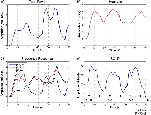
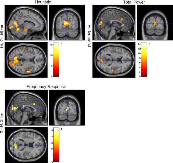
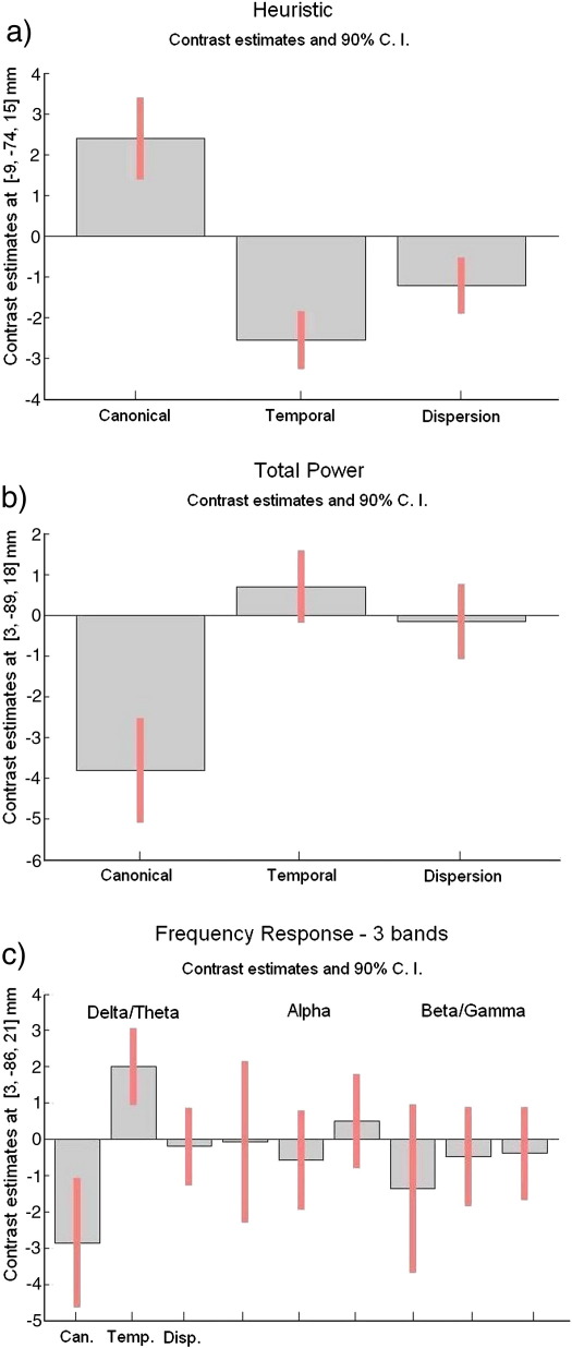
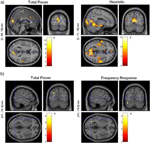
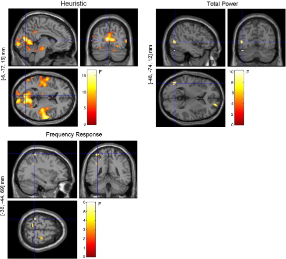
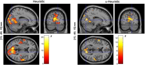
Previous studies using combined electrical and hemodynamic measurements of brain activity, such as EEG and (BOLD) fMRI, have yielded discrepant results regarding the relationship between neuronal activity and the associated BOLD response. In particular, some studies suggest that this link, or transfer function, depends on the frequency content of neuronal activity, while others suggest that total neuronal power accounts for the changes in BOLD. Here we explored this dependency by comparing different frequency-dependent and -independent transfer functions, using simultaneous EEG-fMRI. Our results suggest that changes in BOLD are indeed associated with changes in the spectral profile of neuronal activity and that these changes do not arise from one specific spectral band. Instead they result from the dynamics of the various frequency components together, in particular, from the relative power between high and low frequencies. Understanding the nature of the link between neuronal activity and BOLD plays a crucial role in improving the interpretability of BOLD images as well as on the design of more robust and realistic models for the integration of EEG and fMRI.
Overview
The claims and statistics of this paper haven’t been checked yet.
Run the check — free, no account needed. It reads the open-access text and checks the sources it cites.
Integrity signals
Registry
Cites
Statistics
Figures
Verification
Claims the paper makes
The claims and statistics of this paper haven’t been checked yet.
Run the check — free, no account needed. It reads the open-access text and checks the sources it cites.
Claims other papers make about this one
Each is checked against this paper’s own text: does it say what the citing paper says it does?
In the citing paper
“This is because it is generally assumed that fMRI signals scale with the predominant frequency of neuronal activity (Kilner et al., 2005; Laufs & Duncan 2007; Rosa et al., 2010).”
From Network discovery with DCM (2011) · cites this paper as Rosa et al., 2010
Self-check
Percentages, sums and reported differences re-computed from the paper’s own numbers.
The self-check needs the paper’s full text.
Citations
Cites
Co-Planar Stereotaxic Atlas of the Human Brain
Talairach, J., & Tournoux, P. (1988) · Medical Entomology and Zoology · Cited by 11,798
Statistical parametric maps in functional imaging: A general linear approach
Friston, K. J., Holmes, A. P., Worsley, K. J., Poline, J. ‐., Frith, C. D., & Frackowiak, R. S. J. (1994) · Human Brain Mapping · doi:10.1002/hbm.460020402 · Cited by 9,688
Co-planar stereotaxic atlas of the human brain
Laitinen, L. (1989) · Clinical Neurology and Neurosurgery · doi:10.1016/0303-8467(89)90128-5 · Cited by 7,565
Neurophysiological investigation of the basis of the fMRI signal
Logothetis, N. K., Pauls, J., Augath, M., Trinath, T., & Oeltermann, A. (2001) · Nature · doi:10.1038/35084005 · Cited by 6,551
Hämäläinen, M., Hari, R., Ilmoniemi, R. J., Knuutila, J., & Lounasmaa, O. V. (1993) · Reviews of Modern Physics · doi:10.1103/revmodphys.65.413 · Cited by 4,684
A new SPM toolbox for combining probabilistic cytoarchitectonic maps and functional imaging data
Eickhoff, S. B., Stephan, K. E., Mohlberg, H., Grefkes, C., Fink, G. R., Amunts, K., & Zilles, K. (2005) · NeuroImage · doi:10.1016/j.neuroimage.2004.12.034 · Cited by 4,033
What we can do and what we cannot do with fMRI
Logothetis, N. K. (2008) · Nature · doi:10.1038/nature06976 · Cited by 3,612
Statistical Parametric Mapping: The Analysis of Functional Brain Images
Penny, W. D., Friston, K. J., Ashburner, J., Kiebel, S. J., & Thomas, N. (2007) · UCL Discovery (University College London) · Cited by 3,339
Oscillatory gamma activity in humans and its role in object representation
Tallon‐Baudry, C. (1999) · Trends in Cognitive Sciences · doi:10.1016/s1364-6613(99)01299-1 · Cited by 2,205
Frackowiak, R. S. J. (1997) · Cited by 1,713
Cited by
Sorted by citations
Scheeringa, R., Fries, P., Petersson, K. M., Oostenveld, R., Grothe, I., Norris, D. G., Peter Hagoort, A., & BASTIAANSEN, M. C. M. (2011) · Neuron · doi:10.1016/j.neuron.2010.11.044 · Cited by 479
Magri, C., Schridde, U., Murayama, Y., Panzeri, S., & Logothetis, N. K. (2012) · Journal of Neuroscience · doi:10.1523/jneurosci.3985-11.2012 · Cited by 356
Friston, K. J., Li, B., Daunizeau, J., & Stephan, K. E. (2011) · NeuroImage · doi:10.1016/j.neuroimage.2010.12.039 · Cited by 283
Transfer Function between EEG and BOLD Signals of Epileptic Activity
Leite, M., Leal, A., & Figueiredo, P. (2013) · Frontiers in Neurology · doi:10.3389/fneur.2013.00001 · Cited by 234
Neurovascular coupling in normal aging: A combined optical, ERP and fMRI study
Fabiani, M., Gordon, B. A., Maclin, E. L., Pearson, M. A., Brumback, C. R., Low, K. A., McAuley, E., Sutton, B. P., Kramer, A. F., & Gratton, G. (2013) · NeuroImage · doi:10.1016/j.neuroimage.2013.04.113 · Cited by 230
Altered Resting State Cortico-Striatal Connectivity in Mild to Moderate Stage Parkinson's Disease
Kwak, Y., Peltier, S., Bohnen, N. I., Müller, M. L. T. M., Dayalu, P., & Seidler, R. D. (2010) · Frontiers in Systems Neuroscience · doi:10.3389/fnsys.2010.00143 · Cited by 189
Electrophysiological correlates of the BOLD signal for EEG-informed fMRI
Murta, T., Leite, M., Carmichael, D. W., Figueiredo, P., & Lemieux, L. (2015) · Human Brain Mapping · doi:10.1002/hbm.22623 · Cited by 165
EEG-Informed fMRI: A Review of Data Analysis Methods
Abreu, R., Leal, A., & Figueiredo, P. (2018) · Frontiers in Human Neuroscience · doi:10.3389/fnhum.2018.00029 · Cited by 171
EEG–fMRI integration for the study of human brain function
Jorge, J., van der Zwaag, W., & Figueiredo, P. (2013) · NeuroImage · doi:10.1016/j.neuroimage.2013.05.114 · Cited by 185
Analysis of Multimodal Neuroimaging Data
Bießmann, F., Plis, S., Meinecke, F. C., Eichele, T., & Müller, K. (2011) · IEEE Reviews in Biomedical Engineering · doi:10.1109/rbme.2011.2170675 · Cited by 163
Frontal Brain Asymmetry and Willingness to Pay
Ramsøy, T. Z., Skov, M., Christensen, M. K., & Stahlhut, C. (2018) · Frontiers in Neuroscience · doi:10.3389/fnins.2018.00138 · Cited by 126
EEG-fMRI INTEGRATION: A CRITICAL REVIEW OF BIOPHYSICAL MODELING AND DATA ANALYSIS APPROACHES
Rosa, M. J., Jean Daunizeau, A., & Friston, K. J. (2010) · Journal of Integrative Neuroscience · doi:10.1142/s0219635210002512 · Cited by 116
Impaired auditory sampling in dyslexia: further evidence from combined fMRI and EEG
Lehongre, K., Morillon, B., Giraud, A., & Ramus, F. (2013) · Frontiers in Human Neuroscience · doi:10.3389/fnhum.2013.00454 · Cited by 113
A personalized history of EEG–fMRI integration
Laufs, H. (2012) · NeuroImage · doi:10.1016/j.neuroimage.2012.01.039 · Cited by 103
Identifying robust and sensitive frequency bands for interrogating neural oscillations
Shackman, A. J., McMenamin, B. W., Maxwell, J. S., Greischar, L. L., & Davidson, R. J. (2010) · NeuroImage · doi:10.1016/j.neuroimage.2010.03.037 · Cited by 92
Romei, V., Bauer, M., Brooks, J. L., Economides, M., Penny, W. D., Thut, G., Driver, J., & Bestmann, S. (2015) · NeuroImage · doi:10.1016/j.neuroimage.2015.11.020 · Cited by 91
Vartiainen, J., Liljeström, M., Koskinen, M., Renvall, H., & Salmelin, R. H. (2011) · Journal of Neuroscience · doi:10.1523/jneurosci.3113-10.2011 · Cited by 81
Differential cortical activation during observation and observation-and-imagination
Berends, H. I., Wolkorte, R., IJzerman, M. J., & van Putten, M. J. A. M. (2013) · Experimental Brain Research · doi:10.1007/s00221-013-3571-8 · Cited by 82
Kwak, Y., Peltier, S., Bohnen, N. I., Müller, M. L. T. M., Dayalu, P., & Seidler, R. D. (2012) · Frontiers in Systems Neuroscience · doi:10.3389/fnsys.2012.00052 · Cited by 80
van Houdt, P. J., de Munck, J. C., Leijten, F. S. S., Huiskamp, G. J. M., Colon, A., Boon, P., & Ossenblok, P. (2013) · NeuroImage · doi:10.1016/j.neuroimage.2013.02.033 · Cited by 72
Driving and Driven Architectures of Directed Small-World Human Brain Functional Networks
Yan, C., & Yong He, A. (2011) · PLoS ONE · doi:10.1371/journal.pone.0023460 · Cited by 72
Present and future of simultaneous EEG-fMRI
Rosenkranz, K., & Lemieux, L. (2010) · Magnetic Resonance Materials in Physics Biology and Medicine · doi:10.1007/s10334-009-0196-9 · Cited by 72
Legon, W., Punzell, S., Dowlati, E. P., Adams, S. E., Stiles, A. B., & Moran, R. (2015) · Cerebral Cortex · doi:10.1093/cercor/bhv200 · Cited by 69
Adaptive optimal basis set for BCG artifact removal in simultaneous EEG-fMRI
Marino, M., Liu, Q., Koudelka, V., Porcaro, C., Hlinka, J., Wenderoth, N., & Mantini, D. (2018) · Scientific Reports · doi:10.1038/s41598-018-27187-6 · Cited by 63
Balsters, J. H., O’Connell, R. G., Martin, M. P., Galli, A., Cassidy, S. M., Kilcullen, S. M., Delmonte, S., Brennan, S., Meaney, J. F., Fagan, A. J., et al. (2011) · PLoS ONE · doi:10.1371/journal.pone.0024126 · Cited by 56
Frequency-specific electrocorticographic correlates of working memory delay period fMRI activity
Khursheed, F., Tandon, N., Tertel, K., Pieters, T. A., Disano, M. A., & Ellmore, T. M. (2011) · NeuroImage · doi:10.1016/j.neuroimage.2011.02.062 · Cited by 54
Bentley, W. J., Li, J. M., Snyder, A. Z., Raichle, M. E., & Snyder, L. H. (2014) · Cerebral Cortex · doi:10.1093/cercor/bhu260 · Cited by 46
Lüchinger, R., Michels, L., Martin, E., & Brandeis, D. (2012) · NeuroImage · doi:10.1016/j.neuroimage.2012.01.031 · Cited by 49
Independent Vector Analysis for Gradient Artifact Removal in Concurrent EEG-fMRI Data
Acharjee, P. P., Phlypo, R., Wu, L., Calhoun, V. D., & Adalı, T. (2015) · IEEE Transactions on Biomedical Engineering · doi:10.1109/tbme.2015.2403298 · Cited by 43
Domain-general Stroop Performance and Hemispheric Asymmetries: A Resting-state EEG Study
Ambrosini, E., & Vallesi, A. (2017) · Journal of Cognitive Neuroscience · doi:10.1162/jocn_a_01076 · Cited by 39
Abreu, R., Leite, M., Jorge, J., Grouiller, F., van der Zwaag, W., Leal, A., & Figueiredo, P. (2016) · NeuroImage · doi:10.1016/j.neuroimage.2016.03.034 · Cited by 40
Lei, X., Valdes-Sosa, P. A., & Yao, D. (2012) · Journal of Integrative Neuroscience · doi:10.1142/s0219635212500203 · Cited by 40
A Sparse EEG-Informed fMRI Model for Hybrid EEG-fMRI Neurofeedback Prediction
Cury, C., Maurel, P., Gribonval, R., & Barillot, C. (2020) · Frontiers in Neuroscience · doi:10.3389/fnins.2019.01451 · Cited by 35
Inferring Macroscale Brain Dynamics via Fusion of Simultaneous EEG-fMRI
Philiastides, M. G., Tu, T., & Sajda, P. (2021) · Annual Review of Neuroscience · doi:10.1146/annurev-neuro-100220-093239 · Cited by 41
Simultaneous EEG/fMRI Analysis of the Resonance Phenomena in Steady-State Visual Evoked Responses
Bayram, A., Bayraktaroğlu, Z., Karahan, E., Erdogan, B., Bılgıç, B., Özker, M., Kaşikçi, I., Duru, A. D., Ademoğlu, A., Öztürk, C., et al. (2011) · Clinical EEG and Neuroscience · doi:10.1177/155005941104200210 · Cited by 37
Rothlübbers, S., Relvas, V., Leal, A., Murta, T., Lemieux, L., & Figueiredo, P. (2014) · Brain Topography · doi:10.1007/s10548-014-0408-0 · Cited by 36
Uji, M., Wilson, R., Francis, S., Mullinger, K. J., & Mayhew, S. D. (2018) · Human Brain Mapping · doi:10.1002/hbm.23943 · Cited by 37
Abreu, R., Leal, A., & Figueiredo, P. (2019) · Scientific Reports · doi:10.1038/s41598-018-36976-y · Cited by 33
Abreu, R., Leal, A., Lopes, F. H. D. S., & Figueiredo, P. (2018) · Clinical Neurophysiology · doi:10.1016/j.clinph.2017.12.038 · Cited by 32
Bayesian Comparison of Neurovascular Coupling Models Using EEG-fMRI
Rosa, M. J., Kilner, J. M., & Penny, W. D. (2011) · PLoS Computational Biology · doi:10.1371/journal.pcbi.1002070 · Cited by 32
Giménez, M., Guinea-Izquierdo, A., Villalta‐Gil, V., Martínez‐Zalacaín, I., Segalàs, C., Subirà, M., Real, E., Pujol, J., Harrison, B. J., Haro, J. M., et al. (2016) · Brain Imaging and Behavior · doi:10.1007/s11682-016-9601-y · Cited by 32
Bruyns‐Haylett, M., Harris, S., Boorman, L. W., Zheng, Y., Berwick, J., & Jones, M. (2013) · European Journal of Neuroscience · doi:10.1111/ejn.12295 · Cited by 31
Abreu, R., Nunes, S., Leal, A., & Figueiredo, P. (2016) · NeuroImage · doi:10.1016/j.neuroimage.2016.08.008 · Cited by 29
Zama, T., Takahashi, Y., & Shimada, S. (2019) · Frontiers in Human Neuroscience · doi:10.3389/fnhum.2019.00301 · Cited by 27
Understanding the pathophysiology of reflex epilepsy using simultaneous EEG‐fMRI
Sandhya, M., Bharath, R. D., Panda, R., Chandra, S., Kumar, N., George, L., Thamodharan, A., Gupta, A. K., & Satishchandra, P. (2014) · Epileptic Disorders · doi:10.1684/epd.2014.0632 · Cited by 26
Sclocco, R., Tana, M. G., Visani, E., Gilioli, I., Panzica, F., Franceschetti, S., Cerutti, S., & Bianchi, A. M. (2014) · Frontiers in Human Neuroscience · doi:10.3389/fnhum.2014.00186 · Cited by 24
Do Motor Imagery Performances Depend on the Side of the Lesion at the Acute Stage of Stroke?
Kemlin, C., Moulton, E. J., Samson, Y., & Rosso, C. (2016) · Frontiers in Human Neuroscience · doi:10.3389/fnhum.2016.00321 · Cited by 31
Mayhew, S. D., & Bagshaw, A. P. (2017) · NeuroImage · doi:10.1016/j.neuroimage.2017.04.051 · Cited by 26
An electrophysiological validation of stochastic DCM for fMRI
Jean Daunizeau, A., Lemieux, L., Vaudano, A. E., Friston, K. J., & Stephan, K. E. (2013) · Frontiers in Computational Neuroscience · doi:10.3389/fncom.2012.00103 · Cited by 21
Eyndhoven, S. V., Dupont, P. J., Tousseyn, S., Vervliet, N., Paesschen, W. V., Huffel, S. V., & Hunyadi, B. (2020) · NeuroImage · doi:10.1016/j.neuroimage.2020.117652 · Cited by 20
Dondé, C., Brevet-Aeby, C., Poulet, E., Mondino, M., & Brunelin, J. (2019) · Neurophysiologie Clinique · doi:10.1016/j.neucli.2019.03.002 · Cited by 19
Krishnan, B., Tousseyn, S., Nayak, C. S., Aung, T., Kheder, A., Wang, Z. I., Wu, G., Gonzalez-Martinez, J. A., Nair, D. R., Burgess, R. C., et al. (2021) · NeuroImage · doi:10.1016/j.neuroimage.2021.117838 · Cited by 19
Dowdle, L. T., Ghose, G. M., Chen, C. C. C., Uǧurbil, K., Yacoub, E. S., & Vizioli, L. (2021) · Progress in Neurobiology · doi:10.1016/j.pneurobio.2021.102171 · Cited by 23
Maggioni, E., Arrubla, J., Warbrick, T., Dammers, J., Bianchi, A. M., Reni, G., Tosetti, M., Neuner, I., & Shah, N. J. (2014) · PLoS ONE · doi:10.1371/journal.pone.0112147 · Cited by 18
Correspondence between fMRI and electrophysiology during visual motion processing in human MT+
Gaglianese, A., Vansteensel, M. J., Harvey, B. M., Dumoulin, S. O., Petridou, N., & Ramsey, N. F. (2017) · NeuroImage · doi:10.1016/j.neuroimage.2017.04.007 · Cited by 18
Fuglø, D., Pedersen, H., Rostrup, E., Hansen, A. E., & Larsson, H. B. W. (2011) · Magnetic Resonance in Medicine · doi:10.1002/mrm.23227 · Cited by 17
Davis, B., Jovicich, J., Iacovella, V., & Hasson, U. (2013) · Cerebral Cortex · doi:10.1093/cercor/bhs416 · Cited by 17
Spatial-temporal-spectral EEG patterns of BOLD functional network connectivity dynamics
Lamoš, M., Mareček, R., Slavicek, T., Mikl, M., Rektor, I., & Jan, J. (2018) · Journal of Neural Engineering · doi:10.1088/1741-2552/aab66b · Cited by 17
Prokopiou, P. C., Xifra‐Porxas, A., Kassinopoulos, M., Boudrias, M., & Mitsis, G. D. (2022) · Brain Topography · doi:10.1007/s10548-022-00898-w · Cited by 15
What can be found in scalp EEG spectrum beyond common frequency bands. EEG–fMRI study
Mareček, R., Lamoš, M., Mikl, M., Barton, M., Fajkus, J., Rektor, & Brázdil, M. (2016) · Journal of Neural Engineering · doi:10.1088/1741-2560/13/4/046026 · Cited by 16
Stable Scalp EEG Spatiospectral Patterns Across Paradigms Estimated by Group ICA
Labounek, R., Bridwell, D. A., Mareček, R., Lamoš, M., Mikl, M., Slavicek, T., Bednařík, P., Baštinec, J., Hluštı́k, P., Brázdil, M., et al. (2017) · Brain Topography · doi:10.1007/s10548-017-0585-8 · Cited by 16
Measurement of the Mapping between Intracranial EEG and fMRI Recordings in the Human Brain
Carmichael, D. W., Vulliémoz, S., Murta, T., Chaudhary, U. J., Perani, S., Rodionov, R., Rosa, M. J., Friston, K. J., & Lemieux, L. (2024) · Bioengineering · doi:10.3390/bioengineering11030224 · Cited by 16
Labounek, R., Lamoš, M., Mareček, R., Brázdil, M., & Jan, J. (2015) · Journal of Neuroscience Methods · doi:10.1016/j.jneumeth.2015.02.016 · Cited by 15
Subject-Specific Modeling of EEG-fNIRS Neurovascular Coupling by Task-Related Tensor Decomposition
Lin, J., Lu, J., Shu, Z., Han, J., & Yu, N. (2024) · IEEE Transactions on Neural Systems and Rehabilitation Engineering · doi:10.1109/tnsre.2024.3355121 · Cited by 15
Labounek, R., Bridwell, D. A., Mareček, R., Lamoš, M., Mikl, M., Bednařík, P., Baštinec, J., Slavicek, T., Hluštı́k, P., Brázdil, M., et al. (2019) · Journal of Neuroscience Methods · doi:10.1016/j.jneumeth.2019.02.012 · Cited by 13
Bayram, A., Karahan, E., Bılgıç, B., Ademoğlu, A., & Demıralp, T. (2016) · Vision Research · doi:10.1016/j.visres.2016.09.001 · Cited by 12
Right-lateralized intrinsic brain dynamics predict monitoring abilities
Ambrosini, E., Capizzi, M., Arbula, S., & Vallesi, A. (2020) · Cognitive Affective & Behavioral Neuroscience · doi:10.3758/s13415-020-00769-6 · Cited by 12
Mareček, R., Lamoš, M., Labounek, R., Bartoň, M., Slavicek, T., Mikl, M., Rektor, I., & Brázdil, M. (2017) · Neural Computation · doi:10.1162/neco_a_00933 · Cited by 10
EEG To FMRI Synthesis: Is Deep Learning a Candidate?
Calhas, D., & Henriques, R. (2023) · Proceedings of the International Conference on Information Systems Development · doi:10.62036/isd.2023.26 · Cited by 10
Li, X., Cao, L., Li, H., Cao, L., Liu, J., Gong, Q., Gong, Q., & Huang, X. (2022) · Journal of Psychiatric Research · doi:10.1016/j.jpsychires.2022.03.041 · Cited by 8
Generalized EEG-FMRI spectral and spatiospectral heuristic models
Labounek, R., Janeček, D., Mareček, R., Lamoš, M., Slavicek, T., Mikl, M., Baštinec, J., Bednařík, P., Bridwell, D. A., Brázdil, M., et al. (2016) · IEEE International Symposium on Biomedical Imaging (ISBI) · doi:10.1109/isbi.2016.7493379 · Cited by 7
Using multi-modal neuroimaging to characterise social brain specialisation in infants
SIDDIQUI, M., Pinti, P., Brigadoi, S., Lloyd‐Fox, S., Elwell, C. E., Johnson, M. H., Tachtsidis, I., & Jones, E. J. H. (2023) · eLife · doi:10.7554/elife.84122 · Cited by 7
Semi-automated EEG Enhancement Improves Localization of Ictal Onset Zone With EEG-Correlated fMRI
Eyndhoven, S. V., Hunyadi, B., Dupont, P. J., Paesschen, W. V., & Huffel, S. V. (2019) · Frontiers in Neurology · doi:10.3389/fneur.2019.00805 · Cited by 5
Toward Completely Sampled Extracellular Neural Recording During fMRI
Cruttenden, C. E., Zhu, W., Zhang, Y., Zhu, X.-H., Chen, W., & Rajamani, R. (2022) · IEEE Transactions on Medical Imaging · doi:10.1109/tmi.2022.3149002 · Cited by 5
EEG-informed fMRI analysis during a hand grip task
Sclocco, R., Tana, M. G., Visani, E., Gilioli, I., Panzica, F., Franceschetti, S., Cerutti, S., & Bianchi, A. M. (2012) · Annual International Conference of the IEEE Engineering in Medicine and Biology Society (EMBC) · doi:10.1109/embc.2012.6347019 · Cited by 4
Lei, X. (2019) · Springer eBooks · doi:10.1007/978-981-13-9113-2_18 · Cited by 4
BOLD correlates of Alpha and Beta EEG-rhythm during a motor task
Cooreman, C., Sclocco, R., Tana, M. G., Vanderperren, K., Visani, E., Panzica, F., Franceschetti, S., Huffel, S. V., Cerutti, S., & Bianchi, A. M. (2011) · International IEEE/EMBS Conference on Neural Engineering · doi:10.1109/ner.2011.5910481 · Cited by 4
Gradient artifact removal in concurrently acquired EEG data using independent vector analysis
Acharjee, P. P., Phlypo, R., Wu, L., Calhoun, V. D., & Adalı, T. (2014) · IEEE International Conference on Acoustics Speech and Signal Processing · doi:10.1109/icassp.2014.6854727 · Cited by 3
Labounek, R., Wu, Z., Bridwell, D. A., Brázdil, M., Jan, J., & Nestrašil, I. (2021) · Frontiers in Neurology · doi:10.3389/fneur.2021.644874 · Cited by 3
Williams, J. C., Tubiolo, P. N., Zheng, Z. J., Silver-Frankel, E. B., Pham, D. T., Haubold, N. K., Abeykoon, S. K., Abi‐Dargham, A., Horga, G., & Snellenberg, J. X. V. (2024) · Imaging Neuroscience · doi:10.1162/imag_a_00360 · Cited by 2
Investigating the structure and function of the brain
Thomson, R. H., & Fitzgerald, P. B. (2013) · Advances in consciousness research · doi:10.1075/aicr.90.06tho · Cited by 3
EEG–fMRI Information Fusion: Biophysics and Data Analysis
Trujillo‐Barreto, N. J., Jean Daunizeau, A., Laufs, H., & Friston, K. J. (2022) · Springer eBooks · doi:10.1007/978-3-031-07121-8_28 · Cited by 2
Heugel, N., Liebenthal, E., & Beardsley, S. A. (2019) · Journal of Neuroscience Methods · doi:10.1016/j.jneumeth.2019.108401 · Cited by 2
Oxygen Polarography in the Awake Macaque: Bridging BOLD fMRI and Electrophysiology
Bentley, W. J. (2014) · Open Scholarship Institutional Repository (Washington University in St. Louis) · doi:10.7936/k7rn35wg · Cited by 2
Prokopiou, P. C., Xifra‐Porxas, A., Kassinopoulos, M., Boudrias, M., & Mitsis, G. D. (2020) · bioRxiv (Cold Spring Harbor Laboratory) · doi:10.1101/2020.06.29.178483 · Cited by 2
Tiraboschi, E., Minati, L., Monachino, S., Frasca, M., & Haase, A. (2022) · IEEE Access · doi:10.1109/access.2022.3149822 · Cited by 1
Kasiri, M., Abe, S., Soroushmojdehi, R., Hernández‐Martín, E., Mousavi, S. A. S., & Sanger, T. D. (2025) · Brain Communications · doi:10.1093/braincomms/fcaf336 · Cited by 1
Najafi, T., Jaafar, R., Remli, R., Zaidi, W. A. W., & Chellappan, K. (2023) · doi:10.3390/psf2022005045 · Cited by 1
Estimation of the haemodynamic response to epileptic activity in EEG-fMRI data
Leite, M., Figueiredo, P., & Leal, A. (2012) · IEEE Portuguese Meeting on Bioengineering · doi:10.1109/enbeng.2012.6331398 · Cited by 1
An information theoretic approach to EEG-fMRI integration
Ostwald, D. (2010) · University of Birmingham Institutional Research Archive (University of Birmingham) · Cited by 1
Development and application of model selection methodsfor investigating brain function
Rosa, M. J. (2012) · UCL Discovery (University College London)
Neural Basis of Functional Connectivity MRI
Li, J. (2015) · Open Scholarship Institutional Repository (Washington University in St. Louis) · doi:10.7936/k7cc0xw8
Analysis of Brain Activity for Motor Task Using Simultaneous EEG - fMRI
Mangalore, S., Bharath, R. D., & Panda, R. (2012) · Advances in intelligent systems and computing · doi:10.1007/978-81-322-0740-5_134
Hu, Z., Ni, P., Wan, Q., Zhang, Y., Shi, P., & Lin, Q. (2016) · Scientific Reports · doi:10.1038/srep29426
Functional imaging of symptoms
Papageorgiou, T. D., Valenzuela, J. O., & Jackson, E. F. (2010) · Cambridge University Press eBooks · doi:10.1017/cbo9780511780868.023
Multimodal imaging of language perception
Vartiainen, J. (2010) · Aaltodoc (Aalto University)
Current Awareness in NMR in Biomedicine
(2010) · NMR in Biomedicine · doi:10.1002/nbm.1455
Imaging of brain electric field networks with spatially resolved EEG
Frank, L. R., Galinsky, V. L., Krigolson, O. E., Tapert, S. F., Bickel, S., & Martı́nez, A. (2025) · eLife · doi:10.7554/elife.100123.3
Imaging of brain electric field networks with spatially resolved EEGPreprint
Frank, L. R., Galinsky, V. L., Krigolson, O. E., Tapert, S. F., Bickel, S., & Martı́nez, A. (2024) · eLife · doi:10.7554/elife.100123
Imaging of brain electric field networksPreprint
Frank, L. R., Galinsky, V. L., Krigolson, O. E., Tapert, S. F., Bickel, S., & Martı́nez, A. (2024) · eLife · doi:10.7554/elife.100123.1
Metadata
- Authors
- Rosa, M. J., Kilner, J., Blankenburg, F., Josephs, O., & Penny, W.
- Affiliations
- Wellcome Centre for Human Neuroimaging (GB)
- Bernstein Center for Computational Neuroscience Berlin (DE)
- Corresponding author
- Rosa, M.J.
- Venue
- NeuroImage
- Publisher
- Elsevier BV
- Published
- 2010-01
- Volume
- 49
- Issue
- 2
- Pages
- 1496-1509
- ISSN
- 1053-8119 (ISSN-L)
- PMC
- PMC2793371
- OpenAlex
- W2011544331
- Cited by
- 105
- References
- 59
- Citations by year
- 2026: 2 · 2025: 4 · 2024: 5 · 2023: 3 · 2022: 5 · 2021: 4 · 2020: 4 · 2019: 7 · 2018: 6 · 2017: 4 · 2016: 9 · 2015: 6 · 2014: 8 · 2013: 10 · 2012: 9
- FWCI
- 7.02
- Type
- journal article
- Language
- en
- Open access
- hybrid
- doi.org
- Licence
- Locations
- NeuroImage (publishedVersion, cc-by)
- PubMed Central (publishedVersion, cc-by)
- PubMed (publishedVersion)
- UCL Discovery (University College London) (submittedVersion)
- Research Portal (King's College London) (submittedVersion, other-oa)
- Funders
- Wellcome Trust and the Portuguese Foundation for Science and Technology (FCT)
- Topics
- Health Sciences › Medicine › Radiology, Nuclear Medicine and Imaging › Advanced MRI Techniques and Applications
- Health Sciences › Medicine › Radiology, Nuclear Medicine and Imaging › Optical Imaging and Spectroscopy Techniques
- Health Sciences › Medicine › Radiology, Nuclear Medicine and Imaging › Advanced Neuroimaging Techniques and Applications
- Keywords
- BOLD signal, neuronal activity, transfer function, EEG-fMRI integration, spectral power, hemodynamic response, simultaneous EEG-fMRI
- MeSH terms
- Adult, Algorithms, Brain, Electroencephalography, Humans, Magnetic Resonance Imaging, Male, Models, Theoretical, Neurons, Oxygen, Photic Stimulation, Signal Processing, Computer-Assisted, Time Factors, Visual Perception, Cluster Analysis, Artifacts, Nonlinear Dynamics, Principal Component Analysis
About these checks
Registry, retraction, Cites and statistics results are computed, not judged, with one exception: a printed reference that no index lists is read once by a model before it counts as a citation error. Claims are extracted by a model and kept only when their quote appears verbatim in the text; each cited source is then checked, with a model’s help, for whether it says what the claim relies on. Those verdicts can be wrong — read the claim’s quote and the cited source before relying on one.
Statistics and arithmetic are recomputed, not judged.
Confidence bands are shown only for fields calibrated on our annotated test set. None is calibrated yet, so no band is shown.
What counts as a claim ›How we measure confidence ›How we check this ›
Are you an author, or do you see a problem with this page? Request a review
