

The DOI matches its Crossref record; 60 of the 60 references it cites were found in a registry or an index.
Nanoscale Distribution of Presynaptic Ca2+ Channels and Its Impact on Vesicular Release during Development
Open accessAbstract
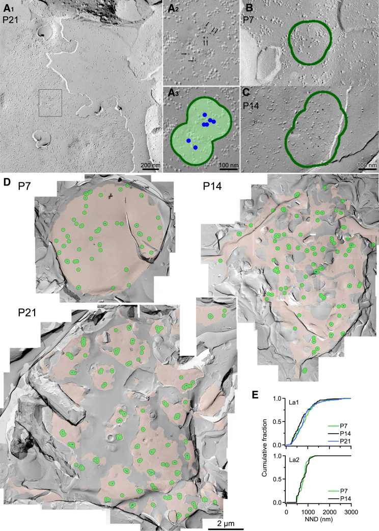
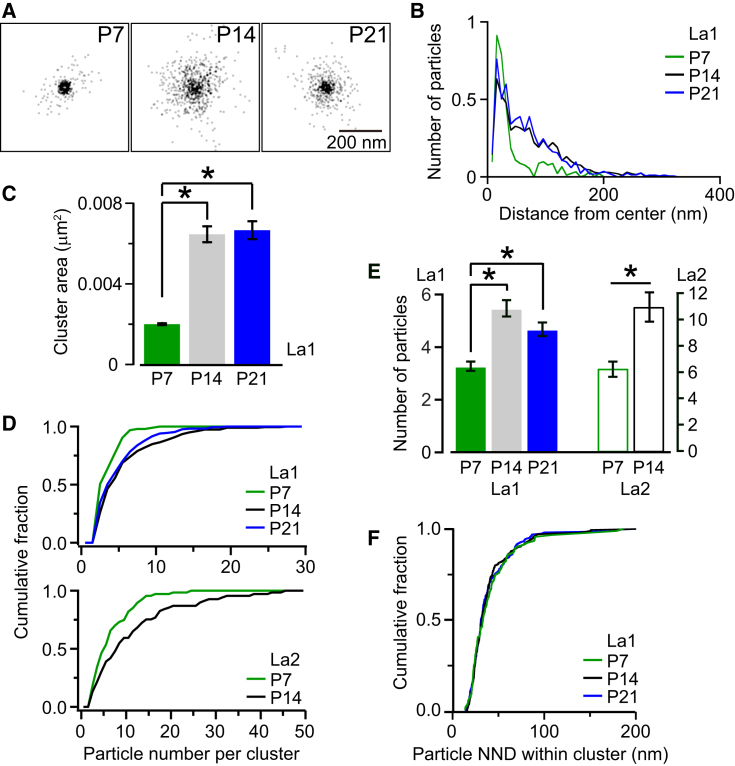
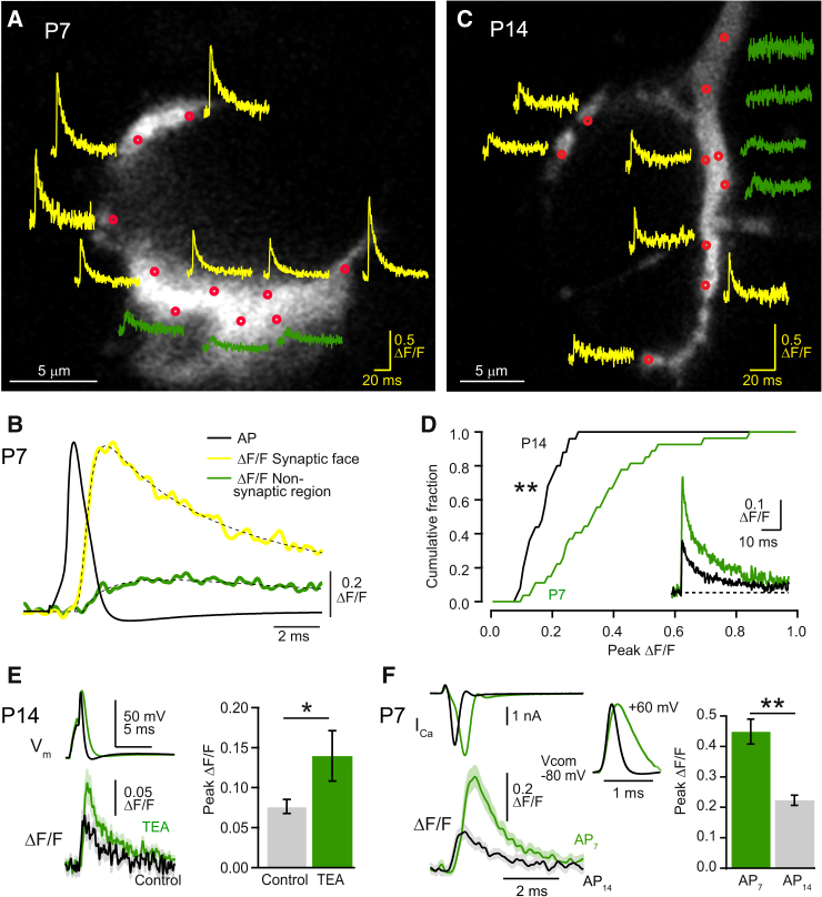
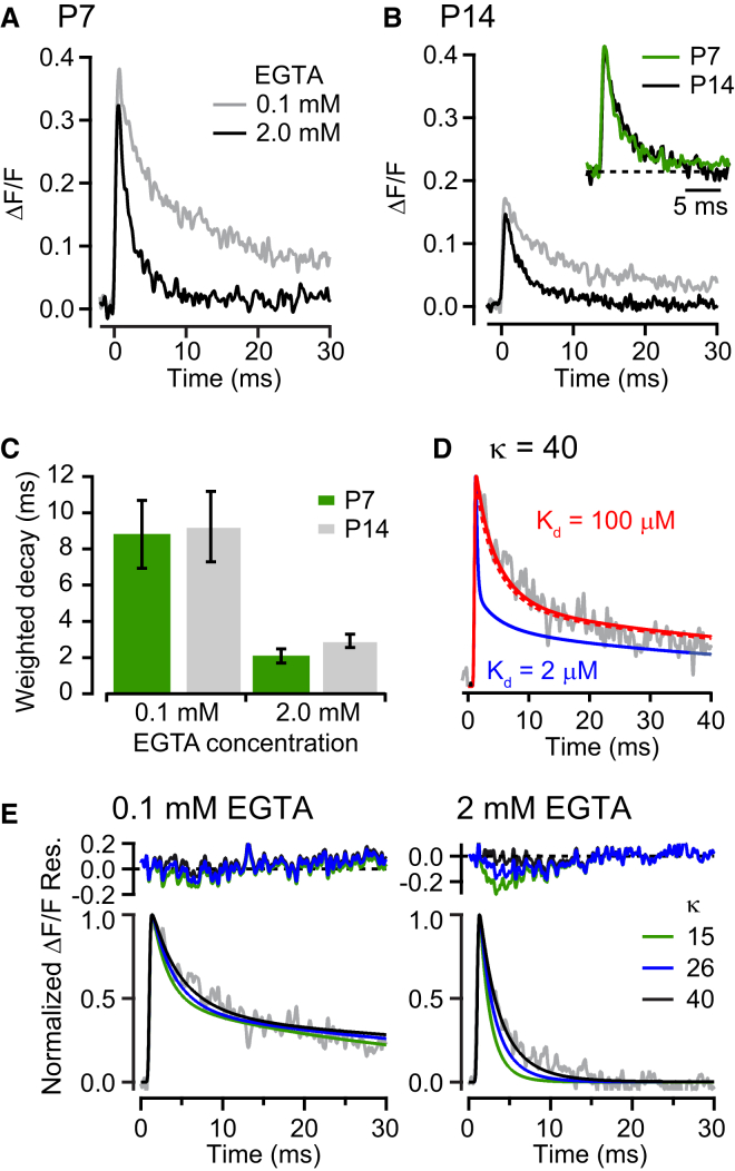
Synaptic efficacy and precision are influenced by the coupling of voltage-gated Ca(2+) channels (VGCCs) to vesicles. But because the topography of VGCCs and their proximity to vesicles is unknown, a quantitative understanding of the determinants of vesicular release at nanometer scale is lacking. To investigate this, we combined freeze-fracture replica immunogold labeling of Cav2.1 channels, local [Ca(2+)] imaging, and patch pipette perfusion of EGTA at the calyx of Held. Between postnatal day 7 and 21, VGCCs formed variable sized clusters and vesicular release became less sensitive to EGTA, whereas fixed Ca(2+) buffer properties remained constant. Experimentally constrained reaction-diffusion simulations suggest that Ca(2+) sensors for vesicular release are located at the perimeter of VGCC clusters (<30 nm) and predict that VGCC number per cluster determines vesicular release probability without altering release time course. This "perimeter release model" provides a unifying framework accounting for developmental changes in both synaptic efficacy and time course.
Overview
The claims and statistics of this paper haven’t been checked yet.
Run the check — free, no account needed. It reads the open-access text and checks the sources it cites.
Integrity signals
Registry
Cites
Statistics
Figures
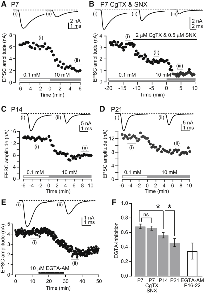
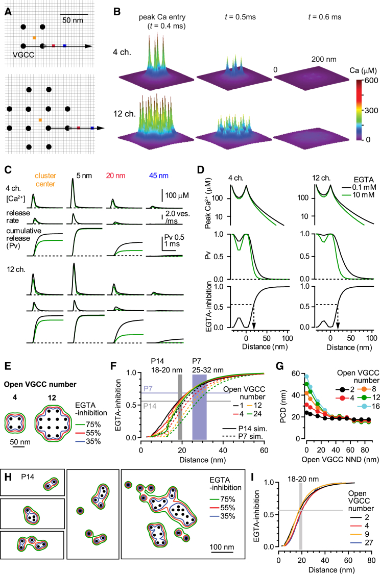
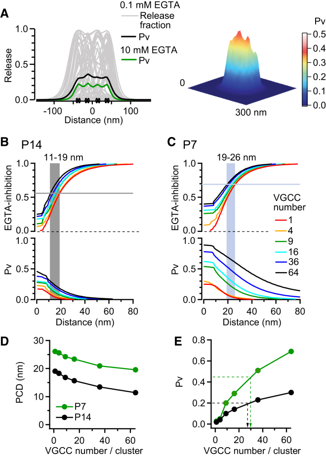
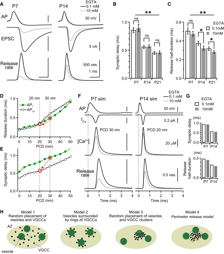
Verification
Claims the paper makes
The claims and statistics of this paper haven’t been checked yet.
Run the check — free, no account needed. It reads the open-access text and checks the sources it cites.
Claims other papers make about this one
Each is checked against this paper’s own text: does it say what the citing paper says it does?
The cited source doesn’t support the claim as written.
In the citing paper
“However, observations in small CNS synapses of both clustered24,25 and random distribution of Ca2+ channels26, and emerging evidence for channel mobility as an equalizer of Pr for vesicles independent of channel positioning27 suggest that their precise distribution may not be the sole determinant of the AZ release likelihood landscape.”
From A trans-synaptic nanocolumn aligns neurotransmitter release to receptors (2016) · cites this paper as [25]
Checked by a reasoning model — may be wrong.
Experimental — accuracy not yet measuredThe source backs part of the claim, not all of it — or backs it for a different population, condition or comparison.
In the citing paper
“Modeling of Ca2+ domains showed that tightening of coupling increases Pr approximately 10-fold per 20 nm change in coupling distance [87,88].”
From Presynaptic cAMP-PKA-mediated potentiation induces reconfiguration of synaptic vesicle pools and channel-vesicle coupling at hippocampal mossy fiber boutons (2024) · cites this paper as [88]
Experimental — accuracy not yet measured
Self-check
Percentages, sums and reported differences re-computed from the paper’s own numbers.
The self-check needs the paper’s full text.
Citations
Cites
Neurotransmitter Release: The Last Millisecond in the Life of a Synaptic Vesicle
Südhof, T. C. (2013) · Neuron · doi:10.1016/j.neuron.2013.10.022 · Cited by 1,197
Calcium gradients and buffers in bovine chromaffin cells.
Neher, E., & Augustine, G. J. (1992) · The Journal of Physiology · doi:10.1113/jphysiol.1992.sp019127 · Cited by 753
Calcium influx and transmitter release in a fast CNS synapse
Borst, J. G. G., & Sakmann, B. J. (1996) · Nature · doi:10.1038/383431a0 · Cited by 632
Calcium control of transmitter release at a cerebellar synapse.
Mintz, I., Sabatini, B., & Regehr, W. (1995) · Neuron · doi:10.1016/0896-6273(95)90155-8 · Cited by 551
Determinants of the Time Course of Facilitation at the Granule Cell to Purkinje Cell Synapse
Atluri, P. P., & Regehr, W. G. (1996) · Journal of Neuroscience · doi:10.1523/jneurosci.16-18-05661.1996 · Cited by 513
Nanodomain coupling between Ca2+ channels and sensors of exocytosis at fast mammalian synapses
Eggermann, E., Bucurenciu, I., Goswami, S. P., & Jonas, P. (2012) · Nature Reviews Neuroscience · doi:10.1038/nrn3125 · Cited by 500
Taschenberger, H., & von Gersdorff, H. (2000) · Journal of Neuroscience · doi:10.1523/jneurosci.20-24-09162.2000 · Cited by 486
Fujimoto, K. (1995) · Journal of Cell Science · doi:10.1242/jcs.108.11.3443 · Cited by 431
Release probability of hippocampal glutamatergic terminals scales with the size of the active zone
Holderith, N., Lorincz, A., Katona, G., Rózsa, B., Kulik, A., Watanabe, M., & Nusser, Z. (2012) · Nature Neuroscience · doi:10.1038/nn.3137 · Cited by 405
Rozov, A., Burnashev, N., Sakmann, B., & Neher, E. (2001) · Journal of Physiology · doi:10.1111/j.1469-7793.2001.0807h.x · Cited by 392
Cited by
Sorted by citations
A trans-synaptic nanocolumn aligns neurotransmitter release to receptors
Tang, A.-H., Chen, H., Li, T. P., Metzbower, S. R., MacGillavry, H. D., & Blanpied, T. A. (2016) · Nature · doi:10.1038/nature19058 · Cited by 677
DOLPHIN, A. C. (2016) · The Journal of Physiology · doi:10.1113/jp272262 · Cited by 367
Liquid-liquid phase separation in biology: mechanisms, physiological functions and human diseases
Zhang, H., Ji, X., Li, P., Liu, C., Lou, J., Wang, Z., Wen, W., Xiao, Y., Zhang, M., & Zhu, X. (2020) · Science China Life Sciences · doi:10.1007/s11427-020-1702-x · Cited by 355
Transcellular Nanoalignment of Synaptic Function
Biederer, T., Kaeser, P. S., & Blanpied, T. A. (2017) · Neuron · doi:10.1016/j.neuron.2017.10.006 · Cited by 352
Presynaptic calcium channels: specialized control of synaptic neurotransmitter release
DOLPHIN, A. C., & Lee, A. (2020) · Nature reviews. Neuroscience · doi:10.1038/s41583-020-0278-2 · Cited by 291
Phase separation at the synapse
Chen, X., Wu, X., Wu, H., & Zhang, M. (2020) · Nature Neuroscience · doi:10.1038/s41593-019-0579-9 · Cited by 261
Rothman, J. S., & Silver, R. A. (2018) · Frontiers in Neuroinformatics · doi:10.3389/fninf.2018.00014 · Cited by 256
Self-citationNeher, E. (2015) · Neuron · doi:10.1016/j.neuron.2015.08.038 · Cited by 236
Genetic disruption of voltage-gated calcium channels in psychiatric and neurological disorders
Heyes, S., Pratt, W. S., Rees, E., Dahimène, S., Ferron, L., Owen, M. J., & DOLPHIN, A. C. (2015) · Progress in Neurobiology · doi:10.1016/j.pneurobio.2015.09.002 · Cited by 241
RIM and RIM-BP Form Presynaptic Active-Zone-like Condensates via Phase Separation
Wu, X., Cai, Q., Shen, Z., Chen, X., Zeng, M., Du, S., & Zhang, M. (2019) · Molecular Cell · doi:10.1016/j.molcel.2018.12.007 · Cited by 242
Active zone scaffolds differentially accumulate Unc13 isoforms to tune Ca2+ channel–vesicle coupling
Böhme, M. A., Beis, C., Reddy‐Alla, S., Reynolds, E., Mampell, M. M., Grasskamp, A. T., Lützkendorf, J., Bergeron, D. D., Driller, J. H., Babikir, H. A., et al. (2016) · Nature Neuroscience · doi:10.1038/nn.4364 · Cited by 219
Dynamically Primed Synaptic Vesicle States: Key to Understand Synaptic Short-Term Plasticity
Neher, E., & Brose, N. (2018) · Neuron · doi:10.1016/j.neuron.2018.11.024 · Cited by 210
Monday, H. R., Younts, T. J., & Castillo, P. E. (2018) · Annual Review of Neuroscience · doi:10.1146/annurev-neuro-080317-062155 · Cited by 175
Synaptic vesicles transiently dock to refill release sites
Kusick, G. F., Chin, M., Raychaudhuri, S., Lippmann, K., Adula, K. P., Hujber, E. J., Vu, T., Davis, M. W., Jorgensen, E. M., & Watanabe, S. (2020) · Nature Neuroscience · doi:10.1038/s41593-020-00716-1 · Cited by 138
Reddy‐Alla, S., Böhme, M. A., Reynolds, E., Beis, C., Grasskamp, A. T., Mampell, M. M., Maglione, M., Jusyte, M., Rey, U., Babikir, H. A., et al. (2017) · Neuron · doi:10.1016/j.neuron.2017.08.016 · Cited by 134
Rebola, N., Reva, M., Kirizs, T., Szoboszlay, M., Lőrincz, A., Moneron, G., Nusser, Z., & DiGregorio, D. A. (2019) · Neuron · doi:10.1016/j.neuron.2019.08.014 · Cited by 136
Self-citationPhysiology of intracellular calcium buffering
Eisner, D. A., Neher, E., Taschenberger, H., & Smith, G. L. (2023) · Physiological Reviews · doi:10.1152/physrev.00042.2022 · Cited by 162
The control of release probability at nerve terminals
Dittman, J. S., & Ryan, T. A. (2019) · Nature reviews. Neuroscience · doi:10.1038/s41583-018-0111-3 · Cited by 127
Quantitative optical nanophysiology of Ca2+ signaling at inner hair cell active zones
Neef, J., Urban, N. T., Ohn, T., Frank, T., Jean, P., Hell, S. W., Willig, K. I., & Moser, T. (2018) · Nature Communications · doi:10.1038/s41467-017-02612-y · Cited by 119
Akbergenova, Y., Zhang, Y. V., Weiss, S., Cunningham, K. L., & Littleton, J. T. (2018) · eLife · doi:10.7554/elife.38268 · Cited by 109
RIM-BPs Mediate Tight Coupling of Action Potentials to Ca 2+ -Triggered Neurotransmitter Release
Acuna, C., Liu, X., Gonzalez-Suarez, A. D., & Südhof, T. C. (2015) · Neuron · doi:10.1016/j.neuron.2015.08.027 · Cited by 113
Approaches and Limitations in the Investigation of Synaptic Transmission and Plasticity
Glasgow, S. D., McPhedrain, R., Madranges, J. F., Kennedy, T. E., & Ruthazer, E. S. (2019) · Frontiers in Synaptic Neuroscience · doi:10.3389/fnsyn.2019.00020 · Cited by 100
Nanoscale Structural Plasticity of the Active Zone Matrix Modulates Presynaptic Function
Glebov, O. O., Jackson, R. E., Winterflood, C. M., Owen, D. M., Barker, E. A., Doherty, P., Ewers, H., & Burrone, J. (2017) · Cell Reports · doi:10.1016/j.celrep.2017.02.064 · Cited by 90
The Nanophysiology of Fast Transmitter Release
Stanley, E. F. (2016) · Trends in Neurosciences · doi:10.1016/j.tins.2016.01.005 · Cited by 89
Synapse and Active Zone Assembly in the Absence of Presynaptic Ca2+ Channels and Ca2+ Entry
Held, R. G., Liu, C., Ma, K., Ramsey, A. M., Tarr, T. B., de Nola, G., Wang, S. S. H., Wang, J., van den Maagdenberg, A. M. J. M., Schneider, T., et al. (2020) · Neuron · doi:10.1016/j.neuron.2020.05.032 · Cited by 84
Transient Confinement of CaV2.1 Ca2+-Channel Splice Variants Shapes Synaptic Short-Term Plasticity
Heck, J., Parutto, P., Ciuraszkiewicz, A., Bikbaev, A. F., Freund, R., Mitlöhner, J., Andres‐Alonso, M., Fejtová, A., Holcman, D., & Heine, M. (2019) · Neuron · doi:10.1016/j.neuron.2019.04.030 · Cited by 84
Chen, Z., Das, B., Nakamura, Y., DiGregorio, D. A., & Young, S. (2015) · Journal of Neuroscience · doi:10.1523/jneurosci.2753-14.2015 · Cited by 89
Self-citationMiki, T., Kaufmann, W. A., Malagón, G., Gomez, L. C., Tabuchi, K., Watanabe, M., Shigemoto, R., & Marty, A. (2017) · Proceedings of the National Academy of Sciences · doi:10.1073/pnas.1704470114 · Cited by 84
Self-citationα-Neurexins Together with α2δ-1 Auxiliary Subunits Regulate Ca 2+ Influx through Ca v 2.1 Channels
Brockhaus, J., Schreitmüller, M., Repetto, D., Klatt, O., Reißner, C., Elmslie, K. S., Heine, M., & Missler, M. (2018) · Journal of Neuroscience · doi:10.1523/jneurosci.0511-18.2018 · Cited by 85
Kubota, Y., Kondo, S., Nomura, M., Hatada, S., Yamaguchi, N., Mohamed, A. A., Karube, F., Lübke, J. H. R., & Kawaguchi, Y. (2015) · eLife · doi:10.7554/elife.07919 · Cited by 83
A C1–C2 Module in Munc13 Inhibits Calcium-Dependent Neurotransmitter Release
Michelassi, F., Liu, H.-W., Hu, Z.-T., & Dittman, J. (2017) · Neuron · doi:10.1016/j.neuron.2017.07.015 · Cited by 72
Dong, W., Radulović, T., Goral, R. O., Thomas, C. I., Montesinos, M. S., Guerrero‐Given, D., Hagiwara, A., Putzke, T., Hida, Y., Abe, M., et al. (2018) · Cell Reports · doi:10.1016/j.celrep.2018.06.024 · Cited by 73
Self-citationMolecular Machines Regulating the Release Probability of Synaptic Vesicles at the Active Zone
Körber, C., & Kuner, T. (2016) · Frontiers in Synaptic Neuroscience · doi:10.3389/fnsyn.2016.00005 · Cited by 77
Fekete, Á., Nakamura, Y., Yang, Y., Herlitze, S., Mark, M. D., DiGregorio, D. A., & Wang, L. (2019) · Nature Communications · doi:10.1038/s41467-019-08452-2 · Cited by 71
Self-citationMiki, T., Nakamura, Y., Malagon, G., Neher, E., & Marty, A. (2018) · Nature Communications · doi:10.1038/s41467-018-06336-5 · Cited by 53
Self-citationCalcium dependence of spontaneous neurotransmitter release
Williams, C. L., & Smith, S. M. (2017) · Journal of Neuroscience Research · doi:10.1002/jnr.24116 · Cited by 71
A sensory cell diversifies its output by varying Ca2+ influx‐release coupling among active zones
Özçete, Ö. D., & Moser, T. (2020) · The EMBO Journal · doi:10.15252/embj.2020106010 · Cited by 66
Chang, S., Reim, K., Pedersen, M., Neher, E., Brose, N., & Taschenberger, H. (2015) · Journal of Neuroscience · doi:10.1523/jneurosci.4841-14.2015 · Cited by 66
Fife organizes synaptic vesicles and calcium channels for high-probability neurotransmitter release
Bruckner, J. J., Zhan, H., Gratz, S. J., Rao, M., Ukken, F. P., Zilberg, G., & O’Connor-Giles, K. M. (2016) · The Journal of Cell Biology · doi:10.1083/jcb.201601098 · Cited by 62
Functions of Presynaptic Voltage-gated Calcium Channels
DOLPHIN, A. C. (2020) · Function · doi:10.1093/function/zqaa027 · Cited by 65
Quantal Fluctuations in Central Mammalian Synapses: Functional Role of Vesicular Docking Sites
Pulido, C., & Marty, A. (2017) · Physiological Reviews · doi:10.1152/physrev.00032.2016 · Cited by 63
Borges-Merjane, C., Kim, O., & Jonas, P. (2020) · Neuron · doi:10.1016/j.neuron.2019.12.022 · Cited by 60
Diversity matters — extending sound intensity coding by inner hair cells via heterogeneous synapses
Moser, T., Karagulyan, N., Neef, J., & Tobón, L. M. J. (2023) · The EMBO Journal · doi:10.15252/embj.2023114587 · Cited by 58
Reduced endogenous Ca 2+ buffering speeds active zone Ca 2+ signaling
Delvendahl, I., Jablonski, L., Baade, C., Matveev, V., Neher, E., & Hallermann, S. (2015) · Proceedings of the National Academy of Sciences · doi:10.1073/pnas.1508419112 · Cited by 63
Johnson, S. L., Olt, J., Cho, S., von Gersdorff, H., & Marcotti, W. (2017) · Journal of Neuroscience · doi:10.1523/jneurosci.2867-16.2017 · Cited by 60
Creating diverse synapses from the same molecules
Nusser, Z. (2018) · Current Opinion in Neurobiology · doi:10.1016/j.conb.2018.01.001 · Cited by 71
Calcium dependence of neurotransmitter release at a high fidelity synapse
Eshra, A., Schmidt, H., Eilers, J., & Hallermann, S. (2021) · eLife · doi:10.7554/elife.70408 · Cited by 56
Lübbert, M., Goral, R. O., Keine, C., Thomas, C. I., Guerrero‐Given, D., Putzke, T., Satterfield, R., Kamasawa, N., & Young, S. (2018) · Neuron · doi:10.1016/j.neuron.2018.11.028 · Cited by 61
Self-citationKeller, D. X., Babai, N. Z., Kochubey, O., Han, Y., Markram, H., Schürmann, F., & Schneggenburger, R. (2015) · PLoS Computational Biology · doi:10.1371/journal.pcbi.1004253 · Cited by 61
Éltes, T., Kirizs, T., Nusser, Z., & Holderith, N. B. (2017) · Journal of Neuroscience · doi:10.1523/jneurosci.2024-16.2017 · Cited by 53
Kobbersmed, J. R., Grasskamp, A. T., Jusyte, M., Böhme, M. A., Ditlevsen, S., Sørensen, J. B., & Walter, A. M. (2020) · eLife · doi:10.7554/elife.51032 · Cited by 54
Molecular logic of synaptic diversity between Drosophila tonic and phasic motoneurons
Jetti, S. K., Crane, A. B., Akbergenova, Y., Aponte-Santiago, N. A., Cunningham, K. L., Whittaker, C. A., & Littleton, J. T. (2023) · Neuron · doi:10.1016/j.neuron.2023.07.019 · Cited by 51
Physical determinants of vesicle mobility and supply at a central synapse
Rothman, J. S., Kocsis, L., Herzog, É., Nusser, Z., & Silver, R. A. (2016) · eLife · doi:10.7554/elife.15133 · Cited by 53
Self-citationLübbert, M., Goral, R. O., Satterfield, R., Putzke, T., van den Maagdenberg, A. M. J. M., Kamasawa, N., & Young, S. M. (2017) · eLife · doi:10.7554/elife.28412 · Cited by 55
Self-citationAldahabi, M., Balint, F., Holderith, N., Lorincz, A., Reva, M., & Nusser, Z. (2022) · Neuron · doi:10.1016/j.neuron.2022.09.035 · Cited by 50
The Cerebellar Mossy Fiber Synapse as a Model for High-Frequency Transmission in the Mammalian CNS
Delvendahl, I., & Hallermann, S. (2016) · Trends in Neurosciences · doi:10.1016/j.tins.2016.09.006 · Cited by 52
Heterogeneity of glutamatergic synapses: cellular mechanisms and network consequences
Wichmann, C., & Kuner, T. (2021) · Physiological Reviews · doi:10.1152/physrev.00039.2020 · Cited by 55
Resolving the molecular architecture of the photoreceptor active zone with 3D-MINFLUX
Grabner, C. P., Jansen, I., Neef, J., Weihs, T., Schmidt, R., Riedel, D., Wurm, C. A., & Moser, T. (2022) · Science Advances · doi:10.1126/sciadv.abl7560 · Cited by 49
Synaptic plasticity in the auditory system: a review
Friauf, E., Fischer, A. U., & Fuhr, M. F. (2015) · Cell and Tissue Research · doi:10.1007/s00441-015-2176-x · Cited by 49
Bornschein, G., & Schmidt, H. (2019) · Frontiers in Molecular Neuroscience · doi:10.3389/fnmol.2018.00494 · Cited by 47
Synaptic Transmission Optimization Predicts Expression Loci of Long-Term Plasticity
Costa, R. P., Padamsey, Z., D’amour, J. A., Emptage, N. J., Froemke, R. C., & Vogels, T. P. (2017) · Neuron · doi:10.1016/j.neuron.2017.09.021 · Cited by 45
Eguchi, K., Velicky, P., Hollergschwandtner, E., Itakura, M., Fukazawa, Y., Danzl, J. G., & Shigemoto, R. (2020) · Frontiers in Cellular Neuroscience · doi:10.3389/fncel.2020.00063 · Cited by 45
Self-citationGhelani, T., Escher, M., Thomas, U., Esch, K., Lützkendorf, J., Depner, H., Maglione, M., Parutto, P., Gratz, S. J., Matkovic, T., et al. (2023) · Science Advances · doi:10.1126/sciadv.ade7804 · Cited by 42
Functional specificity of liquid-liquid phase separation at the synapse
Guzikowski, N. J., & Kavalali, E. T. (2024) · Nature Communications · doi:10.1038/s41467-024-54423-7 · Cited by 45
Lou, X. (2018) · Frontiers in Cellular Neuroscience · doi:10.3389/fncel.2018.00066 · Cited by 39
Coupling the Structural and Functional Assembly of Synaptic Release Sites
Ghelani, T., & Sigrist, S. J. (2018) · Frontiers in Neuroanatomy · doi:10.3389/fnana.2018.00081 · Cited by 40
Montesinos, M. S., Dong, W., Goff, K. M., Das, B., Guerrero‐Given, D., Schmalzigaug, R., Premont, R. T., Satterfield, R., Kamasawa, N., & Young, S. (2015) · Neuron · doi:10.1016/j.neuron.2015.10.042 · Cited by 41
Self-citationVesicle release site organization at synaptic active zones
Walter, A. M., Böhme, M. A., & Sigrist, S. J. (2017) · Neuroscience Research · doi:10.1016/j.neures.2017.12.006 · Cited by 40
The Calyx of Held in the auditory system: structure, function, and development
Baydyuk, M., Xu, J.-H., & Wu, L. (2016) · Hearing Research · doi:10.1016/j.heares.2016.03.009 · Cited by 33
A Well-Defined Readily Releasable Pool with Fixed Capacity for Storing Vesicles at Calyx of Held
Mahfooz, K., Singh, M., Renden, R., & Wesseling, J. F. (2016) · PLoS Computational Biology · doi:10.1371/journal.pcbi.1004855 · Cited by 38
Monday, H. R., Bourdenx, M., Jordan, B. A., & Castillo, P. E. (2020) · eLife · doi:10.7554/elife.54812 · Cited by 39
Althof, D., Baehrens, D., Watanabe, M., 昇, 鈴., Fakler, B., & Kulik, Á. (2015) · Frontiers in Cellular Neuroscience · doi:10.3389/fncel.2015.00315 · Cited by 37
Transmitter release site organization can predict synaptic function at the neuromuscular junction
Laghaei, R., Ma, J., Tarr, T. B., Homan, A. E., Kelly, L. S., Tilvawala, M., Vuocolo, B. S., Rajasekaran, H. P., Meriney, S. D., & Dittrich, M. (2017) · Journal of Neurophysiology · doi:10.1152/jn.00168.2017 · Cited by 33
Han, Y., Cao, R., Qin, L., Chen, L. Y., Tang, A., Südhof, T. C., & Zhang, B. (2022) · Science Advances · doi:10.1126/sciadv.abo4173 · Cited by 38
Damonte, V. M., Rodríguez, S. S., & Raingo, J. (2018) · The Journal of Physiology · doi:10.1113/jp276256 · Cited by 36
Nanoscale Organization of Vesicle Release at Central Synapses
Gramlich, M. W., & Klyachko, V. A. (2019) · Trends in Neurosciences · doi:10.1016/j.tins.2019.03.001 · Cited by 35
First passage time distribution of multiple impatient particles with reversible binding
Lawley, S. D., & Madrid, J. B. (2019) · The Journal of Chemical Physics · doi:10.1063/1.5098312 · Cited by 36
Asymmetry Between Pre- and Postsynaptic Transient Nanodomains Shapes Neuronal Communication
Heine, M., & Holcman, D. (2020) · Trends in Neurosciences · doi:10.1016/j.tins.2020.01.005 · Cited by 36
A theory of synaptic transmission
Wang, B., & Dudko, O. K. (2021) · eLife · doi:10.7554/elife.73585 · Cited by 38
Cunningham, K. L., Sauvola, C. W., Tavana, S. Z., & Littleton, J. T. (2022) · eLife · doi:10.7554/elife.78648 · Cited by 33
CaV1 and CaV2 calcium channels mediate the release of distinct pools of synaptic vesicles
Mueller, B. D., Merrill, S. A., Watanabe, S., Liu, P., Niu, L., Singh, A., Maldonado-Catala, P. J., Cherry, A., Rich, M. S., Silva, M., et al. (2023) · eLife · doi:10.7554/elife.81407 · Cited by 37
The glutamatergic synapse: a complex machinery for information processing
Maio, V. D. (2021) · Cognitive Neurodynamics · doi:10.1007/s11571-021-09679-w · Cited by 38
Emerging Roles of Activity-Dependent Alternative Splicing in Homeostatic Plasticity
Thalhammer, A., Jaudon, F., & Cingolani, L. A. (2020) · Frontiers in Cellular Neuroscience · doi:10.3389/fncel.2020.00104 · Cited by 32
Functional Microstructure of Ca V -Mediated Calcium Signaling in the Axon Initial Segment
Lipkin, A. M., Cunniff, M. M., Spratt, P. W. E., Lemke, S. M., & Bender, K. J. (2021) · Journal of Neuroscience · doi:10.1523/jneurosci.2843-20.2021 · Cited by 32
Böhme, M. A., Grasskamp, A. T., & Walter, A. M. (2018) · FEBS Letters · doi:10.1002/1873-3468.13188 · Cited by 35
Presynaptic development is controlled by the core active zone proteins CAST/ELKS
Radulović, T., Dong, W., Goral, R. O., Thomas, C. I., Veeraraghavan, P., Montesinos, M. S., Guerrero‐Given, D., Goff, K. M., Lübbert, M., Kamasawa, N., et al. (2020) · The Journal of Physiology · doi:10.1113/jp279736 · Cited by 29
Self-citationTimofeeva, Y., & Volynski, K. E. (2015) · Frontiers in Cellular Neuroscience · doi:10.3389/fncel.2015.00239 · Cited by 30
First passage times for multiple particles with reversible target-binding kinetics
Grebenkov, D. S. (2017) · The Journal of Chemical Physics · doi:10.1063/1.4996395 · Cited by 31
The First 100 nm Inside the Pre-synaptic Terminal Where Calcium Diffusion Triggers Vesicular Release
Guerrier, C., & Holcman, D. (2018) · Frontiers in Synaptic Neuroscience · doi:10.3389/fnsyn.2018.00023 · Cited by 30
Nakamura, Y., Reva, M., & DiGregorio, D. A. (2018) · Journal of Neuroscience · doi:10.1523/jneurosci.2061-17.2018 · Cited by 30
Self-citationBornschein, G., Eilers, J., & Schmidt, H. (2019) · Cell Reports · doi:10.1016/j.celrep.2019.07.008 · Cited by 28
Reva, M., DiGregorio, D. A., & Grebenkov, D. S. (2021) · Scientific Reports · doi:10.1038/s41598-021-84340-4 · Cited by 27
Self-citationKrick, N., Ryglewski, S., Pichler, A., Bikbaev, A. F., Götz, T., Kobler, O., Heine, M., Thomas, U., & Duch, C. (2021) · Proceedings of the National Academy of Sciences · doi:10.1073/pnas.2106621118 · Cited by 31
The presynaptic active zone: A dynamic scaffold that regulates synaptic efficacy
Michel, K., Müller, J. A., Oprişoreanu, A., & Schoch, S. (2015) · Experimental Cell Research · doi:10.1016/j.yexcr.2015.02.011 · Cited by 33
Dynamic association of calcium channel subunits at the cellular membrane
Voigt, A., Freund, R., Heck, J., Missler, M., Obermair, G. J., Thomas, U., & Heine, M. (2016) · Neurophotonics · doi:10.1117/1.nph.3.4.041809 · Cited by 30
GABAB receptor auxiliary subunits modulate Cav2.3-mediated release from medial habenula terminals
Bhandari, P., Vandael, D. H. F., Fernández‐Fernández, D., Fritzius, T., Kleindienst, D., Önal, C., Montanaro, J., Gassmann, M., Jónás, P., Kulik, Á., et al. (2021) · eLife · doi:10.7554/elife.68274 · Cited by 28
Self-citationPresynaptic Rac1 controls synaptic strength through the regulation of synaptic vesicle priming
Keine, C., Al-Yaari, M., Radulović, T., Thomas, C. I., Ramos, P. V., Guerrero‐Given, D., Ranjan, M., Taschenberger, H., Kamasawa, N., & Young, S. M. (2022) · eLife · doi:10.7554/elife.81505 · Cited by 23
Self-citationBadawi, Y., & Nishimune, H. (2017) · Neuroscience Research · doi:10.1016/j.neures.2017.11.014 · Cited by 29
Emperador-Melero, J., Andersen, J. W., Metzbower, S. R., Levy, A. D., Dharmasri, P. A., de Nola, G., Blanpied, T. A., & Kaeser, P. S. (2024) · Nature Neuroscience · doi:10.1038/s41593-024-01720-5 · Cited by 27
Proteolytic maturation of α2δ controls the probability of synaptic vesicular release
Ferron, L., Kadurin, I., & DOLPHIN, A. C. (2018) · eLife · doi:10.7554/elife.37507 · Cited by 25
Metadata
- Authors
- Nakamura, Y., Harada, H., Kamasawa, N., Matsui, K., Rothman, J. S., Shigemoto, R., Silver, R. A., DiGregorio, D. A., & Takahashi, T.
- Affiliations
- Centre National de la Recherche Scientifique (FR)
- Institute of Science and Technology Austria (AT)
- National Institute for Physiological Sciences (JP)
- University College London (GB)
- Doshisha University (JP)
- Corresponding author
- DiGregorio, David A.
- Takahashi, Tomoyuki
- ORCID
- Venue
- Neuron
- Publisher
- Elsevier BV
- Published
- 2015-01
- Volume
- 85
- Issue
- 1
- Pages
- 145-158
- ISSN
- 0896-6273 (ISSN-L)
- PMC
- PMC4305191
- OpenAlex
- W2030428080
- Cited by
- 245
- References
- 60
- Citations by year
- 2026: 10 · 2025: 18 · 2024: 26 · 2023: 19 · 2022: 22 · 2021: 23 · 2020: 27 · 2019: 24 · 2018: 22 · 2017: 21 · 2016: 14 · 2015: 18
- FWCI
- 8.23
- Type
- journal article
- Language
- en
- Open access
- hybrid
- doi.org
- Licence
- Locations
- Neuron (publishedVersion, cc-by)
- Europe PMC (PubMed Central) (publishedVersion, cc-by)
- PubMed Central (publishedVersion, cc-by)
- PubMed (publishedVersion)
- UCL Discovery (University College London) (submittedVersion)
- MPG.PuRe (Max Planck Society) (submittedVersion)
- Funders
- Core Research for Evolutional Science and Technology (CREST) of Japan Science and Technology Agency
- Okinawa Institute of Science and Technology (OIST)
- JSPS
- Japanese Ministry of Education, Culture, Sports, Science and Technology (23700474)
- Centre National de la Recherche Scientifique
- Fondation Fyssen
- Fondation pour la Recherche Medicale
- Federation pour la Recherche sur le Cerveau
- Agence Nationale de la Recherche (ANR-2007-Neuro-008-01, ANR-2010-BLAN-1411-01)
- European Commission Coordination Action ENINET (LSHM-CT-2005-19063)
- Wellcome Trust (064413, 095667)
- ERC (294667)
- Topics
- Life Sciences › Neuroscience › Cellular and Molecular Neuroscience › Neuroscience and Neuropharmacology Research
- Life Sciences › Biochemistry, Genetics and Molecular Biology › Molecular Biology › Ion channel regulation and function
- Life Sciences › Biochemistry, Genetics and Molecular Biology › Cell Biology › Cellular transport and secretion
- Keywords
- voltage-gated calcium channels, Cav2.1, presynaptic calcium channels, synaptic vesicle release, release probability, calyx of Held, synaptic development, reaction-diffusion simulation
- MeSH terms
- Action Potentials, Animals, Calcium, Egtazic Acid, Exocytosis, Synapses, Synaptic Vesicles, Presynaptic Terminals, Patch-Clamp Techniques, Calcium Channels, N-Type, Mice, Rats, Calcium Chelating Agents
About these checks
Registry, retraction, Cites and statistics results are computed, not judged, with one exception: a printed reference that no index lists is read once by a model before it counts as a citation error. Claims are extracted by a model and kept only when their quote appears verbatim in the text; each cited source is then checked, with a model’s help, for whether it says what the claim relies on. Those verdicts can be wrong — read the claim’s quote and the cited source before relying on one.
Statistics and arithmetic are recomputed, not judged.
Confidence bands are shown only for fields calibrated on our annotated test set. None is calibrated yet, so no band is shown.
What counts as a claim ›How we measure confidence ›How we check this ›
Are you an author, or do you see a problem with this page? Request a review
