

The DOI matches its Crossref record; 35 of the 35 references it cites were found in a registry or an index.
Optimization of Lipid Nanoparticles for Intramuscular Administration of mRNA Vaccines
Open accessAbstract
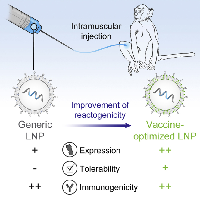
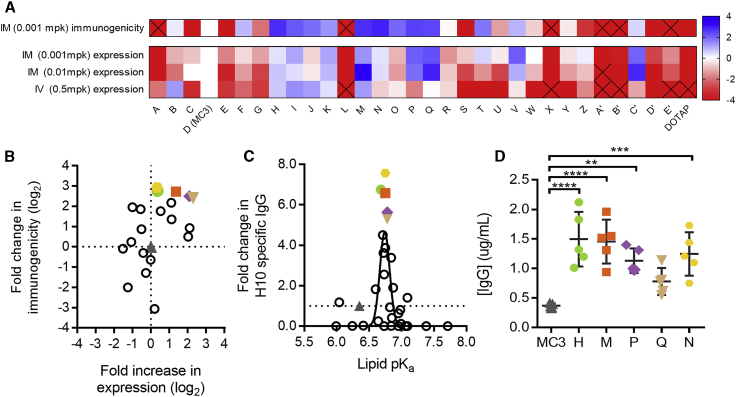
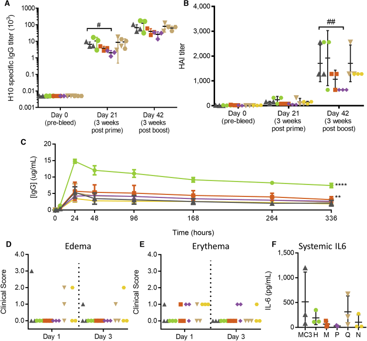
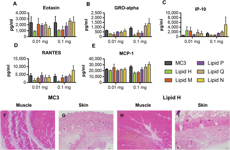
mRNA vaccines have the potential to tackle many unmet medical needs that are unable to be addressed with conventional vaccine technologies. A potent and well-tolerated delivery technology is integral to fully realizing the potential of mRNA vaccines. Pre-clinical and clinical studies have demonstrated that mRNA delivered intramuscularly (IM) with first-generation lipid nanoparticles (LNPs) generates robust immune responses. Despite progress made over the past several years, there remains significant opportunity for improvement, as the most advanced LNPs were designed for intravenous (IV) delivery of siRNA to the liver. Here, we screened a panel of proprietary biodegradable ionizable lipids for both expression and immunogenicity in a rodent model when administered IM. A subset of compounds was selected and further evaluated for tolerability, immunogenicity, and expression in rodents and non-human primates (NHPs). A lead formulation was identified that yielded a robust immune response with improved tolerability. More importantly for vaccines, increased innate immune stimulation driven by LNPs does not equate to increased immunogenicity, illustrating that mRNA vaccine tolerability can be improved without affecting potency.
Overview
The claims and statistics of this paper haven’t been checked yet.
Run the check — free, no account needed. It reads the open-access text and checks the sources it cites.
Integrity signals
Registry
Cites
Statistics
Figures
Verification
Claims the paper makes
The claims and statistics of this paper haven’t been checked yet.
Run the check — free, no account needed. It reads the open-access text and checks the sources it cites.
Claims other papers make about this one
Each is checked against this paper’s own text: does it say what the citing paper says it does?
The source backs part of the claim, not all of it — or backs it for a different population, condition or comparison.
In the citing paper
“For Moderna's LNP formulation, Hassett et al. reported that the SM-102 lipid was selected based on its improved tolerability profile in non-human primates, as evidenced by a reduced local reactogenicity (e.g., edema and erythema) and lowest induction of systemic cytokine responses (e.g., IL-6) [13]”
From The dawn of mRNA vaccines: the COVID-19 case (2021) · cites this paper as [13]
Experimental — accuracy not yet measuredIn the citing paper
“which are known to have significantly higher delivery efficiency in hepatocytes after intravenous (i.v.) injection31 or in muscle cells after intramuscular (i.m.) injection.32”
From COVID-19 mRNA vaccines: Platforms and current developments (2022) · cites this paper as [32]
In the citing paper
“Based on these observations, Moderna has developed an ionizable lipid (namely Lipid H, SM-102), whose tail adopts larger branches which in turn increases potency, and whose introduced ester bonds increase biodegradability (Fig. 7).290,291”
From Advances in COVID-19 mRNA vaccine development (2022) · cites this paper as [291]
In the citing paper
“All three major players in the mRNA vaccine field have previously optimized so-called lipid nanoparticles (LNPs), constructs that typically consist of an ionizable lipid and one or more helper lipids to improve LNP stability and promote endosomal escape upon cellular uptake [12,13]”
From The dawn of mRNA vaccines: the COVID-19 case (2021) · cites this paper as [13]
In the citing paper
“the usage of the SM-102 lipid in Moderna's mRNA-1273 vaccine was found to outperform Onpattro's MC3 LNPs for the intramuscular (i.m.) delivery of mRNA in rodents and non-human primates, given its improved tolerability and higher endosomal escape efficiency [12,13]”
From The dawn of mRNA vaccines: the COVID-19 case (2021) · cites this paper as [13]
In the citing paper
“Endosomal escape can be increased by optimizing the pKa values of ionizable lipids66,76–78,88,100,140.”
From Lipid nanoparticles for mRNA delivery (2021) · cites this paper as [78]
In the citing paper
“To improve the biocompatibility of lipid nanoparticles, biodegradable lipids can be applied76–78,108,179.”
From Lipid nanoparticles for mRNA delivery (2021) · cites this paper as [78]
In the citing paper
“Moreover, the delivery efficacy of mRNA could be improved, for example, by rational design of lipids through modulation of head groups and hydrophobic tails to increase cellular uptake and endosomal escape of lipid nanoparticle–mRNA formulations64,66,76–78,88,97,100,114,140,141.”
From Lipid nanoparticles for mRNA delivery (2021) · cites this paper as [78]
Self-check
Percentages, sums and reported differences re-computed from the paper’s own numbers.
The self-check needs the paper’s full text.
Citations
Cites
Karikó, K., Buckstein, M., Ni, H., & Weissman, D. (2005) · Immunity · doi:10.1016/j.immuni.2005.06.008 · Cited by 2,728
Heterologous Protection Against Influenza by Injection of DNA Encoding a Viral Protein
Ulmer, J. B., Donnelly, J., Parker, S. E., Rhodes, G. H., Felgner, P. L., Dwarki, V., Gromkowski, S. H., Deck, R. R., DeWitt, C. M., Friedman, A., et al. (1993) · Science · doi:10.1126/science.8456302 · Cited by 2,318
Lanciotti, R. S., Kosoy, O. L., Laven, J. J., Velez, J. O., Lambert, A. J., Johnson, A. J., Stanfield, S. M., & Duffy, M. R. (2008) · Emerging infectious diseases · doi:10.3201/eid1408.080287 · Cited by 2,173
Rational design of cationic lipids for siRNA delivery
Semple, S. C., Akinc, A., Chen, J., Sandhu, A. P., Mui, B. L., Cho, C. K., Sah, D. W. Y., Stebbing, D., Crosley, E. J., Yaworski, E., et al. (2010) · Nature Biotechnology · doi:10.1038/nbt.1602 · Cited by 1,822 · cited 1×
“8.(11,13,23) However, the best lipids with respect to protein expression after IV administration generally had lower pKas than the best lipids for protein expression after IM administration.”
Maximizing the potency of siRNA lipid nanoparticles for hepatic gene silencing in vivo
Jayaraman, M., Ansell, S. M., Mui, B. L., Tam, Y. K., Chen, J., Du, X., Butler, D., Eltepu, L., Matsuda, S., Narayanannair, J. K., et al. (2012) · Angewandte Chemie International Edition · doi:10.1002/anie.201203263 · Cited by 1,267
Safety and efficacy of RNAi therapy for transthyretin amyloidosis
Coelho, T., Adams, D., Silva, A., Lozeron, P., Hawkins, P. N., Mant, T., Perez, J., Chiesa, J., Warrington, S., Tranter, E., et al. (2013) · New England Journal of Medicine · doi:10.1056/nejmoa1208760 · Cited by 971
Lipid-like materials for low-dose, in vivo gene silencing
Love, K. T., Mahon, K. P., Levins, C. G., Whitehead, K. A., Querbes, W., Dorkin, J. R., Qin, J., Cantley, W., Qin, L. L., Racie, T., et al. (2010) · Proceedings of the National Academy of Sciences · doi:10.1073/pnas.0910603106 · Cited by 969
Edward Jenner and the History of Smallpox and Vaccination
Riedel, S. F. (2005) · Baylor University Medical Center Proceedings · doi:10.1080/08998280.2005.11928028 · Cited by 965
Modified mRNA vaccines protect against zika virus infection
Richner, J. M., Himansu, S., Dowd, K. A., Butler, S. L., Salazar, V., Fox, J. M., Julander, J. G., Tang, W. W., Shresta, S., Pierson, T. C., et al. (2017) · Cell · doi:10.1016/j.cell.2017.02.017 · Cited by 909
Self-citationZika virus protection by a single low-dose nucleoside-modified mRNA vaccination
Pardi, N., Hogan, M. J., Pelc, R. S., Muramatsu, H., Andersen, H., DeMaso, C. R., Dowd, K. A., Sutherland, L. L., Scearce, R. M., Parks, R., et al. (2017) · Nature · doi:10.1038/nature21428 · Cited by 884
Cited by
Sorted by citations
Items 901–918 of 918
Physical Basis Of pK a Shift For Ionizable Lipids In An LNP-like EnvironmentPreprint
Sharma, A., Sur, S., & Grossfield, A. M. (2026) · bioRxiv (Cold Spring Harbor Laboratory) · doi:10.64898/2026.10.07.757025
Cook, A., Yadav, P. K., Helmy, E. W., Vlasova, K. Y., Chang, H. Y., & Wender, P. A. (2026) · Journal of the American Chemical Society · doi:10.1021/jacs.6c14175
Wang, Y., Gan, R., & Chen, W. (2026) · Advanced Materials · doi:10.1002/adma.75381
Grasso, F., Famà, A., Coppolino, F., Gaetano, G. V. D., Tavella, L., Petrungaro, A., Quartarone, E., Teti, G., Beninati, C., & Lentini, G. (2026) · Molecular Therapy — Nucleic Acids · doi:10.1016/j.omtn.2026.103141
Noury, F., Bonnet, S., Lemaire, L. P., Gimel, J.-C., Pottier, S., Bondon, A., Eliat, P., Mariette, F., Valette, J., & Franconi, F. (2026) · International Journal of Pharmaceutics X · doi:10.1016/j.ijpx.2026.100688
Toward a lab-in-the-loop AI platform for RNA therapeutics
Li, G., & Li, B. (2026) · Cambridge Materials Health · doi:10.1017/s2978170126000038
Singh, B., Sah, A., Gautam, R., Thakur, G., Kumar, D., Jayaswal, J., & Kumar, S. (2026) · DARU Journal of Pharmaceutical Sciences · doi:10.1007/s40199-026-00676-0
Principles, Optimization, Applications, and Future Prospects of mRNA Vaccine Technology
Qin, L., Yang, J., Hu, Y., Li, Q., Guo, T., Tang, J., & Zhao, W. (2026) · SynBio · doi:10.3390/synbio4040021
Haque, M. A., Shrestha, A., & Mattheolabakis, G. (2026) · Future Pharmacology · doi:10.3390/futurepharmacol6040058
Qiao, N., Ishikawa, J., Yasui, F., Sano, K., Miyakawa, K., Hayashi, A., Amiry, A. F., Haonan, L., Zhu, X., Takeda-Miyata, N., et al. (2026) · Journal of Clinical Investigation · doi:10.1172/jci204021
Espinar, A. L., Ru, E. C. L., Smith, K., & Darby, B. L. (2026) · Analytical Chemistry · doi:10.1021/acs.analchem.6c04173
Nanotechnology for next-generation CAR T-Cell manufacturing
Schvartzman, M., & Ghassemi, S. (2026) · Frontiers in Immunology · doi:10.3389/fimmu.2026.1896088
Ionizable lipid impact on mRNA vaccine formulation properties and immunogenicity
Alsaiari, S. K., Dyett, B., Daristotle, J., Tostanoski, L. H., Yu, H., Zhai, J., Nadeef, S., Forster, T. A., Mursalova, S., Drummond, C. J., et al. (2026) · Journal of Controlled Release · doi:10.1016/j.jconrel.2026.115467
Severe Acute Respiratory Syndrome Coronavirus-2 (SARS-CoV-2)
Modjarrad, K., & Swanson, K. A. (2026) · Springer eBooks · doi:10.1007/978-3-032-37993-1_12
Ross, T. (2025) · ImmPort · doi:10.21430/m3upgipx6o
Protective activity of mRNA vaccines
Krammer, F. (2024) · ImmPort · doi:10.21430/m3nt600qa9
Engineered extracellular vesicles as next-generation platforms for mRNA therapeutics delivery
Xiao, Y., Tong, X., Wang, Z., Zhang, Y., Liu, S., Tian, M., Chen, Y., Gan, L., Yang, X., & Yong, T. (2026) · Biomaterials Science · doi:10.1039/d6bm00986g
Miyazaki, T., Kubara, K., Kondo, K., Kamiyama, H., Hara, S., Moriya, J., Ishihara, H., & Suzuki, Y. (2026) · International Journal of Pharmaceutics · doi:10.1016/j.ijpharm.2026.127599
Metadata
- Authors
- Hassett, K. J., Benenato, K. E., Jacquinet, E., Lee, A., Woods, A., Yuzhakov, O., Himansu, S., Deterling, J., Geilich, B. M., Ketova, T., Mihai, C., Lynn, A., McFadyen, I., Moore, M. J., Senn, J. J., Stanton, M. G., Almarsson, Ö., Ciaramella, G., & Brito, L. A.
- Affiliations
- Moderna Therapeutics (United States) (US)
- Corresponding author
- Brito, Luis A.
- ORCID
- Venue
- Molecular Therapy — Nucleic Acids
- Publisher
- Elsevier BV
- Published
- 2019-04
- Volume
- 15
- Pages
- 1-11
- ISSN
- 2162-2531 (ISSN-L)
- PMC
- PMC6383180
- OpenAlex
- W2914060533
- Cited by
- 927
- References
- 35
- Citations by year
- 2026: 141 · 2025: 202 · 2024: 162 · 2023: 140 · 2022: 128 · 2021: 114 · 2020: 35 · 2019: 5
- FWCI
- 23.20
- Type
- journal article
- Language
- en
- Open access
- gold
- doi.org
- Licence
- Locations
- Molecular Therapy — Nucleic Acids (publishedVersion, cc-by-nc-nd)
- Europe PMC (PubMed Central) (publishedVersion, cc-by-nc-nd)
- PubMed Central (publishedVersion, cc-by-nc-nd)
- PubMed (publishedVersion)
- DOAJ (DOAJ: Directory of Open Access Journals) (submittedVersion, cc-by-sa)
- Funders
- Moderna Therapeutics
- Topics
- Life Sciences › Biochemistry, Genetics and Molecular Biology › Molecular Biology › RNA Interference and Gene Delivery
- Life Sciences › Immunology and Microbiology › Immunology › Immunotherapy and Immune Responses
- Physical Sciences › Materials Science › Biomaterials › Nanoparticle-Based Drug Delivery
- Keywords
- lipid nanoparticles, mRNA vaccines, intramuscular administration, ionizable lipids, biodegradable lipids, immunogenicity, tolerability, innate immune activation, non-human primates
About these checks
Registry, retraction, Cites and statistics results are computed, not judged, with one exception: a printed reference that no index lists is read once by a model before it counts as a citation error. Claims are extracted by a model and kept only when their quote appears verbatim in the text; each cited source is then checked, with a model’s help, for whether it says what the claim relies on. Those verdicts can be wrong — read the claim’s quote and the cited source before relying on one.
Statistics and arithmetic are recomputed, not judged.
Confidence bands are shown only for fields calibrated on our annotated test set. None is calibrated yet, so no band is shown.
What counts as a claim ›How we measure confidence ›How we check this ›
Are you an author, or do you see a problem with this page? Request a review
