

The DOI matches its Crossref record; 1 of the 1 references it cites were found in a registry or an index.
Quantitative analysis of mitochondrial morphologies in human induced pluripotent stem cells for Leigh syndrome
Overview
The claims and statistics of this paper haven’t been checked yet.
Run the check — free, no account needed. It reads the open-access text and checks the sources it cites.
Integrity signals
Registry
Cites
Statistics
Figures
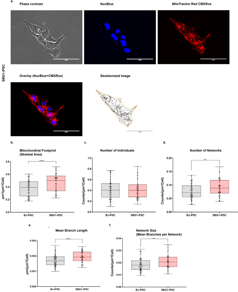
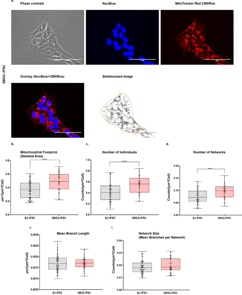
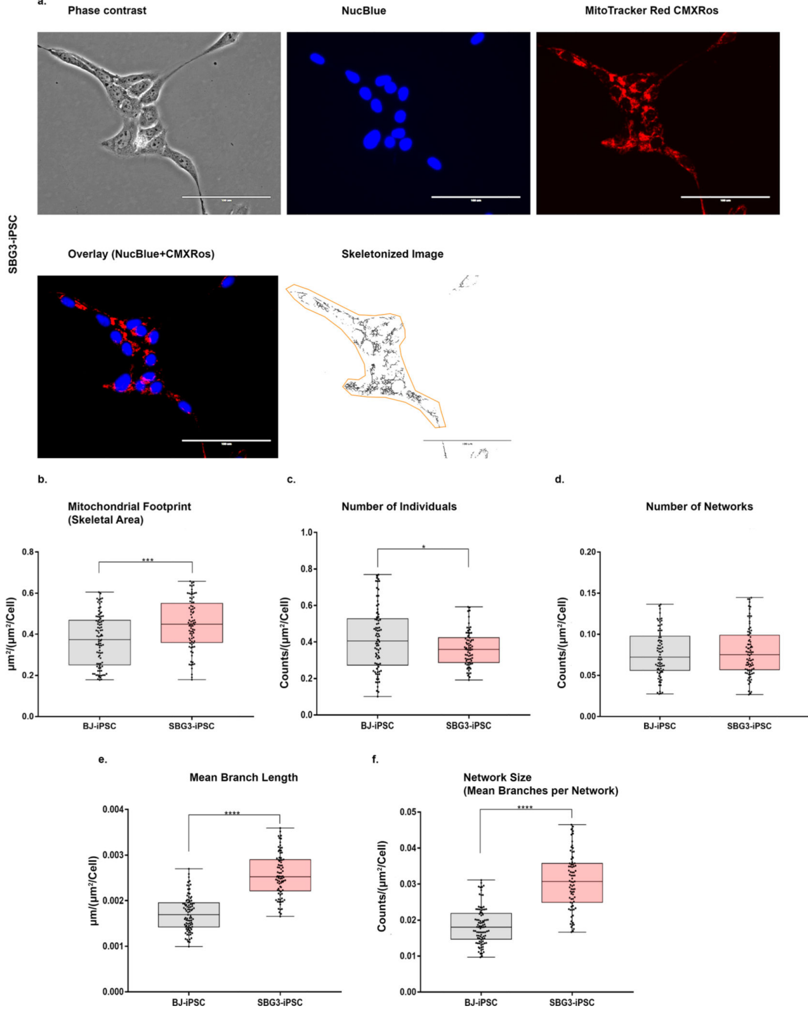
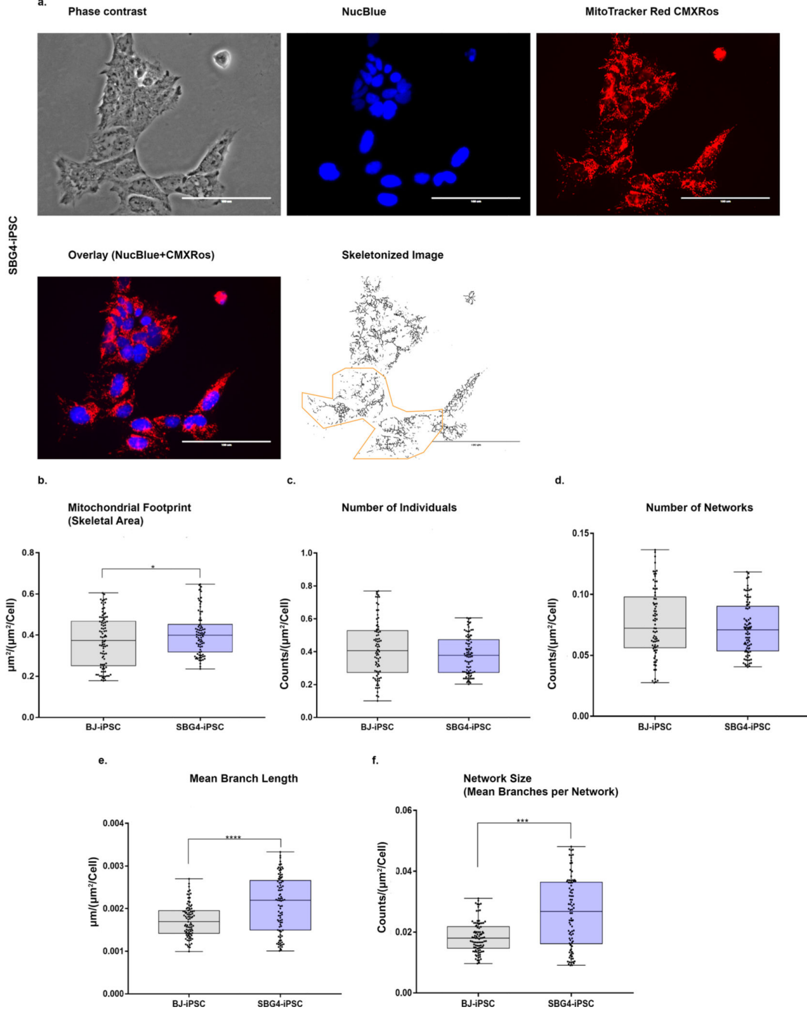
Verification
Claims the paper makes
The claims and statistics of this paper haven’t been checked yet.
Run the check — free, no account needed. It reads the open-access text and checks the sources it cites.
Claims other papers make about this one
Each is checked against this paper’s own text: does it say what the citing paper says it does?
The cited source says what the claim attributes to it.
In the citing paper
“Recent advances in generating induced pluripotent stem cells (iPSCs) from primary cells of affected patients has also led to the creation of model cell lines with specific mtDNA mutations [160,161,162].”
From The Mitochondrial Genome in Aging and Disease and the Future of Mitochondrial Therapeutics (2022) · cites this paper as [161]
Self-check
Percentages, sums and reported differences re-computed from the paper’s own numbers.
The self-check needs the paper’s full text.
Citations
Cites
Mitochondrial dynamics: overview of molecular mechanisms
Tilokani, L., Nagashima, S., Paupe, V., & Prudent, J. (2018) · Essays in Biochemistry · doi:10.1042/ebc20170104 · Cited by 1,397
Research works printed but not linked: 88
- Armstrong, 2010, Human induced pluripotent stem cell lines show stress defense mechanisms and mitochondrial regulation similar to those of human embryonic stem cells, Stem Cells, 28, 661, 10.1002/stem.307 · DOI
- Bakare, 2021, Leigh syndrome: a tale of two genomes, Front. Physiol., 12, 10.3389/fphys.2021.693734 · DOI
- Bakare, 2021, Quantifying mitochondrial dynamics in patient fibroblasts with multiple developmental defects and mitochondrial disorders, Int. J. Mol. Sci., 22, 10.3390/ijms22126263 · DOI
- Baracca, 2007, Biochemical phenotypes associated with the mitochondrial ATP6 gene mutations at nt8993, Biochim. Biophys. Acta, Mol. Cell. Biol. Lipids, 1767, 913, 10.1016/j.bbabio.2007.05.005 · DOI
- Castagna, 2007, Late onset leigh syndrome and ataxia due to a T to C mutation at bp 9,185 of mitochondrial DNA, Am. J. Med. Genet.Part A, 143A, 808, 10.1002/ajmg.a.31637 · DOI
- Chan, 2006, Mitochondria: dynamic organelles in disease, aging, and development, Cell, 125, 1241, 10.1016/j.cell.2006.06.010 · DOI · Paper page
- Chen, 2017, Mitochondrial dynamics in regulating the unique phenotypes of cancer and stem cells, Cell Metab., 26, 39, 10.1016/j.cmet.2017.05.016 · DOI
- Cherry, 2013, Induced pluripotent stem cells with a mitochondrial DNA deletion, Stem cells (Dayton, Ohio), 31, 1287, 10.1002/stem.1354 · DOI
- Choi, 2015, Mitochondrial and metabolic remodeling during reprogramming and differentiation of the reprogrammed cells, Stem Cells Dev., 24, 1366, 10.1089/scd.2014.0561 · DOI
- Cottet-Rousselle, 2011, Cytometric assessment of mitochondria using fluorescent probes, Cytometry A, 79, 405, 10.1002/cyto.a.21061 · DOI
- Dahan, 2019, Metabolism in pluripotency: both driver and passenger?, J. Biol. Chem., 294, 5420, 10.1074/jbc.TM117.000832 · DOI
- D'Aurelio, 2010, Mitochondrial DNA background modifies the bioenergetics of NARP/MILS ATP6 mutant cells, Hum. Mol. Genet., 19, 374, 10.1093/hmg/ddp503 · DOI
- Debray, 2007, Long-term outcome of Leigh syndrome caused by the NARP-T8993C mtDNA mutation, Am. J. Med. Genet. A, 143A, 2046, 10.1002/ajmg.a.31880 · DOI
- Duchen, 2004, Roles of mitochondria in health and disease, Diabetes, 53, S96, 10.2337/diabetes.53.2007.S96 · DOI
- Esteras, 2020, Visualization of mitochondrial membrane potential in mammalian cells, Methods Cell Biol., 155, 221, 10.1016/bs.mcb.2019.10.003 · DOI
- Folmes, 2011, Somatic oxidative bioenergetics transitions into pluripotency-dependent glycolysis to facilitate nuclear reprogramming, Cell Metab., 14, 264, 10.1016/j.cmet.2011.06.011 · DOI
- Folmes, 2013, Disease-causing mitochondrial heteroplasmy segregated within induced pluripotent stem cell clones derived from a patient with MELAS, Stem Cells, 31, 1298, 10.1002/stem.1389 · DOI
- Galera, 2016, Generation of a human iPSC line from a patient with Leigh syndrome, Stem Cell Res., 16, 63, 10.1016/j.scr.2015.12.005 · DOI
- Galera-Monge, 2016, Generation of a human iPSC line from a patient with Leigh syndrome caused by a mutation in the MT-ATP6 gene, Stem Cell Res., 16, 766, 10.1016/j.scr.2016.04.012 · DOI
- Galera-Monge, 2020, Mitochondrial dysfunction and calcium dysregulation in leigh syndrome induced pluripotent stem cell derived neurons, Int. J. Mol. Sci., 21, 10.3390/ijms21093191 · DOI
- Gao, 2017, Abnormalities of mitochondrial dynamics in neurodegenerative diseases, Antioxidants (Basel), 6
- Grace, 2019, mRNA reprogramming of T8993G Leigh's syndrome fibroblast cells to create induced pluripotent stem cell models for mitochondrial disorders, Stem Cells Dev., 28, 846, 10.1089/scd.2019.0045 · DOI
- Harwig, 2018, Methods for imaging mammalian mitochondrial morphology: a prospective on MitoGraph, Anal. Biochem., 552, 81, 10.1016/j.ab.2018.02.022 · DOI
- Hattori, 2016, Metabolic signature of MELAS/leigh overlap syndrome in patient-specific induced pluripotent stem cells model, Osaka City Med. J., 62, 69
- Hoppel, 2009, Dynamic organization of mitochondria in human heart and in myocardial disease, Int. J. Biochem. Cell Biol., 41, 1949, 10.1016/j.biocel.2009.05.004 · DOI
- Iannetti, 2015, Toward high-content screening of mitochondrial morphology and membrane potential in living cells, Int. J. Biochem. Cell Biol., 63, 66, 10.1016/j.biocel.2015.01.020 · DOI
- Inak, 2021, Defective metabolic programming impairs early neuronal morphogenesis in neural cultures and an organoid model of Leigh syndrome, Nat. Commun., 12, 1929, 10.1038/s41467-021-22117-z · DOI
- Iyer, 2009, Stem cell-based models and therapies for neurodegenerative diseases, Crit. Rev. Biomed. Eng., 37, 321, 10.1615/CritRevBiomedEng.v37.i4-5.20 · DOI
- Khacho, 2017, Mitochondrial dysfunction underlies cognitive defects as a result of neural stem cell depletion and impaired neurogenesis, Hum. Mol. Genet., 26, 3327, 10.1093/hmg/ddx217 · DOI
- Kirby, 2003, Low mutant load of mitochondrial DNA G13513A mutation can cause Leigh's disease, Ann. Neurol., 54, 473, 10.1002/ana.10687 · DOI
- Kiryu-Seo, 2016, Mitochondrial fission is an acute and adaptive response in injured motor neurons, Sci. Rep., 6, 28331, 10.1038/srep28331 · DOI
- Koopman, 2005, Mitochondrial network complexity and pathological decrease in complex I activity are tightly correlated in isolated human complex I deficiency, Am. J. Physiol. Cell Physiol., 289, C881, 10.1152/ajpcell.00104.2005 · DOI
- Kori, 2019, Transition from Leigh syndrome to MELAS syndrome in a patient with heteroplasmic MT-ND3 m.10158T>C, Brain Dev., 41, 803, 10.1016/j.braindev.2019.05.006 · DOI
- Lake, 2016, Leigh syndrome: one disorder, more than 75 monogenic causes, Ann. Neurol., 79, 190, 10.1002/ana.24551 · DOI
- Legros, 2002, Mitochondrial fusion in human cells is efficient, requires the inner membrane potential, and is mediated by mitofusins, Mol. Biol. Cell, 13, 4343, 10.1091/mbc.e02-06-0330 · DOI
- Leigh, 1951, Subacute necrotizing encephalomyelopathy in an infant, J. Neurol. Neurosurg. Psychiatry, 14, 216, 10.1136/jnnp.14.3.216 · DOI
- Liu, 2009, Ca2+-dependent regulation of mitochondrial dynamics by the Miro-Milton complex, Int. J. Biochem. Cell Biol., 41, 1972, 10.1016/j.biocel.2009.05.013 · DOI
- Loeffen, 2000, Isolated complex I deficiency in children: clinical, biochemical and genetic aspects, Hum. Mutat., 15, 123, 10.1002/(SICI)1098-1004(200002)15:2<123::AID-HUMU1>3.0.CO;2-P · DOI
- Ma, 2015, Metabolic rescue in pluripotent cells from patients with mtDNA disease, Nature, 524, 234, 10.1038/nature14546 · DOI
- Mah, 2011, Molecular insights into reprogramming-initiation events mediated by the OSKM gene regulatory network, PLoS ONE, 6, 10.1371/journal.pone.0024351 · DOI
- McCarron, 2013, From structure to function: mitochondrial morphology, motion and shaping in vascular smooth muscle, J. Vasc. Res., 50, 357, 10.1159/000353883 · DOI
- McFarland, 2004, De novo mutations in the mitochondrial ND3 gene as a cause of infantile mitochondrial encephalopathy and complex I deficiency, Ann. Neurol., 55, 58, 10.1002/ana.10787 · DOI
- Mitchell, 1966, Chemiosmotic coupling in oxidative and photosynthetic phosphorylation, Biol. Rev. Camb. Philos. Soc., 41, 445, 10.1111/j.1469-185X.1966.tb01501.x · DOI
- Mitra, 2009, A hyperfused mitochondrial state achieved at G1-S regulates cyclin E buildup and entry into S phase, Proc. Natl. Acad. Sci. U.S.A., 106, 11960, 10.1073/pnas.0904875106 · DOI
- Mortiboys, 2008, Mitochondrial function and morphology are impaired in parkin-mutant fibroblasts, Ann. Neurol., 64, 555, 10.1002/ana.21492 · DOI
- Nakada, 2001, Inter-mitochondrial complementation: Mitochondria-specific system preventing mice from expression of disease phenotypes by mutant mtDNA, Nat. Med., 7, 934, 10.1038/90976 · DOI
- Nesbitt, 2012, The clinical spectrum of the m.10191T>C mutation in complex I-deficient Leigh syndrome, Dev. Med. Child Neurol., 54, 500, 10.1111/j.1469-8749.2012.04224.x · DOI
- Ni, 2015, Mitochondrial dynamics and mitochondrial quality control, Redox Biol., 4, 6, 10.1016/j.redox.2014.11.006 · DOI · Paper page
- Ogawa, 2003, Starvation induces the formation of giant mitochondria in gastric parietal cells of guinea pigs, J. Electron. Microsc. (Tokyo), 52, 217, 10.1093/jmicro/52.2.217 · DOI
- Picard, 2013, Mitochondrial morphology transitions and functions: implications for retrograde signaling?, Am. J. Physiol. Regul. Integr. Comp. Physiol., 304, R393, 10.1152/ajpregu.00584.2012 · DOI
- Piekutowska-Abramczuk, 2018, Leigh syndrome in individuals bearing m.9185T>C MTATP6 variant. Is hyperventilation a factor which starts its development?, Metab. Brain Dis., 33, 191, 10.1007/s11011-017-0122-1 · DOI
- Prigione, 2010, The senescence-related mitochondrial/oxidative stress pathway is repressed in human induced pluripotent stem cells, Stem Cells, 28, 721, 10.1002/stem.404 · DOI
- Prigione, 2011, Mitochondrial-associated cell death mechanisms are reset to an embryonic-like state in aged donor-derived iPS cells harboring chromosomal aberrations, PLoS ONE, 6, 10.1371/journal.pone.0027352 · DOI
- S. Rahman, D. Thorburn, Nuclear Gene-Encoded Leigh Syndrome Spectrum Overview, in: M.P. Adam, H.H. Ardinger, R.A. Pagon, S.E. Wallace, L.J.H. Bean, G. Mirzaa, A. Amemiya (Eds.) GeneReviews((R)), Seattle (WA), 1993.
- Rambold, 2011, Tubular network formation protects mitochondria from autophagosomal degradation during nutrient starvation, Proc. Natl. Acad. Sci. U.S.A., 108, 10190, 10.1073/pnas.1107402108 · DOI
- Rasmussen, 2018, A non-apoptotic function of MCL-1 in promoting pluripotency and modulating mitochondrial dynamics in stem cells, Stem Cell Rep., 10, 684, 10.1016/j.stemcr.2018.01.005 · DOI
- Rastogi, 2019, Remodeling of mitochondrial morphology and function: an emerging hallmark of cellular reprogramming, Cell Stress, 3, 181, 10.15698/cst2019.06.189 · DOI
- Rolland, 2013, Impaired complex IV activity in response to loss of LRPPRC function can be compensated by mitochondrial hyperfusion, Proc. Natl. Acad. Sci. U.S.A., 110, E2967, 10.1073/pnas.1303872110 · DOI
- Salisbury-Ruf, 2018, Bid maintains mitochondrial cristae structure and function and protects against cardiac disease in an integrative genomics study, Elife, 7, 10.7554/eLife.40907 · DOI
- Scaduto, 1999, Measurement of mitochondrial membrane potential using fluorescent rhodamine derivatives, Biophys. J., 76, 469, 10.1016/S0006-3495(99)77214-0 · DOI
- Schieke, 2008, Mitochondrial metabolism modulates differentiation and teratoma formation capacity in mouse embryonic stem cells, J. Biol. Chem., 283, 28506, 10.1074/jbc.M802763200 · DOI
- Seo, 2018, Mitochondrial dynamics in stem cells and differentiation, Int. J. Mol. Sci., 19, 10.3390/ijms19123893 · DOI
- Sequiera, 2020, Generation of human induced pluripotent stem cell (hiPSC) line UOMi001-A from a patient with Leigh-like syndrome harbouring compound heterozygous variants in ECHS1 gene, Stem Cell Res., 48
- Sequiera, 2020, Induced pluripotent stem cell line UOMi002-A from a patient with Leigh syndrome with compound heterozygous mutations in the NDUFV1 gene, Stem Cell Res., 48
- Son, 2013, Unveiling the critical role of REX1 in the regulation of human stem cell pluripotency, Stem Cells, 31, 2374, 10.1002/stem.1509 · DOI
- Son, 2015, Mitofusins deficiency elicits mitochondrial metabolic reprogramming to pluripotency, Cell Death Differ., 22, 1957, 10.1038/cdd.2015.43 · DOI
- Son, 2021, Self-replicative mRNA-mediated generation of induced pluripotent stem cell line from a 1-year-old Leigh syndrome patient with mitochondrial DNA cytochrome b mutation, Stem Cell Res., 54, 10.1016/j.scr.2021.102392 · DOI
- Sprenger, 2019, The good and the bad of mitochondrial breakups, Trends Cell Biol., 29, 888, 10.1016/j.tcb.2019.08.003 · DOI
- Suhr, 2010, Mitochondrial rejuvenation after induced pluripotency, PLoS ONE, 5, 10.1371/journal.pone.0014095 · DOI
- Taylor, 2002, Leigh disease associated with a novel mitochondrial DNA ND5 mutation, Eur. J. Hum. Genet., 10, 141, 10.1038/sj.ejhg.5200773 · DOI
- Tokuyama, 2020, Mitochondrial dynamics regulation in skin fibroblasts from mitochondrial disease patients, Biomolecules, 10, 10.3390/biom10030450 · DOI
- Tsogtbaatar, 2020, Energy metabolism regulates stem cell pluripotency, Front. Cell Dev. Biol., 8, 87, 10.3389/fcell.2020.00087 · DOI
- Uziel, 1997, Mitochondrial disease associated with the T8993G mutation of the mitochondrial ATPase 6 gene: a clinical, biochemical, and molecular study in six families, J. Neurol. Neurosurg. Psychiatry, 63, 16, 10.1136/jnnp.63.1.16 · DOI
- Valente, 2017, A simple ImageJ macro tool for analyzing mitochondrial network morphology in mammalian cell culture, Acta Histochem., 119, 315, 10.1016/j.acthis.2017.03.001 · DOI
- Varum, 2011, Energy metabolism in human pluripotent stem cells and their differentiated counterparts, PLoS ONE, 6, 10.1371/journal.pone.0020914 · DOI · Paper page
- Vodopivec, 2016, Mitochondrial encephalopathy and optic neuropathy due to m.10158 MT-ND3 complex i mutation presenting in an adult patient: case report and review of the literature, Neurologist, 21, 61, 10.1097/NRL.0000000000000084 · DOI
- Wai, 2016, Mitochondrial dynamics and metabolic regulation, Trends Endocrinol. Metab., 27, 105, 10.1016/j.tem.2015.12.001 · DOI
- Wallace, 1999, Mitochondrial diseases in man and mouse, Science, 283, 1482, 10.1126/science.283.5407.1482 · DOI · Paper page
- Wang, 2017, Fatty acid synthesis is critical for stem cell pluripotency via promoting mitochondrial fission, EMBO J., 36, 1330, 10.15252/embj.201695417 · DOI
- Willems, 2015, Redox homeostasis and mitochondrial dynamics, Cell Metab., 22, 207, 10.1016/j.cmet.2015.06.006 · DOI
- Yoneda, 1994, Complementation of mutant and wild-type human mitochondrial DNAs coexisting since the mutation event and lack of complementation of DNAs introduced separately into a cell within distinct organelles, Mol. Cell. Biol., 14, 2699
- Youle, 2012, Mitochondrial fission, fusion, and stress, Science, 337, 1062, 10.1126/science.1219855 · DOI · Paper page
- Zemirli, 2018, Mitochondrial Dynamics in Basal and Stressful Conditions, Int. J. Mol. Sci., 19, 10.3390/ijms19020564 · DOI
- Zhadanov, 2007, Fatal manifestation of a de novo ND5 mutation: insights into the pathogenetic mechanisms of mtDNA ND5 gene defects, Mitochondrion, 7, 260, 10.1016/j.mito.2007.01.003 · DOI
- Zhang, 2011, UCP2 regulates energy metabolism and differentiation potential of human pluripotent stem cells, EMBO J., 30, 4860, 10.1038/emboj.2011.401 · DOI
- Zhang, 2018, Metabolism in pluripotent stem cells and early mammalian development, Cell Metab., 27, 332, 10.1016/j.cmet.2018.01.008 · DOI
- Zinovkin, 2016, Mitochondrial genome and longevity, Biochemistry (Mosc), 81, 1401, 10.1134/S0006297916120014 · DOI
- Zorova, 2018, Mitochondrial membrane potential, Anal. Biochem., 552, 50, 10.1016/j.ab.2017.07.009 · DOI
Cited by
Sorted by citations
The Mitochondrial Genome in Aging and Disease and the Future of Mitochondrial Therapeutics
Saravanan, S., Lewis, C. J., Dixit, B., O’Connor, M. S., Stolzing, A., & Boominathan, A. (2022) · Biomedicines · doi:10.3390/biomedicines10020490 · Cited by 13
Metadata
- Authors
- Meshrkey, F., Cabrera Ayuso, A., Rao, R. R., & Iyer, S.
- Venue
- Stem Cell Research
- Publisher
- Elsevier BV
- Published
- 2021-12
- Volume
- 57
- Pages
- 102572
- ISSN
- 1873-5061
- Cited by
- 24
- References
- 1
- Language
- en
- Licence
- Funders
- Arkansas Biosciences Institute
- Medizinische Universität Innsbruck
- U.S. Department of Defense (W81XWH-16-1-0181)
About these checks
Registry, retraction, Cites and statistics results are computed, not judged, with one exception: a printed reference that no index lists is read once by a model before it counts as a citation error. Claims are extracted by a model and kept only when their quote appears verbatim in the text; each cited source is then checked, with a model’s help, for whether it says what the claim relies on. Those verdicts can be wrong — read the claim’s quote and the cited source before relying on one.
Statistics and arithmetic are recomputed, not judged.
Confidence bands are shown only for fields calibrated on our annotated test set. None is calibrated yet, so no band is shown.
What counts as a claim ›How we measure confidence ›How we check this ›
Are you an author, or do you see a problem with this page? Request a review
