

The DOI matches its Crossref record; 47 of the 49 references it cites were found in a registry or an index.
Patient-Specific iPSC-Derived Astrocytes Contribute to Non-Cell-Autonomous Neurodegeneration in Parkinson's Disease
Open accessAbstract
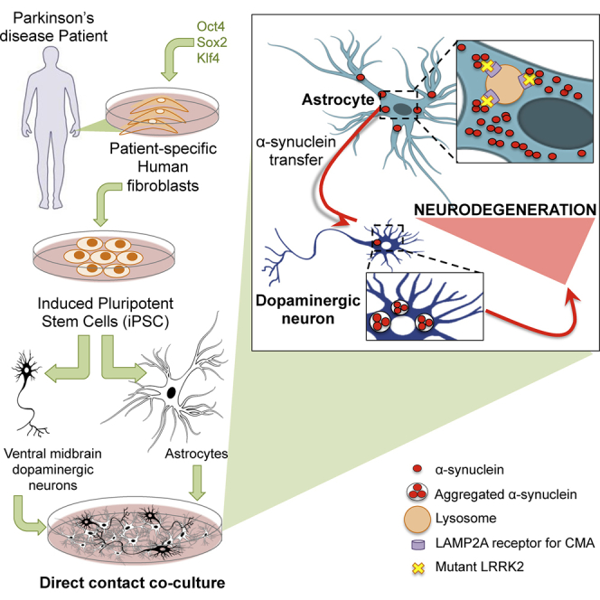
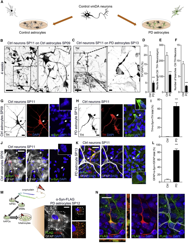
Parkinson's disease (PD) is associated with the degeneration of ventral midbrain dopaminergic neurons (vmDAns) and the accumulation of toxic α-synuclein. A non-cell-autonomous contribution, in particular of astrocytes, during PD pathogenesis has been suggested by observational studies, but remains to be experimentally tested. Here, we generated induced pluripotent stem cell-derived astrocytes and neurons from familial mutant LRRK2 G2019S PD patients and healthy individuals. Upon co-culture on top of PD astrocytes, control vmDAns displayed morphological signs of neurodegeneration and abnormal, astrocyte-derived α-synuclein accumulation. Conversely, control astrocytes partially prevented the appearance of disease-related phenotypes in PD vmDAns. We additionally identified dysfunctional chaperone-mediated autophagy (CMA), impaired macroautophagy, and progressive α-synuclein accumulation in PD astrocytes. Finally, chemical enhancement of CMA protected PD astrocytes and vmDAns via the clearance of α-synuclein accumulation. Our findings unveil a crucial non-cell-autonomous contribution of astrocytes during PD pathogenesis, and open the path to exploring novel therapeutic strategies aimed at blocking the pathogenic cross talk between neurons and glial cells.
Overview
The claims and statistics of this paper haven’t been checked yet.
Run the check — free, no account needed. It reads the open-access text and checks the sources it cites.
Integrity signals
Registry
Cites
Statistics
Figures
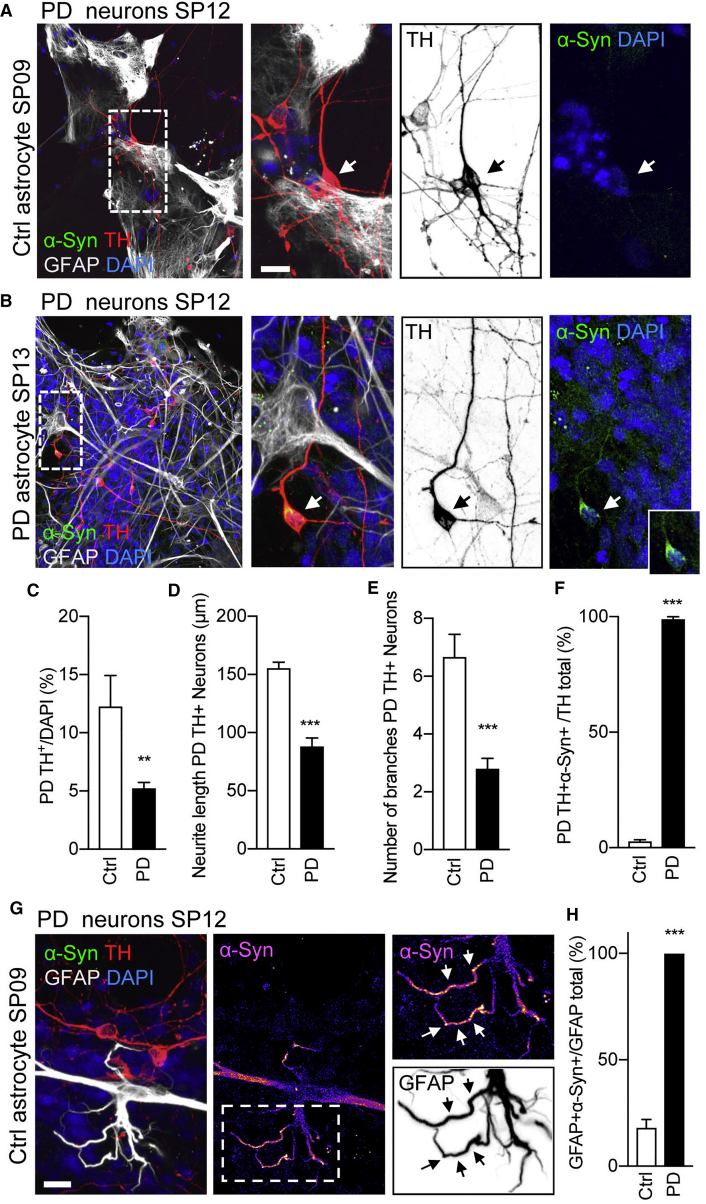
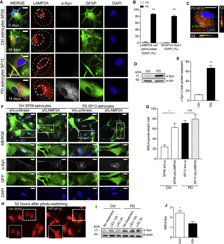
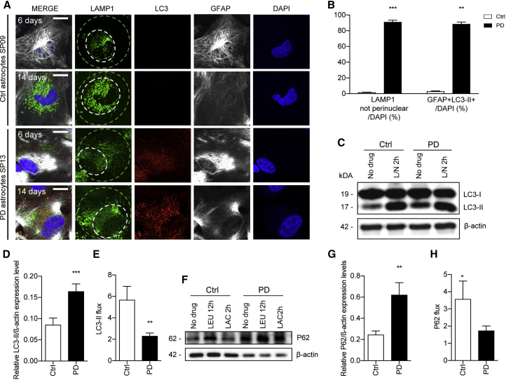
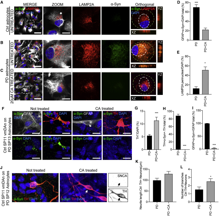
Verification
Claims the paper makes
The claims and statistics of this paper haven’t been checked yet.
Run the check — free, no account needed. It reads the open-access text and checks the sources it cites.
Claims other papers make about this one
Each is checked against this paper’s own text: does it say what the citing paper says it does?
The source backs part of the claim, not all of it — or backs it for a different population, condition or comparison.
In the citing paper
“in contrast to neuron-specific cell autonomous mechanisms that affect individual DA neurons within the SNc, non-cell autonomous mechanisms can cause dysfunction across the entire SNc, thereby leading to rapid DA neurodegeneration”
From Mitochondrial DNA Damage in Substantia Nigra Pars Compacta Astrocytes Exacerbates Dopaminergic Neuron Loss in a 6-Hydroxydopamine Mouse Model of Parkinsonism (2026) · cites this paper as di Domenico et al. 2019
Experimental — accuracy not yet measuredThe cited source says what the claim attributes to it.
In the citing paper
“Interestingly, astrocytes generated from hiPSC derived from fibroblasts obtained from patients with CNS diseases (usually with a genetic mutation causative of disease or a risk polymorphism) show pathological phenotypes, including dysregulation of lipid metabolism11, alteration in the contents of the extracellular vesicles released by astrocytes80, reduced autophagy81, or altered STAT3 signaling82”
From Reactive astrocyte nomenclature, definitions, and future directions (2021) · cites this paper as [81]
Self-check
Percentages, sums and reported differences re-computed from the paper’s own numbers.
The self-check needs the paper’s full text.
Citations
Cites
Vos, T., Barber, R. M., Bell, B., Bertozzi-Villa, A., Biryukov, S., Bolliger, I., Charlson, F., Davis, A., Degenhardt, L., Dicker, D., et al. (2015) · The Lancet · doi:10.1016/s0140-6736(15)60692-4 · Cited by 6,414
Highly efficient neural conversion of human ES and iPS cells by dual inhibition of SMAD signaling
Chambers, S. M., Fasano, C. A., Papapetrou, E. P., Tomishima, M., Sadelain, M., & Studer, L. (2009) · Nature Biotechnology · doi:10.1038/nbt.1529 · Cited by 3,620
Impaired Degradation of Mutant α-Synuclein by Chaperone-Mediated Autophagy
Cuervo, A. M., Stefanis, L., Fredenburg, R., Lansbury, P. T., & Sulzer, D. (2004) · Science · doi:10.1126/science.1101738 · Cited by 2,024 · cited 2×
“…in a-syn turnover in these cells. a-syn has previously been shown to undergo degradation both by the ubiquitin/proteasome system and by autophagy (Cuervo et al., 2004; Webb et al., 2003); therefore a-syn flux in the presence of lysosomal and proteasome inhibitors (leupeptin and lactacystin,…”
BackgroundSelf-citationKriks, S., Shim, J., Piao, J., Ganat, Y. M., Wakeman, D. R., Xie, Z., Carrillo‐Reid, L., Auyeung, G., Antonacci, C. L., Buch, A., et al. (2011) · Nature · doi:10.1038/nature10648 · Cited by 1,951
α-Synuclein Is Degraded by Both Autophagy and the Proteasome
Webb, J. L., Ravikumar, B., Atkins, J., Skepper, J. N., & Rubinsztein, D. C. (2003) · Journal of Biological Chemistry · doi:10.1074/jbc.m300227200 · Cited by 1,429
Lee, H.-J., Suk, J.-E., Patrick, C., Bae, E.-J., Cho, J.-H., Rho, S., Hwang, D., Masliah, E., & Lee, S.-J. (2010) · Journal of Biological Chemistry · doi:10.1074/jbc.m109.081125 · Cited by 877
α-Synuclein impairs macroautophagy: implications for Parkinson’s disease
Winslow, A. R., Chen, C.-W., Corrochano, S., Acevedo-Arozena, A., Gordon, D. E., Peden, A. A., Lichtenberg, M., Menzies, F. M., Ravikumar, B., Imarisio, S., et al. (2010) · Journal of Cell Biology · doi:10.1083/jcb.201003122 · Cited by 825 · cited 2×
“Degradation of a-syn has been shown to occur by both proteasome and autophagic pathways, and conversely, high levels of a-syn have been demonstrated to be toxic for both systems (Tanaka et al., 2001; Webb et al., 2003; Winslow et al., 2010).”
BackgroundA common LRRK2 mutation in idiopathic Parkinson's disease
Gilks, W. P., Abou-Sleiman, P. M., Gandhi, S., Jain, S., Singleton, A., Lees, A. J., Shaw, K., Bhatia, K. P., Bonifati, V., Quinn, N. P., et al. (2005) · The Lancet · doi:10.1016/s0140-6736(05)17830-1 · Cited by 774
LRRK2 Mutant iPSC-Derived DA Neurons Demonstrate Increased Susceptibility to Oxidative Stress
Nguyen, H. N., Byers, B., Cord, B. J., Shcheglovitov, A., Byrne, J., Gujar, P., Kee, K., Schüle, B., Dolmetsch, R. E., Langston, J. W., et al. (2011) · Cell stem cell · doi:10.1016/j.stem.2011.01.013 · Cited by 768
A novel TALE nuclease scaffold enables high genome editing activity in combination with low toxicity
Mussolino, C., Morbitzer, R., Lütge, F., Dannemann, N., Lahaye, T., & Cathomen, T. (2011) · Nucleic Acids Research · doi:10.1093/nar/gkr597 · Cited by 714
Cited by
Sorted by citations
Guidelines for the use and interpretation of assays for monitoring autophagy (4th edition) 1
Klionsky, D. J., Abdel-Aziz, A. K., Abdelfatah, S., Abdellatif, M., Abdoli, A., Abel, S., Abeliovich, H., Abildgaard, M. H., Abudu, Y. P., Acevedo-Arozena, A., et al. (2021) · Autophagy · doi:10.1080/15548627.2020.1797280 · Cited by 2,897
Self-citationReactive astrocyte nomenclature, definitions, and future directions
Escartin, C., Galea, E., Lakatos, A., O’Callaghan, J. P., Petzold, G. C., Serrano-Pozo, A., Steinhäuser, C., Volterra, A., Carmignoto, G., Agarwal, A., et al. (2021) · Nature Neuroscience · doi:10.1038/s41593-020-00783-4 · Cited by 2,513
Functional roles of reactive astrocytes in neuroinflammation and neurodegeneration
Patani, R., Hardingham, G. E., & Liddelow, S. A. (2023) · Nature Reviews Neurology · doi:10.1038/s41582-023-00822-1 · Cited by 531
The different autophagy degradation pathways and neurodegeneration
Fleming, A., Bourdenx, M., Fujimaki, M., Karabiyik, C., Krause, G. J., Lopez, A., Martín‐Segura, A., Puri, C., Scrivo, A., Skidmore, J., et al. (2022) · Neuron · doi:10.1016/j.neuron.2022.01.017 · Cited by 500
Self-citationCai, Y., Song, W., Li, J., Jing, Y., Liang, C., Zhang, L., Zhang, X., Zhang, W., Liu, B., An, Y., et al. (2022) · Science China Life Sciences · doi:10.1007/s11427-022-2161-3 · Cited by 450
Metadata
- Authors
- di Domenico, A., Carola, G., Calatayud, C., Pons-Espinal, M., Muñoz, J. P., Richaud-Patin, Y., Fernandez-Carasa, I., Gut, M., Faella, A., Parameswaran, J., Soriano, J., Ferrer, I., Tolosa, E., Zorzano, A., Cuervo, A. M., Raya, A., & Consiglio, A.
- Affiliations
- Bellvitge University Hospital (ES)
- Institute for Research in Biomedicine (ES)
- Centro Nacional de Análisis Genómico (ES)
- Universitat de Barcelona (ES)
- Biomedical Research Networking Center on Neurodegenerative Diseases (ES)
- Corresponding author
- Consiglio, Antonella
- ORCID
- Venue
- Stem Cell Reports
- Publisher
- Elsevier BV
- Published
- 2019-02
- Volume
- 12
- Issue
- 2
- Pages
- 213-229
- ISSN
- 2213-6711 (ISSN-L)
- PMC
- PMC6372974
- OpenAlex
- W2911036111
- Cited by
- 372
- References
- 49
- Citations by year
- 2026: 32 · 2025: 38 · 2024: 44 · 2023: 45 · 2022: 65 · 2021: 69 · 2020: 53 · 2019: 26
- FWCI
- 36.04
- Type
- journal article
- Language
- en
- Open access
- gold
- doi.org
- Licence
- Locations
- Stem Cell Reports (publishedVersion, cc-by)
- Europe PMC (PubMed Central) (publishedVersion, cc-by)
- PubMed Central (publishedVersion, cc-by)
- PubMed (publishedVersion)
- Dipòsit Digital de la Universitat de Barcelona (Universitat de Barcelona) (publishedVersion, cc-by)
- DOAJ (DOAJ: Directory of Open Access Journals) (submittedVersion, cc-by-sa)
- Institutional Research Information System (Università degli Studi di Brescia) (submittedVersion)
- Repositori digital de la UPF (Universitat Pompeu Fabra) (submittedVersion, cc-by)
- Funders
- Agència de Gestió d'Ajuts Universitaris i de Recerca (2014-SGR-878, 2017-SGR-899)
- Ministerio de Educación, Cultura y Deporte (BES-2014-069603, FPU12/03332)
- Ministerio de Economía y Competitividad (BFU2016-80870-P, FIS2016-78507-C2-2-P, SAF2015-69706-R)
- Instituto de Salud Carlos III
- European Regional Development Fund (RD16/0011/0024)
- Generalitat de Catalunya
- European Research Council (2012-StG-311736-PD-HUMMODEL)
- Topics
- Health Sciences › Medicine › Clinical Neurology › Parkinson's Disease Mechanisms and Treatments
- Life Sciences › Biochemistry, Genetics and Molecular Biology › Molecular Biology › Pluripotent Stem Cells Research
- Life Sciences › Neuroscience › Neurology › Neuroinflammation and Neurodegeneration Mechanisms
- Keywords
- Parkinson's disease, astrocytes, non-cell autonomous neurodegeneration, induced pluripotent stem cell-derived astrocytes, dopaminergic neurons, ventral midbrain, LRRK2 G2019S, familial Parkinson's disease, alpha-synuclein accumulation, chaperone-mediated autophagy, macroautophagy, neuron-glia interactions
- MeSH terms
- Leucine-Rich Repeat Serine-Threonine Protein Kinase-2, Astrocytes, Autophagy, Cell Differentiation, Cells, Cultured, Humans, Mesencephalon, Neuroglia, Parkinson Disease, Phenotype, Coculture Techniques, alpha-Synuclein, Induced Pluripotent Stem Cells, Dopaminergic Neurons
About these checks
Registry, retraction, Cites and statistics results are computed, not judged, with one exception: a printed reference that no index lists is read once by a model before it counts as a citation error. Claims are extracted by a model and kept only when their quote appears verbatim in the text; each cited source is then checked, with a model’s help, for whether it says what the claim relies on. Those verdicts can be wrong — read the claim’s quote and the cited source before relying on one.
Statistics and arithmetic are recomputed, not judged.
Confidence bands are shown only for fields calibrated on our annotated test set. None is calibrated yet, so no band is shown.
What counts as a claim ›How we measure confidence ›How we check this ›
Are you an author, or do you see a problem with this page? Request a review
