

The DOI matches its Crossref record; 117 of the 118 references it cites were found in a registry or an index.
Eye evolution and its functional basis
Open accessAbstract
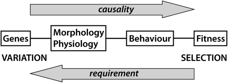
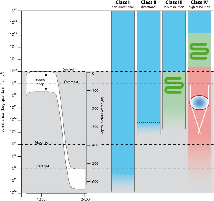
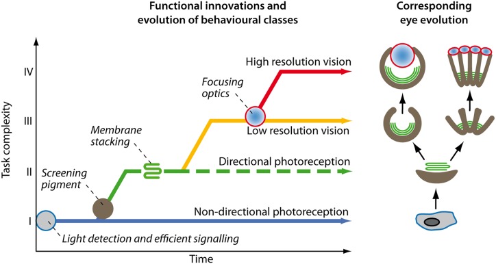
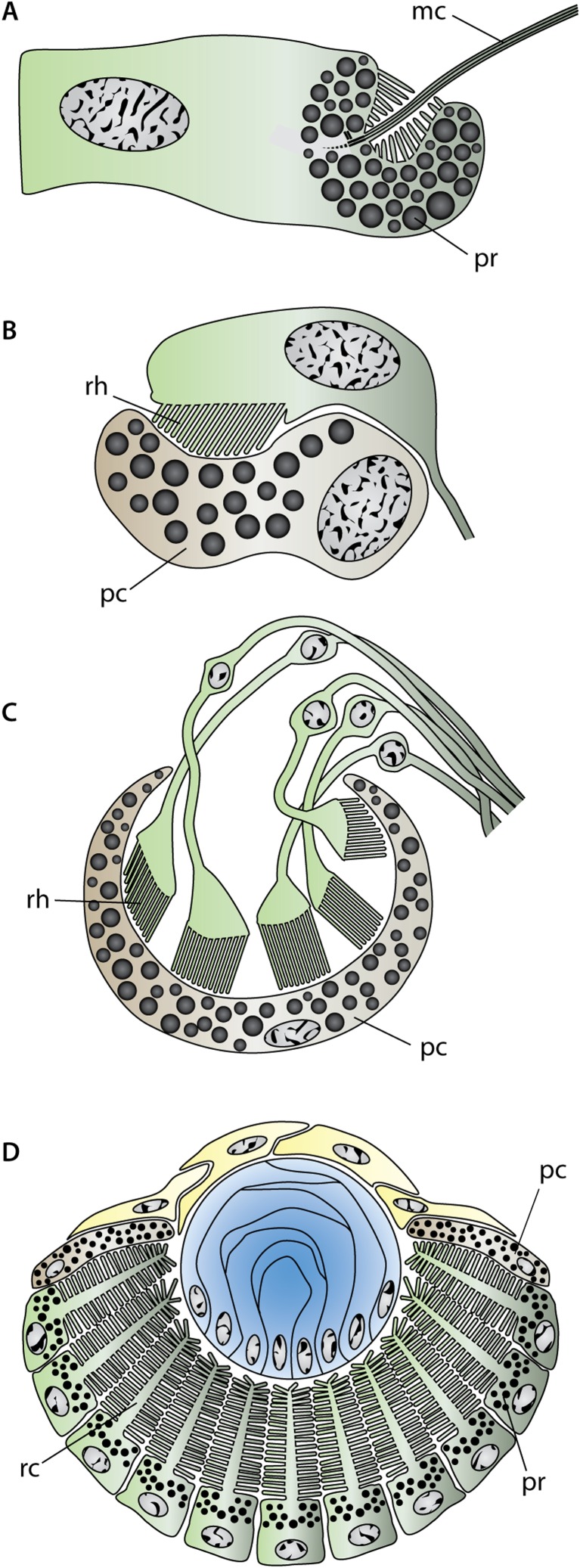
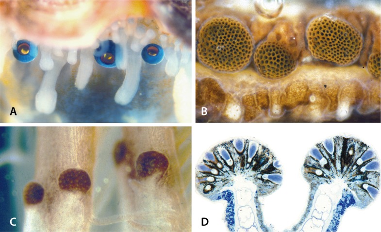
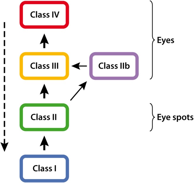
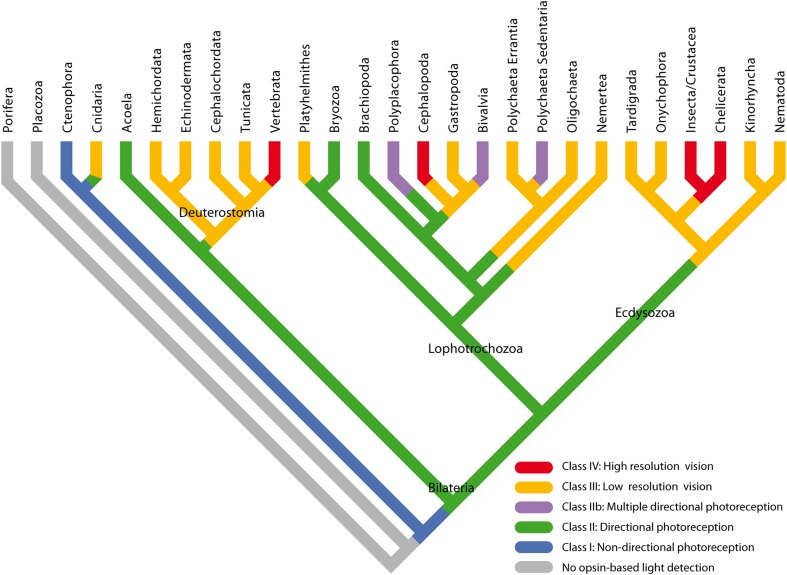
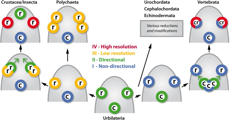
Eye evolution is driven by the evolution of visually guided behavior. Accumulation of gradually more demanding behaviors have continuously increased the performance requirements on the photoreceptor organs. Starting with nondirectional photoreception, I argue for an evolutionary sequence continuing with directional photoreception, low-resolution vision, and finally, high-resolution vision. Calculations of the physical requirements for these four sensory tasks show that they correlate with major innovations in eye evolution and thus work as a relevant classification for a functional analysis of eye evolution. Together with existing molecular and morphological data, the functional analysis suggests that urbilateria had a simple set of rhabdomeric and ciliary receptors used for directional photoreception, and that organ duplications, positional shifts and functional shifts account for the diverse patterns of eyes and photoreceptors seen in extant animals. The analysis also suggests that directional photoreception evolved independently at least twice before the last common ancestor of bilateria and proceeded several times independently to true vision in different bilaterian and cnidarian groups. This scenario is compatible with Pax -gene expression in eye development in the different animal groups. The whole process from the first opsin to high-resolution vision took about 170 million years and was largely completed by the onset of the Cambrian, about 530 million years ago. Evolution from shadow detectors to multiple directional photoreceptors has further led to secondary cases of eye evolution in bivalves, fan worms, and chitons.
Overview
The claims and statistics of this paper haven’t been checked yet.
Free: it reads the open-access text and checks the first 5 claims that cite a source we can check. Create a free account to check every claim.
Integrity signals
Registry
Cites
Statistics
Figures
Verification
Claims the paper makes
The claims and statistics of this paper haven’t been checked yet.
Free: it reads the open-access text and checks the first 5 claims that cite a source we can check. Create a free account to check every claim.
Claims other papers make about this one
Each is checked against this paper’s own text: does it say what the citing paper says it does?
The cited source doesn’t support the claim as written.
In the citing paper
“Take convergent evolution: the photoreceptor system has evolved several times independently [59]”
From The Umwelt Representation Hypothesis: rethinking Universality (2026) · cites this paper as [59]
Checked by a reasoning model — may be wrong.
Experimental — accuracy not yet measuredThe source backs part of the claim, not all of it — or backs it for a different population, condition or comparison.
In the citing paper
“This also aligns with proposed explanations that suggest that lens development coincided with an increase in eye size (59, 60).”
From What if eye...? Computationally recreating vision evolution (2025) · cites this paper as [60]
Experimental — accuracy not yet measuredThe cited source says what the claim attributes to it.
In the citing paper
“Early visual systems faced a fundamental trade-off between light collection and acuity, progressing from simple light-sensitive patches to cup-shaped eyes with smaller apertures (59, 60).”
From What if eye...? Computationally recreating vision evolution (2025) · cites this paper as [60]
The cited source says what the claim attributes to it.
In the citing paper
“The structure and function of such visual systems in non-bilaterians provides further support to the view [72] that camera-type eyes evolved several times.”
From Back to the Basics: Cnidarians Start to Fire (2017) · cites this paper as [72]
Self-check
Percentages, sums and reported differences re-computed from the paper’s own numbers.
The self-check needs the paper’s full text.
Citations
Cites
Handbook of Sensory Physiology
Sanders, M. D. (1975) · British Journal of Ophthalmology · doi:10.1136/bjo.59.2.111-c · Cited by 7,166
Fredriksson, R., Lagerström, M. C., Lundin, L.-G., & Schiöth, H. B. (2003) · Molecular Pharmacology · doi:10.1124/mol.63.6.1256 · Cited by 2,941
Signals, Signal Conditions, and the Direction of Evolution
Endler, J. A. (1992) · The American Naturalist · doi:10.1086/285308 · Cited by 1,960
(1945) · Annals of the Entomological Society of America · doi:10.1093/aesa/38.3.396 · Cited by 1,852
(2005) · Applying Evolutionary Archaeology · doi:10.1007/0-306-47468-9_7 · Cited by 1,644
Erwin, D. H., Laflamme, M., Tweedt, S. M., Sperling, E. A., Pisani, D., & Peterson, K. J. (2011) · Science · doi:10.1126/science.1206375 · Cited by 1,336 · cited 1×
“7 is compared with timing data for the branching points (Peterson et al., 2008 ; Erwin et al., 2011 ), the baffl ing conclusion is that nearly all fall in a period from 700 to 530 million years ago.”
LYTHGOE, J. N. (1979) · Medical Entomology and Zoology · Cited by 1,161
Predictive coding: a fresh view of inhibition in the retina
Srinivasan, M. V., Laughlin, S. B., & Dubs, A. (1982) · Proceedings of the Royal Society of London. Series B. Biological Sciences · doi:10.1098/rspb.1982.0085 · Cited by 1,042
Handbook of Sensory Physiology
Newell, F. W. (1973) · American Journal of Ophthalmology · doi:10.1016/0002-9394(73)90115-3 · Cited by 986
Melanopsin: An opsin in melanophores, brain, and eye
Provencio, I., Jiang, G., De Grip, W. J., Hayes, W. P., & Rollag, M. D. (1998) · Proceedings of the National Academy of Sciences · doi:10.1073/pnas.95.1.340 · Cited by 975
Research works printed but not linked: 3
- 10.1562/0031-8655(2004)079 · doi:10.1562/0031-8655(2004)079
- Bennett, 1979, Handbook of Sensory Phyiology, VII/6A, 641
- Land, 2006, Invertebrate Vision, 167
Cited by
Evolution of phototransduction, vertebrate photoreceptors and retina
Lamb, T. D. (2013) · Progress in Retinal and Eye Research · doi:10.1016/j.preteyeres.2013.06.001 · Cited by 397
Williams, S. T. (2017) · Biological Reviews · doi:10.1111/brv.12268 · Cited by 256
Bioinspired Artificial Eyes: Optic Components, Digital Cameras, and Visual Prostheses
Lee, G. J., Choi, C., Kim, D., & Song, Y. M. (2018) · Advanced Functional Materials · doi:10.1002/adfm.201705202 · Cited by 252
The foundations of plant intelligence
TREWAVAS, A. J. (2017) · Interface Focus · doi:10.1098/rsfs.2016.0098 · Cited by 190
The last common ancestor of most bilaterian animals possessed at least 9 opsins
Ramirez, M. D., Pairett, A., Pankey, M. S., Serb, J., Speiser, D. I., Swafford, A., & Oakley, T. (2016) · Genome Biology and Evolution · doi:10.1093/gbe/evw248 · Cited by 181
Metadata
- Authors
- NILSSON, D.-E.
- Affiliations
- Lund University (SE)
- Corresponding author
- NILSSON, DAN-E.
- Venue
- Visual Neuroscience
- Publisher
- Maximum Academic Press
- Published
- 2013-03
- Volume
- 30
- Issue
- 1-2
- Pages
- 5-20
- ISSN
- 0952-5238 (ISSN-L)
- 1469-8714
- PMC
- PMC3632888
- OpenAlex
- W2151644530
- Cited by
- 267
- References
- 118
- Citations by year
- 2026: 15 · 2025: 24 · 2024: 33 · 2023: 23 · 2022: 27 · 2021: 29 · 2020: 21 · 2019: 13 · 2018: 14 · 2017: 18 · 2016: 23 · 2015: 12 · 2014: 13 · 2013: 2
- FWCI
- 11.29
- Type
- journal article
- Language
- en
- Open access
- hybrid
- doi.org
- Licence
- Locations
- Visual Neuroscience (publishedVersion, cc-by-nc-sa)
- Europe PMC (PubMed Central) (publishedVersion, cc-by-nc-sa)
- PubMed Central (publishedVersion, cc-by-nc-sa)
- PubMed (publishedVersion)
- Lund University Publications (Lund University) (submittedVersion)
- Funders
- Knut och Alice Wallenbergs Stiftelse
- Vetenskapsrådet
- Topics
- Life Sciences › Agricultural and Biological Sciences › Animal Science and Zoology › Marine Invertebrate Physiology and Ecology
- Life Sciences › Biochemistry, Genetics and Molecular Biology › Molecular Biology › Developmental Biology and Gene Regulation
- Life Sciences › Neuroscience › Cognitive Neuroscience › Visual perception and processing mechanisms
- Keywords
- eye evolution, visual perception, rhabdomeric photoreceptors, ciliary photoreceptors, opsin, low-resolution vision, Urbilateria, Pax genes, eye development, Bivalvia, Sabellidae, chitons, Cambrian, bilaterians, Cnidaria
- MeSH terms
- Animals, Biological Evolution, Eye, Humans, Photoreceptor Cells, Vision, Ocular, Visual Pathways, Signal Detection, Psychological
About these checks
Registry, retraction, Cites and statistics results are computed, not judged, with one exception: a printed reference that no index lists is read once by a model before it counts as a citation error. Claims are extracted by a model and kept only when their quote appears verbatim in the text; each cited source is then checked, with a model’s help, for whether it says what the claim relies on. Those verdicts can be wrong — read the claim’s quote and the cited source before relying on one.
Statistics and arithmetic are recomputed, not judged.
Confidence bands are shown only for fields calibrated on our annotated test set. None is calibrated yet, so no band is shown.
What counts as a claim ›How we measure confidence ›How we check this ›
Are you an author, or do you see a problem with this page? Request a review
How to cite
Cite CoChat for the checks on this page, and the paper for its own findings.
- This page
- CoChat. (2026). Citation check: Eye evolution and its functional basis. https://app.cochat.ai/papers/10.1017/s0952523813000035
- The paper
- NILSSON, D.-E. (2013). Eye evolution and its functional basis. Visual Neuroscience. https://doi.org/10.1017/s0952523813000035
Data sources
Bibliographic data: Semantic Scholar, OpenAlex, Crossref.
Contains information from the Semantic Scholar Academic Graph Datasets, made available under the ODC Attribution License (ODC-BY 1.0).
Semantic Scholar’s data is described in: Kinney, R., et al. (2023). The Semantic Scholar Open Data Platform. arXiv:2301.10140.
