

The DOI matches its Crossref record; 10 of the 10 references it cites were found in a registry or an index.
Novel PET Imaging Probe for Quantitative Detection of Senescence In Vivo
Overview
The claims and statistics of this paper haven’t been checked yet.
Run the check — free, no account needed. It reads the open-access text and checks the sources it cites.
Integrity signals
Registry
Cites
Statistics
Figures
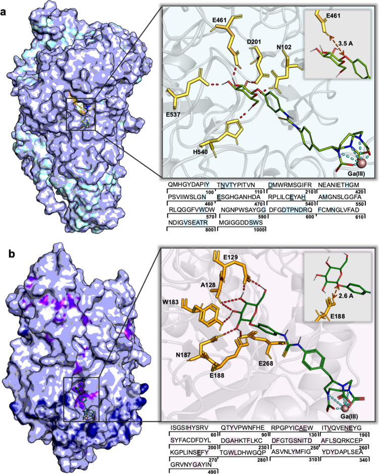
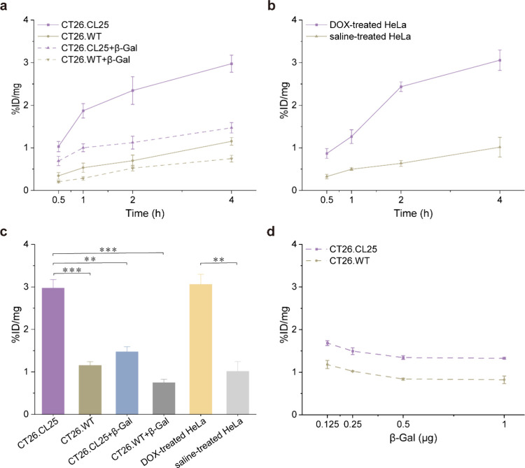
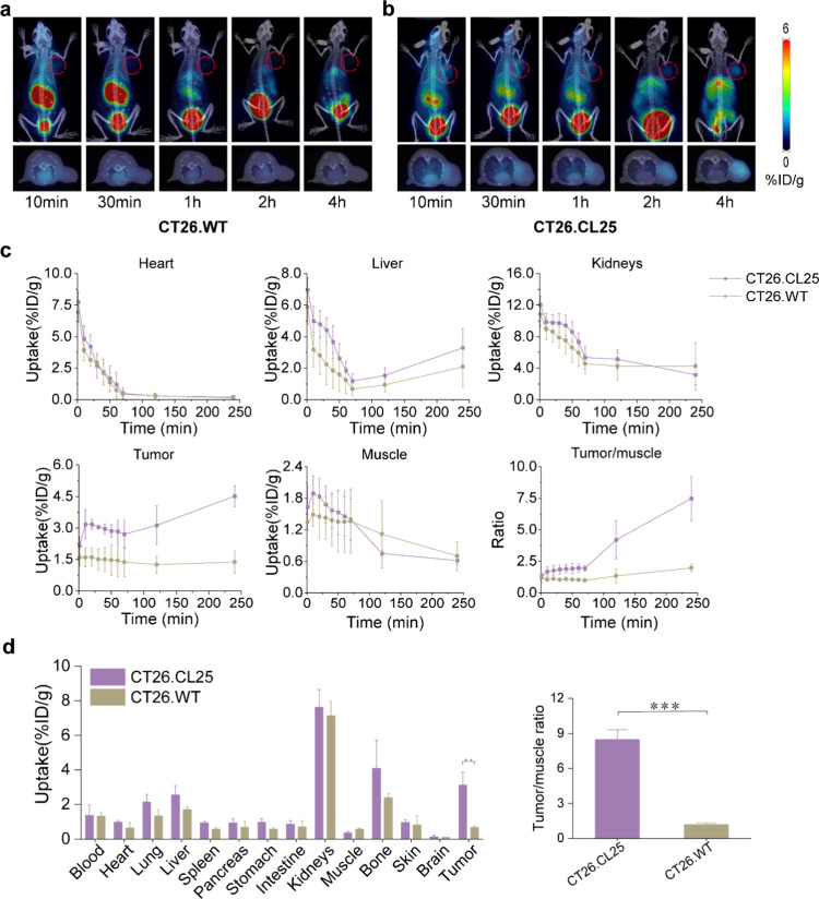
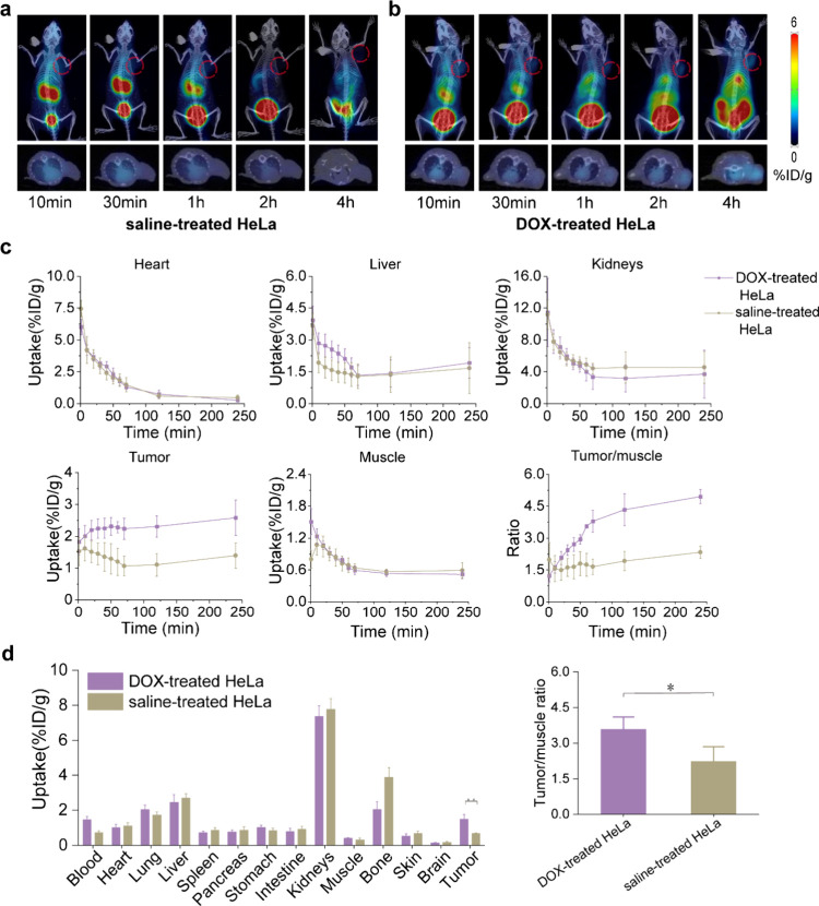
Verification
Claims the paper makes
The claims and statistics of this paper haven’t been checked yet.
Run the check — free, no account needed. It reads the open-access text and checks the sources it cites.
Claims other papers make about this one
Each is checked against this paper’s own text: does it say what the citing paper says it does?
The cited source says what the claim attributes to it.
In the citing paper
“For example, probes targeting SA-β-Gal, such as [68Ga]Ga-BGal, have demonstrated efficacy in preclinical models112,113.”
From Emerging strategies in senotherapeutics: from broad-spectrum senolysis to precision reprogramming (2026) · cites this paper as [112]
Self-check
Percentages, sums and reported differences re-computed from the paper’s own numbers.
The self-check needs the paper’s full text.
Citations
Cites
Cellular Senescence: Defining a Path Forward
Gorgoulis, V., Adams, P. D., Alimonti, A., Bennett, D. C., Bischof, O., Bishop, C., Campisi, J., Collado, M., Evangelou, K., Ferbeyre, G., et al. (2019) · Cell · doi:10.1016/j.cell.2019.10.005 · Cited by 3,225
Cellular senescence: from physiology to pathology
Muñoz-Espín, D., & Serrano, M. (2014) · Nature Reviews Molecular Cell Biology · doi:10.1038/nrm3823 · Cited by 2,818
Cellular senescence in aging and age-related disease: from mechanisms to therapy
Childs, B. G., Durik, M., Baker, D. J., & van Deursen, J. M. (2015) · Nature Medicine · doi:10.1038/nm.4000 · Cited by 2,363
Cellular senescence in ageing: from mechanisms to therapeutic opportunities
Di Micco, R., Krizhanovsky, V., Baker, D., & d’Adda di Fagagna, F. (2021) · Nature Reviews Molecular Cell Biology · doi:10.1038/s41580-020-00314-w · Cited by 2,277
Senescence surveillance of pre-malignant hepatocytes limits liver cancer development
Kang, T.-W., Yevsa, T., Woller, N., Hoenicke, L., Wuestefeld, T., Dauch, D., Hohmeyer, A., Gereke, M., Rudalska, R., Potapova, A., et al. (2011) · Nature · doi:10.1038/nature10599 · Cited by 1,621
Burley, S. K., Bhikadiya, C., Bi, C., Bittrich, S., Chen, L., Crichlow, G. V., Christie, C. H., Dalenberg, K., Di Costanzo, L., Duarte, J. M., et al. (2021) · Nucleic Acids Research · doi:10.1093/nar/gkaa1038 · Cited by 1,581
Cellular senescence and senolytics: the path to the clinic
Chaib, S., Tchkonia, T., & Kirkland, J. L. (2022) · Nature Medicine · doi:10.1038/s41591-022-01923-y · Cited by 1,134
Unmasking senescence: context-dependent effects of SASP in cancer
Faget, D. V., Ren, Q., & Stewart, S. A. (2019) · Nature Reviews Cancer · doi:10.1038/s41568-019-0156-2 · Cited by 863
Cellular senescence and Alzheimer disease: the egg and the chicken scenario
Saez-Atienzar, S., & Masliah, E. (2020) · Nature Reviews Neuroscience · doi:10.1038/s41583-020-0325-z · Cited by 257
Hoenicke, L., & Zender, L. (2012) · Carcinogenesis · doi:10.1093/carcin/bgs124 · Cited by 248
Research works printed but not linked: 36
- 10.1038/s41581-022-00601-z · DOI · Paper page
- 10.1038/s41570-019-0108-0 · DOI · Paper page
- 10.1038/nature05529 · DOI · Paper page
- 10.1016/S0092-8674(00)81023-5 · DOI
- 10.1016/j.cell.2017.05.015 · DOI · Paper page
- 10.1152/physrev.00020.2018 · DOI · Paper page
- 10.1016/j.canlet.2021.07.002 · DOI
- 10.1002/aac2.12067 · DOI
- 10.1016/j.tcb.2018.02.001 · DOI · Paper page
- 10.3390/ijms23084168 · DOI
- 10.1111/j.1474-9726.2006.00199.x · DOI · Paper page
- 10.1073/pnas.92.20.9363 · DOI · Paper page
- 10.3390/biom13020362 · DOI
- 10.1021/acs.bioconjchem.9b00391 · DOI
- 10.1039/D1SC02069B · DOI
- 10.1007/s00216-020-03111-8 · DOI
- 10.1016/j.enzmictec.2021.109885 · DOI
- 10.1039/D0QM00683A · DOI
- 10.1002/cbic.202200364 · DOI
- 10.1038/s41598-019-38511-z · DOI
- 10.1021/acs.jmedchem.1c01313 · DOI
- 10.1152/physrev.00049.2010 · DOI
- 10.1109/TMI.2004.824150 · DOI
- 10.1158/1538-7445.AM2019-1146 · DOI
- 10.1016/j.bbagen.2021.129898 · DOI
- 10.1007/s004240050843 · DOI
- 10.1146/annurev-pathol-121808-102144 · DOI · Paper page
- 10.1016/j.semcancer.2021.03.021 · DOI
- 10.1038/s41569-020-0431-7 · DOI
- 10.1038/s41574-020-0335-y · DOI · Paper page
- 10.1038/s41418-020-0564-6 · DOI
- 10.1093/nar/28.1.235 · DOI · Paper page
- 10.1021/bi011727i · DOI
- 2021, Schrödinger Release 2021-3: LigPrep
- 10.1007/s10822-007-9133-z · DOI
- 10.1021/jm0306430 · DOI · Paper page
Cited by
Sorted by citations
Emerging strategies in senotherapeutics: from broad-spectrum senolysis to precision reprogramming
Zhang, W., Song, S., Zhang, Y., Pan, Y., Hu, D., & Wang, Y. (2026) · npj Aging · doi:10.1038/s41514-026-00355-z · Cited by 6
Self-citation
Metadata
- Authors
- Xiang, X., Dong, C., Zhou, L., Liu, J., Rabinowitz, Z. M., Zhang, Y., Guo, H., He, F., Chen, X., Wang, Y., Cui, L., & Ma, X.
- Venue
- Journal of Medicinal Chemistry
- Publisher
- American Chemical Society (ACS)
- Published
- 2024-03-20
- Volume
- 67
- Issue
- 7
- Pages
- 5924-5934
- ISSN
- 0022-2623
- 1520-4804
- PMC
- PMC11017977
- Cited by
- 41
- References
- 10
- Language
- en
- Funders
- Fundamental Research Funds for Central Universities of the Central South University (2022ZZTS0856)
- National Institute on Aging (R41AG081007)
- Key Research and Development Program of Hunan Province of China (2022SK2035)
About these checks
Registry, retraction, Cites and statistics results are computed, not judged, with one exception: a printed reference that no index lists is read once by a model before it counts as a citation error. Claims are extracted by a model and kept only when their quote appears verbatim in the text; each cited source is then checked, with a model’s help, for whether it says what the claim relies on. Those verdicts can be wrong — read the claim’s quote and the cited source before relying on one.
Statistics and arithmetic are recomputed, not judged.
Confidence bands are shown only for fields calibrated on our annotated test set. None is calibrated yet, so no band is shown.
What counts as a claim ›How we measure confidence ›How we check this ›
Are you an author, or do you see a problem with this page? Request a review
