Cited by 8Enzyme Structure and FunctionEnzyme Catalysis and ImmobilizationProtein Structure and Dynamics

The DOI matches its Crossref record; 1 of the 1 references it cites were found in a registry or an index.

Structural Basis for the Catalysis and Substrate Specificity of a LarA Racemase with a Broad Substrate Spectrum

Open access
Gatreddi, Santhosh, Urdiain-Arraiza, Julian, Desguin, Benoit, Hausinger, Robert P., Hu, Jian·2025·ACS Catalysis

Published version of a preprint

Abstract

Abstract The LarA family consists of diverse racemases/epimerases that interconvert the diastereomers of a variety of α-hydroxyacids by using a nickel-pincer nucleotide (NPN) cofactor. The hidden redox reaction catalyzed by the NPN cofactor makes LarA enzymes attractive engineering targets for applications. However, how a LarA enzyme binds its natural substrate and recognizes different α-hydroxyacids has not been elucidated. Here, we report three high-resolution structures of the enzyme-substrate complexes of a broad-spectrum LarA enzyme from Isosphaera pallida (LarA Ip ). The substrate binding mode reveals an optimal orientation and distance between the hydride donor and acceptor, strongly supporting the proposed proton-coupled hydride transfer mechanism. The experimentally solved structures, together with the structural models of other LarA enzymes, allow us to identify the residues/structural elements critically involved in the interactions with different α-hydroxyacid substrates. Collectively, this work provides a critical structural basis for catalysis and substrate recognition of the diverse enzymes in the LarA family, thus building a foundation for enzyme engineering.

Overview

The claims and statistics of this paper haven’t been checked yet.

Run the check — free, no account needed. It reads the open-access text and checks the sources it cites.

A few free checks a day · papers already verified open instantly · results are public

Integrity signals

Registry

Crossref · 10.1021/acscatal.4c07804

Cites

1 of 1 references verified0 citation errors · 0 not checkable

Statistics

Figures

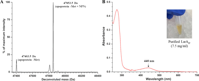
Figure 1 Characterization of the purified LarAIp. (A) ESI-MS of LarAIp. (B) UV–visible spectrum of LarAIp. The inset shows the appearance of the purified protein.
Figure 1 Characterization of the purified LarAIp. (A) ESI-MS of LarAIp. (B) UV–visible spectrum of LarAIp. The inset shows the appearance of the purified protein.CC BY · PubMed Central
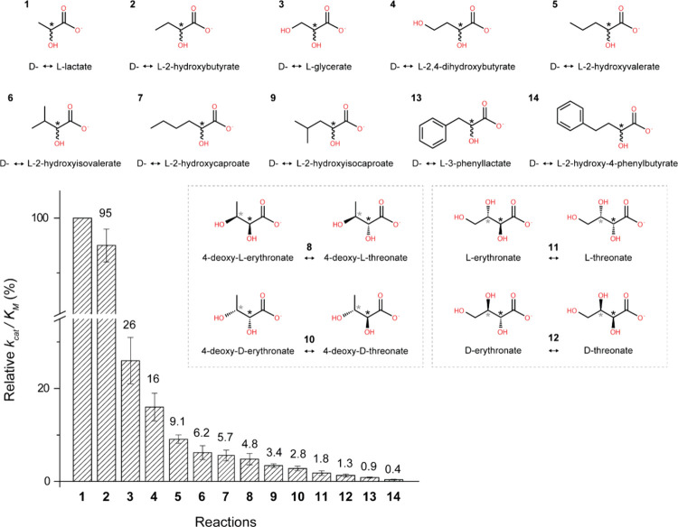
Figure 2 Relative kcat/KM values of LarAIp for racemization/epimerization of α-hydroxyacids. The kcat/KM value for lactate was set to 100%. For racemization reactions, the chiral carbons of enantiomers are depicted achiral for simplification. For epimerization reactions, both epimers are depicted, with diastereomers of the same molecule boxed together. Black asterisks indicate stereoinversion sites, and gray asterisks indicate other stereocenters in the molecule. The error bars indicate standard deviations (n = 3).
Figure 2 Relative kcat/KM values of LarAIp for racemization/epimerization of α-hydroxyacids. The kcat/KM value for lactate was set to 100%. For racemization reactions, the chiral carbons of enantiomers are depicted achiral for simplification. For epimerization reactions, both epimers are depicted, with diastereomers of the same molecule boxed together. Black asterisks indicate stereoinversion sites, and gray asterisks indicate other stereocenters in the molecule. The error bars indicate standard deviations (n = 3).CC BY · PubMed Central
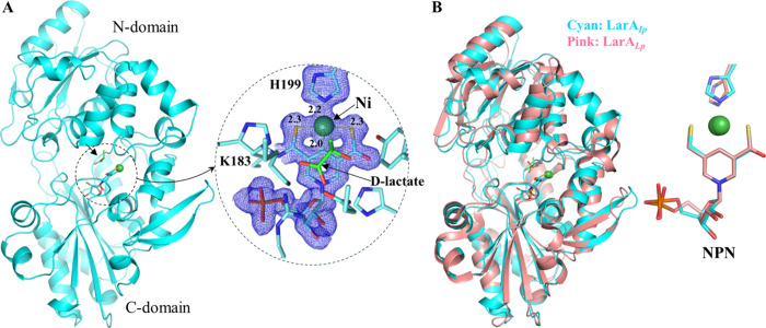
Figure 3 Structure of LarAIp as purified. (A) Overall structure of LarAIp (left) and the zoom-in view of the active site (right). The blue meshes are the electron densities of the NPN cofactor and His199 (2Fo – Fc, σ = 1). (B) Structural comparison of LarAIp (cyan, chain B) and LarALp (pink; PDB: 6C1W; chain B).
Figure 3 Structure of LarAIp as purified. (A) Overall structure of LarAIp (left) and the zoom-in view of the active site (right). The blue meshes are the electron densities of the NPN cofactor and His199 (2Fo – Fc, σ = 1). (B) Structural comparison of LarAIp (cyan, chain B) and LarALp (pink; PDB: 6C1W; chain B).CC BY · PubMed Central
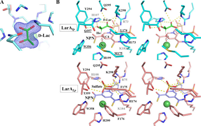
Figure 4 Binding of d-lactate in LarAIp. (A) 2Fo – Fc map (σ = 1) of d-lactate (green) in the active site of LarAIp as purified. (B) Stereo view of the active sites of LarAIp and LarALp. Top: LarAIp with bound d-lactate. NPN is shown in pink, d-lactate in green, and protein in cyan. The yellow dashed lines indicate polar interactions with d-lactate, and the red dashed lines indicate that the distances between the hydrogen atom on Cα (Hα) and C4 or Ni are 2.8 and 2.9 Å, respectively. The underlined residues are involved in the interactions with the 2-methyl group (Cα substituent). Bottom: LarALp with bound sulfate (PDB: 6C1W; chain B).
Figure 4 Binding of d-lactate in LarAIp. (A) 2Fo – Fc map (σ = 1) of d-lactate (green) in the active site of LarAIp as purified. (B) Stereo view of the active sites of LarAIp and LarALp. Top: LarAIp with bound d-lactate. NPN is shown in pink, d-lactate in green, and protein in cyan. The yellow dashed lines indicate polar interactions with d-lactate, and the red dashed lines indicate that the distances between the hydrogen atom on Cα (Hα) and C4 or Ni are 2.8 and 2.9 Å, respectively. The underlined residues are involved in the interactions with the 2-methyl group (Cα substituent). Bottom: LarALp with bound sulfate (PDB: 6C1W; chain B).CC BY · PubMed Central
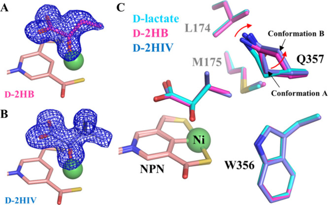
Figure 5 Binding of additional α-hydroxyacids at the active site of LarAIp. (A) 2Fo – Fc map (σ = 1, blue mesh) of D-2HB. The relatively poor density of the terminal methyl group suggests that it is more dynamic than the rest of the ligand. (B) 2Fo – Fc map (σ = 1) of D-2HIV. (C) Structural comparison of the residues contacting the Cα substituents of the different substrates. The red arrow indicates the structural change of Q357 in response to the replacement of d-lactate with 2HB and 2HIV. The NPN cofactor is shown in pink, and Ni is shown as a green sphere.
Figure 5 Binding of additional α-hydroxyacids at the active site of LarAIp. (A) 2Fo – Fc map (σ = 1, blue mesh) of D-2HB. The relatively poor density of the terminal methyl group suggests that it is more dynamic than the rest of the ligand. (B) 2Fo – Fc map (σ = 1) of D-2HIV. (C) Structural comparison of the residues contacting the Cα substituents of the different substrates. The red arrow indicates the structural change of Q357 in response to the replacement of d-lactate with 2HB and 2HIV. The NPN cofactor is shown in pink, and Ni is shown as a green sphere.CC BY · PubMed Central
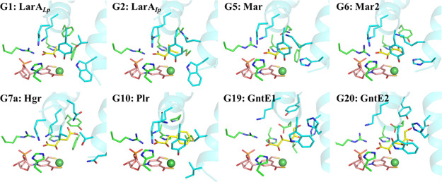
Figure 6 Structural models of the enzyme–substrate complexes for the LarA family members with known substrates. A total of 25 groups of LarA enzymes were identified in a bioinformatics analysis (ref (24)), among which eight groups (G1, G2, G5, G6, G7a, G10, G19, and G20) were shown to process at least one α-hydroxyacid. The structures of the enzyme–substrate complexes for representative group members in G5, G6, G7a, G10, G19, and G20 were generated using the AlphaFold-predicted models and the d-substrate binding mode revealed in this work. d-Lactate was modeled into the structure of LarALp (PDB: 6C1W; chain B) after the removal of the bound sulfate from the active site. G1, lactate racemase; G2, short-chain aliphatic α-hydroxyacid racemase; G5, malate racemase (Mar); G6, malate racemase 2 (Mar2); G7a, hydroxyglutarate racemase (Hgr); G10, phenyllactic acid racemase (Plr); G19, gluconate 2-epimerase 1 (GntE1); G20, gluconate 2-epimerase 2 (GntE2). The substrates are shown in yellow, the NPN cofactors in pink, and Ni in dark green. Only the residues involved in direct interaction with the substrates are shown in stick mode. The residues from the N-terminal domain are in green, and those from the C-terminal domain are in cyan. The two central α-helices are shown in cartoon mode.
Figure 6 Structural models of the enzyme–substrate complexes for the LarA family members with known substrates. A total of 25 groups of LarA enzymes were identified in a bioinformatics analysis (ref (24)), among which eight groups (G1, G2, G5, G6, G7a, G10, G19, and G20) were shown to process at least one α-hydroxyacid. The structures of the enzyme–substrate complexes for representative group members in G5, G6, G7a, G10, G19, and G20 were generated using the AlphaFold-predicted models and the d-substrate binding mode revealed in this work. d-Lactate was modeled into the structure of LarALp (PDB: 6C1W; chain B) after the removal of the bound sulfate from the active site. G1, lactate racemase; G2, short-chain aliphatic α-hydroxyacid racemase; G5, malate racemase (Mar); G6, malate racemase 2 (Mar2); G7a, hydroxyglutarate racemase (Hgr); G10, phenyllactic acid racemase (Plr); G19, gluconate 2-epimerase 1 (GntE1); G20, gluconate 2-epimerase 2 (GntE2). The substrates are shown in yellow, the NPN cofactors in pink, and Ni in dark green. Only the residues involved in direct interaction with the substrates are shown in stick mode. The residues from the N-terminal domain are in green, and those from the C-terminal domain are in cyan. The two central α-helices are shown in cartoon mode.CC BY · PubMed Central
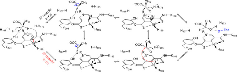
Figure 7 Updated PCHT mechanism of lactate racemization. Two hydride transfer pathways are proposed. The hydride from the d-substrate can be transferred to C4 of the pyridinium ring (black arrow, upper pathway) or to Ni (red arrow, lower pathway). After rotation of the acetyl group of the pyruvate (blue curved arrows) to expose the alternative side of the ketone to the reduced NPN cofactor, the hydride returns to the intermediate to complete racemization. The 2-OH of the product (or l-substrate) is proposed to be stabilized by an unidentified hydrogen donor (capital D in blue), which is equivalent to Y294, in an unresolved conformation of the enzyme.
Figure 7 Updated PCHT mechanism of lactate racemization. Two hydride transfer pathways are proposed. The hydride from the d-substrate can be transferred to C4 of the pyridinium ring (black arrow, upper pathway) or to Ni (red arrow, lower pathway). After rotation of the acetyl group of the pyruvate (blue curved arrows) to expose the alternative side of the ketone to the reduced NPN cofactor, the hydride returns to the intermediate to complete racemization. The 2-OH of the product (or l-substrate) is proposed to be stabilized by an unidentified hydrogen donor (capital D in blue), which is equivalent to Y294, in an unresolved conformation of the enzyme.CC BY · PubMed Central

Verification

Claims the paper makes

The claims and statistics of this paper haven’t been checked yet.

Run the check — free, no account needed. It reads the open-access text and checks the sources it cites.

A few free checks a day · papers already verified open instantly · results are public

Self-check

Percentages, sums and reported differences re-computed from the paper’s own numbers.

The self-check needs the paper’s full text.

Citations

Cites

Research works printed but not linked: 48

  1. 10.2323/jgam.48.91 · DOI
  2. 10.1128/JB.187.19.6750-6761.2005 · DOI
  3. 10.1038/ncomms4615 · DOI
  4. 10.1126/science.aab2272 · DOI
  5. 10.1073/pnas.1600486113 · DOI
  6. 10.1073/pnas.2106202118 · DOI
  7. 10.1021/acs.biochem.3c00242 · DOI
  8. 10.1073/pnas.1704967114 · DOI
  9. 10.1016/j.jbc.2022.102131 · DOI
  10. 10.1021/acs.biochem.8b00601 · DOI
  11. 10.1002/pro.4874 · DOI
  12. 10.1093/mtomcs/mfac014 · DOI
  13. 10.1074/jbc.RA118.003741 · DOI
  14. 10.1016/j.cbpa.2018.06.019 · DOI
  15. 10.1002/cctc.202000575 · DOI
  16. 10.1042/BST20220490 · DOI
  17. 10.1016/bs.mie.2023.03.006 · DOI
  18. 10.1021/acs.biochem.8b00100 · DOI
  19. 10.1021/acscatal.2c05461 · DOI
  20. 10.1002/chem.201700405 · DOI
  21. 10.1002/chem.201604893 · DOI
  22. 10.1039/C7CC06416K · DOI
  23. 10.1002/anie.201612065 · DOI
  24. 10.1038/s41598-020-74802-6 · DOI
  25. 10.1016/j.jbc.2024.108069 · DOI
  26. 10.1007/s10534-022-00372-x · DOI
  27. 10.1007/BF00463488 · DOI
  28. 10.1107/S1744309108007252 · DOI
  29. 10.1021/acscatal.6b03056 · DOI
  30. 10.1074/jbc.M201415200 · DOI
  31. 10.1016/j.jmb.2009.06.022 · DOI
  32. 10.1002/chem.201905826 · DOI
  33. 10.1021/ja056490i · DOI
  34. 10.1038/nature05689 · DOI
  35. 10.1021/ja049579h · DOI
  36. 10.1021/bi00009a007 · DOI
  37. 10.1107/S1744309107033957 · DOI
  38. 10.1021/acs.jpclett.5b00346 · DOI
  39. 10.1073/pnas.94.24.12797 · DOI
  40. 10.1021/acs.organomet.2c00319 · DOI
  41. 10.1073/pnas.1616038114 · DOI
  42. 10.1002/anie.201910490 · DOI
  43. 10.1016/j.chroma.2023.464611 · DOI
  44. 10.1016/j.tibtech.2007.03.010 · DOI
  45. 10.1016/S0076-6879(97)76066-X · DOI · Paper page
  46. 10.1107/S0907444909047337 · DOI · Paper page
  47. 10.1107/S0907444910007493 · DOI · Paper page
  48. 10.1107/S2059798319011471 · DOI · Paper page

Metadata

Authors
Gatreddi, S., Urdiain-Arraiza, J., Desguin, B., Hausinger, R. P., & Hu, J.
Affiliations
  • Michigan State University (US)
  • Louvain Institute of Biomolecular Science and Technology (LIBST)
Corresponding author
  • Hausinger, Robert P.
  • Hu, Jian
Venue
ACS Catalysis
Published
2025-02-03
Volume
15
Issue
4
Pages
2857-2866
ISSN
2155-5435 (ISSN-L)
OpenAlex
W4407108673
Cited by
8
References
1
Citations by year
2026: 2 · 2025: 5
FWCI
1.48
Type
journal article
Language
en
Open access
Locations
  • ACS Catalysis (publishedVersion, cc-by)
  • bioRxiv (Cold Spring Harbor Laboratory) (acceptedVersion, cc-by)
  • PubMed Central (publishedVersion, cc-by)
  • PubMed (publishedVersion)
  • PubMed Central (submittedVersion, cc-by)
  • Digital Access to Libraries (Université catholique de Louvain (UCL), l'Université de Namur (UNamur) and the Université Saint-Louis (USL-B)) (submittedVersion, other-oa)
Funders
  • Universit? Catholique de Louvain
  • National Institute of General Medical Sciences (GM128959, GM140931)
Topics
  • Life Sciences › Biochemistry, Genetics and Molecular Biology › Structural Biology › Enzyme Structure and Function
  • Life Sciences › Biochemistry, Genetics and Molecular Biology › Molecular Biology › Enzyme Catalysis and Immobilization
  • Life Sciences › Biochemistry, Genetics and Molecular Biology › Molecular Biology › Protein Structure and Dynamics
Keywords
enzyme-substrate complex structure, substrate specificity, hydride transfer mechanism, enzyme catalysis, substrate binding mode

About these checks

Registry, retraction, Cites and statistics results are computed, not judged, with one exception: a printed reference that no index lists is read once by a model before it counts as a citation error. Claims are extracted by a model and kept only when their quote appears verbatim in the text; each cited source is then checked, with a model’s help, for whether it says what the claim relies on. Those verdicts can be wrong — read the claim’s quote and the cited source before relying on one.

Statistics and arithmetic are recomputed, not judged.

Confidence bands are shown only for fields calibrated on our annotated test set. None is calibrated yet, so no band is shown.

What counts as a claim ›How we measure confidence ›How we check this ›

Are you an author, or do you see a problem with this page? Request a review