

The DOI matches its Crossref record; 44 of the 44 references it cites were found in a registry or an index.
Development of a human three-dimensional organotypic skin-melanoma spheroid model for in vitro drug testing
Open accessAbstract
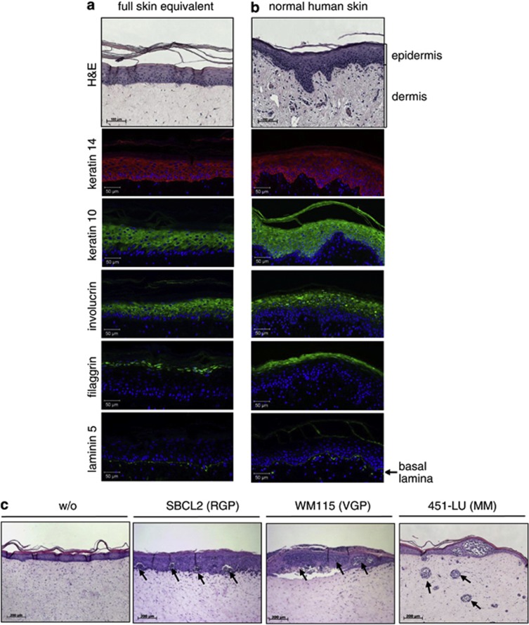
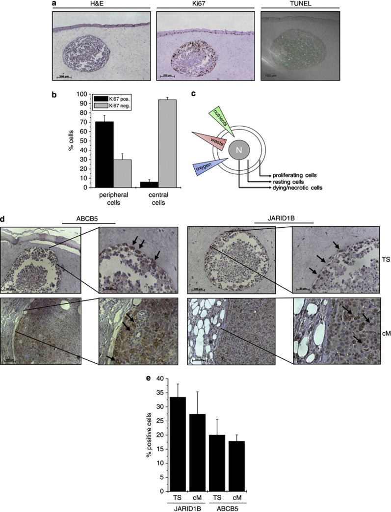
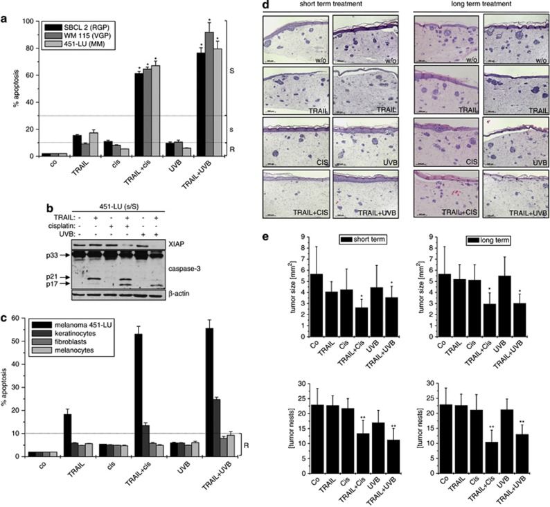
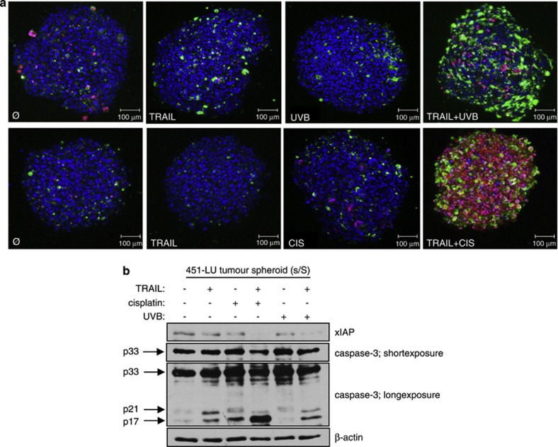
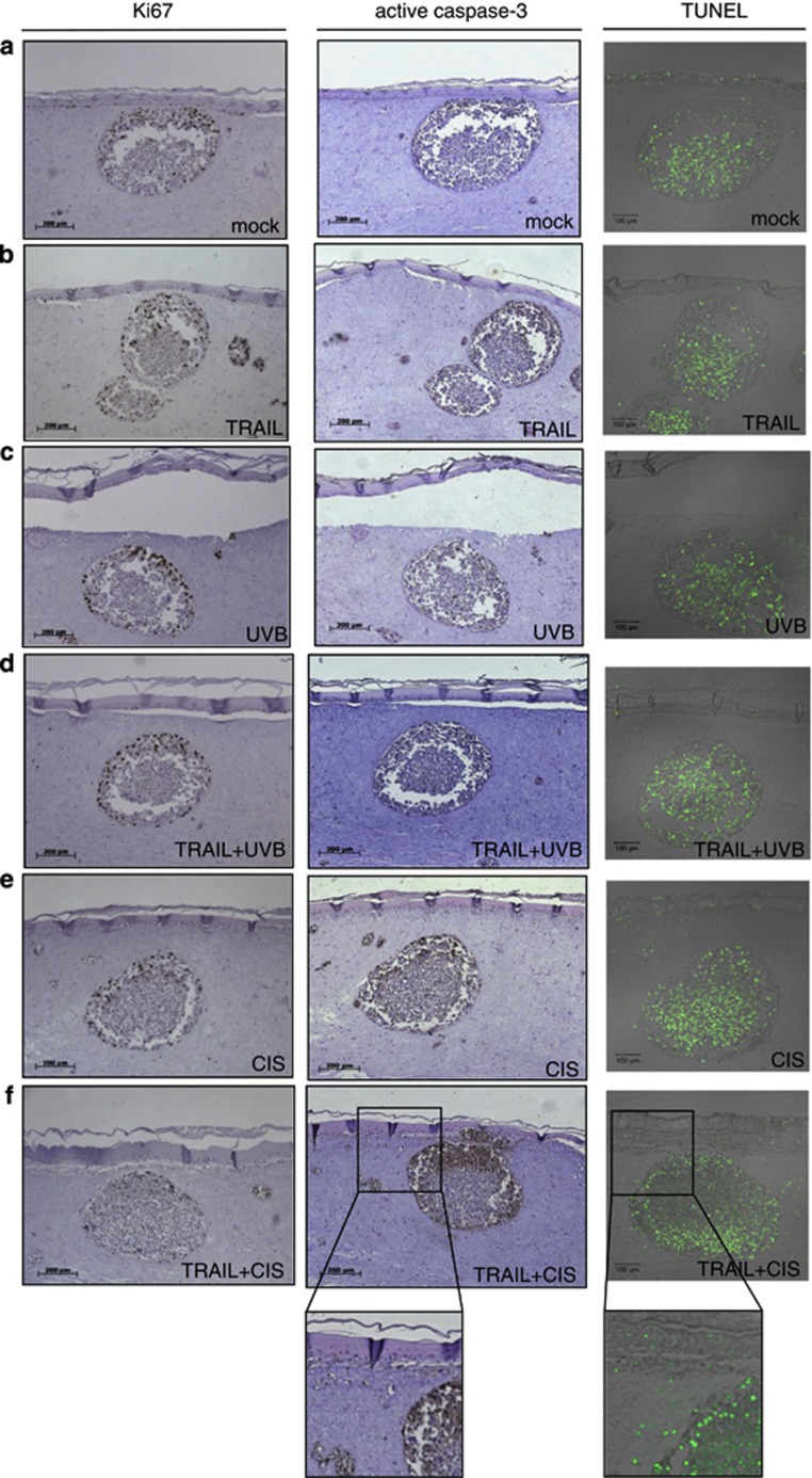
Despite remarkable efforts, metastatic melanoma (MM) still presents with significant mortality. Recently, mono-chemotherapies are increasingly replenished by more cancer-specific combination therapies involving death ligands and drugs interfering with cell signaling. Still, MM remains a fatal disease because tumors rapidly develop resistance to novel therapies thereby regaining tumorigenic capacity. Although genetically engineered mouse models for MM have been developed, at present no model is available that reliably mimics the human disease and is suitable for studying mechanisms of therapeutic obstacles including cell death resistance. To improve the increasing requests on new therapeutic alternatives, reliable human screening models are demanded that translate the findings from basic cellular research into clinical applications. By developing an organotypic full skin equivalent, harboring melanoma tumor spheroids of defined sizes we have invented a cell-based model that recapitulates both the 3D organization and multicellular complexity of an organ/tumor in vivo but at the same time accommodates systematic experimental intervention. By extending our previous findings on melanoma cell sensitization toward TRAIL (tumor necrosis factor-related apoptosis-inducing ligand) by co-application of sublethal doses of ultraviolet-B radiation (UVB) or cisplatin, we show significant differences in the therapeutical outcome to exist between regular two-dimensional (2D) and complex in vivo-like 3D models. Of note, while both treatment combinations killed the same cancer cell lines in 2D culture, skin equivalent-embedded melanoma spheroids are potently killed by TRAIL+cisplatin treatment but remain almost unaffected by the TRAIL+UVB combination. Consequently, we have established an organotypic human skin-melanoma model that will facilitate efforts to improve therapeutic outcomes for malignant melanoma by providing a platform for the investigation of cytotoxic treatments and tailored therapies in a more physiological setting.
Overview
The claims and statistics of this paper haven’t been checked yet.
Run the check — free, no account needed. It reads the open-access text and checks the sources it cites.
Integrity signals
Registry
Cites
Statistics
Figures
Verification
Claims the paper makes
The claims and statistics of this paper haven’t been checked yet.
Run the check — free, no account needed. It reads the open-access text and checks the sources it cites.
Claims other papers make about this one
Each is checked against this paper’s own text: does it say what the citing paper says it does?
The check wasn’t confident either way, so it gives no verdict — never disputed.
In the citing paper
“In melanoma spheroids, for example, fibroblasts have been shown to accumulate in low-oxygen regions and upregulate genes associated with ECM production and wound response [288].”
From Modeling Tumor Microenvironment Complexity In Vitro: Spheroids as Physiologically Relevant Tumor Models and Strategies for Their Analysis (2025) · cites this paper as [288]
Experimental — accuracy not yet measured
Self-check
Percentages, sums and reported differences re-computed from the paper’s own numbers.
The self-check needs the paper’s full text.
Citations
Cites
Improved survival with vemurafenib in melanoma with BRAF V600E mutation
Chapman, P. B., Hauschild, A., Robert, C., Haanen, J. B., Ascierto, P., Larkin, J., Dummer, R., Garbe, C., Testori, A., Maio, M., et al. (2011) · New England Journal of Medicine · doi:10.1056/nejmoa1103782 · Cited by 7,720
Final Version of 2009 AJCC Melanoma Staging and Classification
Balch, C. M., Gershenwald, J. E., Soong, S.-J., Thompson, J. F., Atkins, M. B., Byrd, D. R., Buzaid, A. C., Cochran, A. J., Coit, D. G., Ding, S., et al. (2009) · Journal of Clinical Oncology · doi:10.1200/jco.2009.23.4799 · Cited by 4,653 · cited 1×
“metastatic melanoma (MM) is particularly poor with stage IV patients having a median survival of B8±2 months.(1) Malignant transformation of melanocytes involves stepwise tumor progression.”
BackgroundInhibition of Mutated, Activated BRAF in Metastatic Melanoma
Flaherty, K. T., Puzanov, I., Kim, K. B., Ribas, A., McArthur, G. A., Sosman, J. A., O'Dwyer, P. J., Lee, R. J., Grippo, J. F., Nolop, K., et al. (2010) · New England Journal of Medicine · doi:10.1056/nejmoa1002011 · Cited by 3,103 · cited 1×
“Therefore, alternative therapeutic strategies are required, and the targeting of specific activating mutations in melanoma has been of particular interest, yet almost invariably, responders develop therapy resistance.(11)”
BackgroundGenomic responses in mouse models poorly mimic human inflammatory diseases
Seok, J., Warren, H. S., Cuenca, A. G., Mindrinos, M. N., Baker, H. V., Xu, W., Richards, D. R., McDonald-Smith, G. P., Gao, H., Hennessy, L., et al. (2013) · Proceedings of the National Academy of Sciences · doi:10.1073/pnas.1222878110 · Cited by 3,005
Multicellular tumor spheroids: An underestimated tool is catching up again
Hirschhaeuser, F., Menne, H., Dittfeld, C., West, J., Mueller-Klieser, W., & Kunz-Schughart, L. A. (2010) · Journal of Biotechnology · doi:10.1016/j.jbiotec.2010.01.012 · Cited by 1,674 · cited 2×
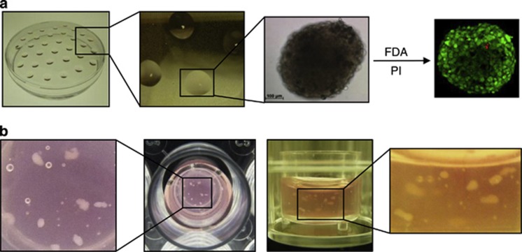
“nodes, micrometastasis or inter-capillary micro regions of solid tumors (Figure 2a).(16,17) We next inserted a defined number of these spheroids (10)”
BackgroundMethodRecent advances in three‐dimensional multicellular spheroid culture for biomedical research
Lin, R., & Chang, H. (2008) · Biotechnology Journal · doi:10.1002/biot.200700228 · Cited by 1,264 · cited 1×
“Only spheroids with sizes of/above 500mm represent typical features of nonvascularized tumor tissue.(16,31) To date, conventional chemotherapy-based cancer treat-”
BackgroundRoesch, A., Fukunaga‐Kalabis, M., Schmidt, E. C., Zabierowski, S. E., Brafford, P. A., Vultur, A., Basu, D., Gimotty, P. A., VOGT, T. M., & Herlyn, M. (2010) · Cell · doi:10.1016/j.cell.2010.04.020 · Cited by 1,177
Widespread potential for growth-factor-driven resistance to anticancer kinase inhibitors
Wilson, T. R., Fridlyand, J., Yan, Y., Penuel, E., Burton, L., Chan, E., Peng, J., Lin, E., Wang, Y., Sosman, J., et al. (2012) · Nature · doi:10.1038/nature11249 · Cited by 1,134
Chapman, P. B., Einhorn, L. H., Meyers, M. L., Saxman, S. B., Destro, A. N., Panageas, K. S., Begg, C. B., Agarwala, S. S., Schuchter, L. M., Ernstoff, M. S., et al. (1999) · Journal of Clinical Oncology · doi:10.1200/jco.1999.17.9.2745 · Cited by 763
A Simple Hanging Drop Cell Culture Protocol for Generation of 3D Spheroids
Foty, R. (2011) · Journal of Visualized Experiments · doi:10.3791/2720 · Cited by 612 · cited 1×
“Melanoma spheroids were generated via the ‘hanging drop’ method incorporating 250 melanoma cells per 25ml RPMI placed on the lid of a non-adhesive PBS containing petri dish.(15) Spheroids were incubated for 15 days at 371C, 5% CO2.”
Method
Cited by
Sorted by citations
3D tumor spheroids: an overview on the tools and techniques used for their analysis
Costa, E. C., Moreira, A. F., de Melo-Diogo, D., Gaspar, V. M., Carvalho, M. P., & Correia, I. J. (2016) · Biotechnology Advances · doi:10.1016/j.biotechadv.2016.11.002 · Cited by 929
Matrigel: From discovery and ECM mimicry to assays and models for cancer research
Benton, G., Arnaoutova, I., George, J., Kleinman, H. K., & Koblinski, J. (2014) · Advanced Drug Delivery Reviews · doi:10.1016/j.addr.2014.06.005 · Cited by 477
Skin models for the testing of transdermal drugs
Abd, E., Yousuf, S. A., Pastore, M. N., Telaprolu, K. C., Mohammed, Y., Namjoshi, S., Grice, J. E., & Roberts, M. S. (2016) · Clinical Pharmacology Advances and Applications · doi:10.2147/cpaa.s64788 · Cited by 430
“Using combination therapies, Vörsmann et al 118 showed significant advantages in using their 3-”
Neutrophil-Derived Proteases Escalate Inflammation through Activation of IL-36 Family Cytokines
Henry, C. M., Sullivan, G. P., Clancy, D. M., Afonina, I. S., Kulms, D., & Martin, S. J. (2016) · Cell Reports · doi:10.1016/j.celrep.2015.12.072 · Cited by 330
“Active IL-36 Perturbs Differentiation in a 3D Organotypic Skin Model To explore the biological effects of active IL-36 in a more physiologically relevant setting, we utilized a well established model of reconstituted human skin with primary keratinocytes and dermal fibroblasts (Vörsmann et al., 2013).”
MethodThe use of skin models in drug development
Mathes, S. H., Ruffner, H., & Graf‐Hausner, U. (2014) · Advanced Drug Delivery Reviews · doi:10.1016/j.addr.2013.12.006 · Cited by 328
“and UVB or cisplatin [112]”
Metadata
- Authors
- Vörsmann, H., Groeber, F., Walles, H., Busch, S., Beissert, S., Walczak, H., & Kulms, D.
- Affiliations
- University of Stuttgart (DE)
- Fraunhofer Institute for Interfacial Engineering and Biotechnology (DE)
- Technische Universität Dresden (DE)
- University College London (GB)
- Corresponding author
- Kulms, D
- Venue
- Cell Death & Disease
- Published
- 2013-07-11
- Volume
- 4
- Issue
- 7
- Pages
- e719-e719
- ISSN
- 2041-4889 (ISSN-L)
- PMC
- PMC3730422
- OpenAlex
- W2042222655
- Cited by
- 168
- References
- 44
- Citations by year
- 2026: 6 · 2025: 10 · 2024: 14 · 2023: 8 · 2022: 18 · 2021: 12 · 2020: 15 · 2019: 14 · 2018: 16 · 2017: 14 · 2016: 16 · 2015: 13 · 2014: 11 · 2013: 1
- FWCI
- 6.12
- Type
- journal article
- Language
- en
- Open access
- gold
- doi.org
- Licence
- cc-by-nc-nd
- www.springer.com/tdm (unspecified)
- Locations
- Cell Death and Disease (publishedVersion, cc-by-nc-nd)
- Europe PMC (PubMed Central) (publishedVersion, cc-by-nc-nd)
- PubMed Central (publishedVersion, cc-by-nc-nd)
- PubMed (publishedVersion)
- UCL Discovery (University College London) (submittedVersion)
- Fraunhofer-Publica (Fraunhofer-Gesellschaft) (submittedVersion)
- Funders
- Deutsche Krebshilfe
- Topics
- Life Sciences › Biochemistry, Genetics and Molecular Biology › Molecular Biology › Melanoma and MAPK Pathways
- Life Sciences › Biochemistry, Genetics and Molecular Biology › Molecular Biology › Cell death mechanisms and regulation
- Health Sciences › Medicine › Oncology › Cutaneous Melanoma Detection and Management
- Keywords
- metastatic melanoma, 3D tumor models, TRAIL sensitization, cisplatin, UV-B radiation, combination therapy, drug resistance, in vitro drug testing, tumor necrosis factor-related apoptosis-inducing ligand
- MeSH terms
- Antineoplastic Agents, Cisplatin, Drug Screening Assays, Antitumor, Drug Synergism, Humans, Melanoma, Skin Neoplasms, Skin, Artificial, Spheroids, Cellular, Coculture Techniques, Cell Line, Tumor, TNF-Related Apoptosis-Inducing Ligand
About these checks
Registry, retraction, Cites and statistics results are computed, not judged, with one exception: a printed reference that no index lists is read once by a model before it counts as a citation error. Claims are extracted by a model and kept only when their quote appears verbatim in the text; each cited source is then checked, with a model’s help, for whether it says what the claim relies on. Those verdicts can be wrong — read the claim’s quote and the cited source before relying on one.
Statistics and arithmetic are recomputed, not judged.
Confidence bands are shown only for fields calibrated on our annotated test set. None is calibrated yet, so no band is shown.
What counts as a claim ›How we measure confidence ›How we check this ›
Are you an author, or do you see a problem with this page? Request a review
