Cited by 339Immune Response and InflammationCardiac Fibrosis and RemodelingInflammasome and immune disorders

The DOI matches its Crossref record; 89 of the 89 references it cites were found in a registry or an index.

The emerging role of Toll-like receptor 4 in myocardial inflammation

Open access
Yang, Y, Lv, J, Jiang, S, Ma, Z, Wang, D, Hu, W, Deng, C, Fan, C, Di, S, Sun, Y, Yi, W·2016·Cell Death & Disease

Abstract

Toll-like receptors (TLRs) are a family of pattern recognition receptors involved in cardiovascular diseases. Notably, numerous studies have demonstrated that TLR4 activates the expression of several of pro-inflammatory cytokine genes that play pivotal roles in myocardial inflammation, particularly myocarditis, myocardial infarction, ischemia-reperfusion injury, and heart failure. In addition, TLR4 is an emerging target for anti-inflammatory therapies. Given the significance of TLR4, it would be useful to summarize the current literature on the molecular mechanisms and roles of TLR4 in myocardial inflammation. Thus, in this review, we first introduce the basic knowledge of the TLR4 gene and describe the activation and signaling pathways of TLR4 in myocardial inflammation. Moreover, we highlight the recent progress of research on the involvement of TLR4 in myocardial inflammation. The information reviewed here may be useful to further experimental research and to increase the potential of TLR4 as a therapeutic target.

Overview

The claims and statistics of this paper haven’t been checked yet.

Run the check — free, no account needed. It reads the open-access text and checks the sources it cites.

A few free checks a day · papers already verified open instantly · results are public

Integrity signals

Registry

Crossref · 10.1038/cddis.2016.140

Cites

89 of 89 references verified0 citation errors · 0 not checkable

Statistics

Figures

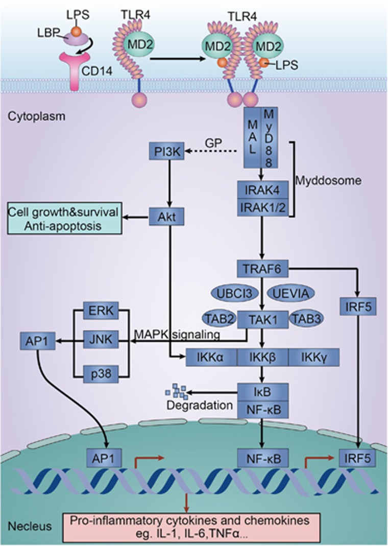
Figure 1 MyD88-dependent pathway. TLR4 uses CD14 to respond to LPS. LBP binds to LPS and presents it to CD14. MD2 is necessary for TLR4 to bind to LPS. MAL connects TLR4 and MyD88. Afterwards, MyD88 binds to IRAK4 and IRAK1/2. The IRAK complex then dissociates from MyD88 and interacts with TRAF6. TRAF6 forms a complex with TAK1, TAB1, and TAB2. Later, the complex binds to ubiquitin ligases, including UBC13 and UEV1A. TAK1 then activates the IKKα/IKKβ/IKKγ complex and induces IκB phosphorylation. Phosphorylated IκB dissociates from the complex and is directly targeted for ubiquitination and degradation by proteasomes, thus activating the transcription factor NF-κB. The released NF-κB translocates into the nucleus and mediates the expression of a number of pro-inflammatory cytokine genes. In addition to the activation of the IKK complex, TAK1 can activate MAPK signaling pathways, such as ERK, JNK, and the p38 pathway. MAPK signaling pathways can activate the transcription factor AP-1. Activation of NF-κB and AP-1 contributes to the expression of pro-inflammatory cytokines, such as IL-1, IL-6, and TNF-α. Notably, GP leads to TLR4 disassociating from MyD88, binding PI3K, further phosphorylating Akt and promoting survival and inhibits cardiac myocyte apoptosis. Gene names: ERK, extracellular signal-regulated kinase; GP, glucan phosphate; IKK, inhibitory κB (IκB) kinase; JNK, c-Jun N-terminal kinase; MAL, MyD88-adapter like; UBC13, ubiquitin-conjugating enzyme 13; UEV1A, ubiquitin-conjugating enzyme variant 1A
Figure 1 MyD88-dependent pathway. TLR4 uses CD14 to respond to LPS. LBP binds to LPS and presents it to CD14. MD2 is necessary for TLR4 to bind to LPS. MAL connects TLR4 and MyD88. Afterwards, MyD88 binds to IRAK4 and IRAK1/2. The IRAK complex then dissociates from MyD88 and interacts with TRAF6. TRAF6 forms a complex with TAK1, TAB1, and TAB2. Later, the complex binds to ubiquitin ligases, including UBC13 and UEV1A. TAK1 then activates the IKKα/IKKβ/IKKγ complex and induces IκB phosphorylation. Phosphorylated IκB dissociates from the complex and is directly targeted for ubiquitination and degradation by proteasomes, thus activating the transcription factor NF-κB. The released NF-κB translocates into the nucleus and mediates the expression of a number of pro-inflammatory cytokine genes. In addition to the activation of the IKK complex, TAK1 can activate MAPK signaling pathways, such as ERK, JNK, and the p38 pathway. MAPK signaling pathways can activate the transcription factor AP-1. Activation of NF-κB and AP-1 contributes to the expression of pro-inflammatory cytokines, such as IL-1, IL-6, and TNF-α. Notably, GP leads to TLR4 disassociating from MyD88, binding PI3K, further phosphorylating Akt and promoting survival and inhibits cardiac myocyte apoptosis. Gene names: ERK, extracellular signal-regulated kinase; GP, glucan phosphate; IKK, inhibitory κB (IκB) kinase; JNK, c-Jun N-terminal kinase; MAL, MyD88-adapter like; UBC13, ubiquitin-conjugating enzyme 13; UEV1A, ubiquitin-conjugating enzyme variant 1ACC BY · PubMed Central
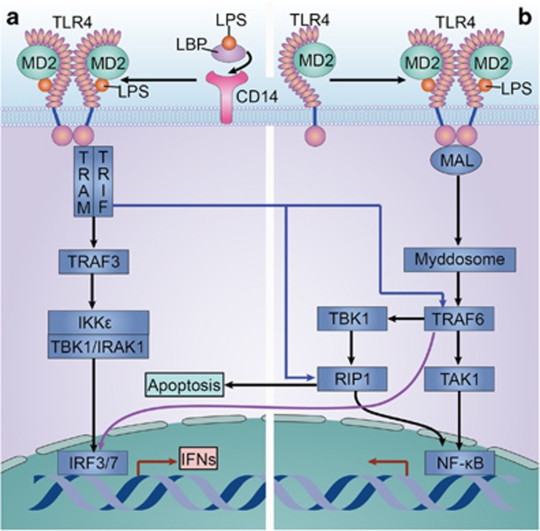
Figure 2 (a) The MyD88-independent pathway. (b) Crosstalk between the MyD88-dependent and MyD88-independent pathways. The MyD88-independent pathway can lead to the activation of both IRF and NF-kB and the subsequent expression of IFNs and pro-inflammatory cytokines, respectively. The MyD88-independent pathway is initiated by TRAM and TRIF. After being recruited, TRIF interacts with TBK and IKK-ɛ to phosphorylate the transcription factor IRF3. TRIF can also interact with IRAK1 and IKK-ɛ to activate the transcription factor IRF7. Activated IRFs then translocate into nucleus, bind with DNA, and produce such antiviral molecules as IFN-β. In addition, TRIF can also promote MyD88-independent NF-κB activation in TLR4 signaling pathways. Similar to the MyD88-dependent pathway, TRIF recruits TRAF6 and activates TAK1 through ubiquitination-dependent mechanisms, which in turn activates the NF-κB and MAPK pathways. Moreover, TRIF also activates MyD88-independent NF-κB activation by recruiting RIP1. Thus, TRIF activates IRFs by interacting with TBK1 and IRAK1 and activates NF-κB by interacting with RIP1. Furthermore, RIP1 can interact with FADD, which initiates caspase cascades and then induces cell apoptosis. Gene names: IKK-ɛ, inhibitory κB (IκB) kinase ɛ; IRAK1, interleukin (IL)-1 receptor-associated kinase 1; IRF, IFN-regulatory factor; TANK, TRAF family member-associated NF-κB activator; TRIF, TIR-domain-containing adapter-inducing interferon-β; TLR4, Toll-like receptor 4
Figure 2 (a) The MyD88-independent pathway. (b) Crosstalk between the MyD88-dependent and MyD88-independent pathways. The MyD88-independent pathway can lead to the activation of both IRF and NF-kB and the subsequent expression of IFNs and pro-inflammatory cytokines, respectively. The MyD88-independent pathway is initiated by TRAM and TRIF. After being recruited, TRIF interacts with TBK and IKK-ɛ to phosphorylate the transcription factor IRF3. TRIF can also interact with IRAK1 and IKK-ɛ to activate the transcription factor IRF7. Activated IRFs then translocate into nucleus, bind with DNA, and produce such antiviral molecules as IFN-β. In addition, TRIF can also promote MyD88-independent NF-κB activation in TLR4 signaling pathways. Similar to the MyD88-dependent pathway, TRIF recruits TRAF6 and activates TAK1 through ubiquitination-dependent mechanisms, which in turn activates the NF-κB and MAPK pathways. Moreover, TRIF also activates MyD88-independent NF-κB activation by recruiting RIP1. Thus, TRIF activates IRFs by interacting with TBK1 and IRAK1 and activates NF-κB by interacting with RIP1. Furthermore, RIP1 can interact with FADD, which initiates caspase cascades and then induces cell apoptosis. Gene names: IKK-ɛ, inhibitory κB (IκB) kinase ɛ; IRAK1, interleukin (IL)-1 receptor-associated kinase 1; IRF, IFN-regulatory factor; TANK, TRAF family member-associated NF-κB activator; TRIF, TIR-domain-containing adapter-inducing interferon-β; TLR4, Toll-like receptor 4CC BY · PubMed Central
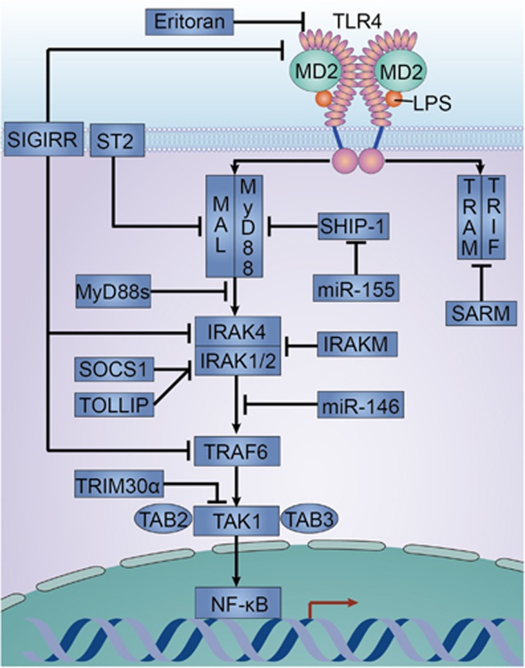
Figure 3 Negative regulation of TLR4 signaling pathways. The TLR4 antagonist eritoran inhibits TLR4 recognition of LPS. SHIP-1 inhibits the interaction between TLR4 and MyD88. ST2 sequesters MAL and MyD88. MyD88s can block the association between IRAK4 and MyD88. TOLLIP and SOCS1 can associate with IRAK1 and inhibit its activity. IRAKM inhibits the dissociation of IRAKs from MyD88 and prevents the formation of the IRAKs-TRAF6 complex. SIGIRR negatively regulates TLR4 signaling pathways by interacting transiently with TLR4, IRAK4, and TRAF6. TRIM30α destabilizes the TAK1 complex by promoting the degradation of TAB2 and TAB3. SARM specifically blocks the MyD88-independent signaling. miR-146 targets TRAF6 and IRAK1 and thus negatively regulates TLR4 signaling pathways. In addition, miR-155 can target SHIP-1 and thus suppress the negative regulation on TLR4 signaling pathways. Gene names: MAL, MyD88-adapter like; miR, microRNA; MyD88s, MyD88 short; SARM, Sterile α- and armadillo-motif-containing protein; SHIP-1, Src homology 2 domain-containing inositol 5-phosphatase 1; SOCS1, suppressor of cytokine signaling 1; ST2, suppression of tumorigenicity 2; TOLLIP, Toll-interacting protein
Figure 3 Negative regulation of TLR4 signaling pathways. The TLR4 antagonist eritoran inhibits TLR4 recognition of LPS. SHIP-1 inhibits the interaction between TLR4 and MyD88. ST2 sequesters MAL and MyD88. MyD88s can block the association between IRAK4 and MyD88. TOLLIP and SOCS1 can associate with IRAK1 and inhibit its activity. IRAKM inhibits the dissociation of IRAKs from MyD88 and prevents the formation of the IRAKs-TRAF6 complex. SIGIRR negatively regulates TLR4 signaling pathways by interacting transiently with TLR4, IRAK4, and TRAF6. TRIM30α destabilizes the TAK1 complex by promoting the degradation of TAB2 and TAB3. SARM specifically blocks the MyD88-independent signaling. miR-146 targets TRAF6 and IRAK1 and thus negatively regulates TLR4 signaling pathways. In addition, miR-155 can target SHIP-1 and thus suppress the negative regulation on TLR4 signaling pathways. Gene names: MAL, MyD88-adapter like; miR, microRNA; MyD88s, MyD88 short; SARM, Sterile α- and armadillo-motif-containing protein; SHIP-1, Src homology 2 domain-containing inositol 5-phosphatase 1; SOCS1, suppressor of cytokine signaling 1; ST2, suppression of tumorigenicity 2; TOLLIP, Toll-interacting proteinCC BY · PubMed Central
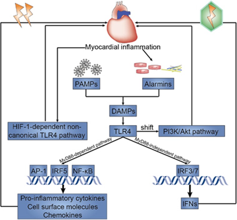
Figure 4 Graphical abstract. TLR4 recognizes and responds to DAMPs, including exogenous PAMPs and endogenous alarmins. The most common PAMPs in myocardial inflammation are thought to be virus, whereas alarmins released in response to myocardial inflammation can be necrotic cells and damaged matrix. TLR4 activates many transcription factors through the MyD88-dependent signaling pathway, subsequently inducing the production of pro-inflammatory cytokines, cell surface molecules, and chemokines and eventually exacerbating myocardial inflammation. TLR4 can also activate IRFs through the MyD88-independent signaling pathway, which further induce IFN production and ultimately exert an antiviral effect on myocardial inflammation. Moreover, a shift from predominant TLR4-NF-κB signaling pathway to the PI3K/Akt signaling pathway may serve a protective role in myocardial inflammation. Furthermore, there might be a novel HIF-1-dependent non-canonical TLR4 signaling pathway during myocardial inflammation. IRF, IFN-regulatory factors; PI3K, phosphatidylinositol 3-kinase
Figure 4 Graphical abstract. TLR4 recognizes and responds to DAMPs, including exogenous PAMPs and endogenous alarmins. The most common PAMPs in myocardial inflammation are thought to be virus, whereas alarmins released in response to myocardial inflammation can be necrotic cells and damaged matrix. TLR4 activates many transcription factors through the MyD88-dependent signaling pathway, subsequently inducing the production of pro-inflammatory cytokines, cell surface molecules, and chemokines and eventually exacerbating myocardial inflammation. TLR4 can also activate IRFs through the MyD88-independent signaling pathway, which further induce IFN production and ultimately exert an antiviral effect on myocardial inflammation. Moreover, a shift from predominant TLR4-NF-κB signaling pathway to the PI3K/Akt signaling pathway may serve a protective role in myocardial inflammation. Furthermore, there might be a novel HIF-1-dependent non-canonical TLR4 signaling pathway during myocardial inflammation. IRF, IFN-regulatory factors; PI3K, phosphatidylinositol 3-kinaseCC BY · PubMed Central

Verification

Claims the paper makes

The claims and statistics of this paper haven’t been checked yet.

Run the check — free, no account needed. It reads the open-access text and checks the sources it cites.

A few free checks a day · papers already verified open instantly · results are public

Claims other papers make about this one

Each is checked against this paper’s own text: does it say what the citing paper says it does?

Self-check

Percentages, sums and reported differences re-computed from the paper’s own numbers.

The self-check needs the paper’s full text.

Citations

Cites

1 retracted

Show all 89 references

Cited by

Sorted by citations

See all 339

Metadata

Authors
Yang, Y., Lv, J., Jiang, S., Ma, Z., Wang, D., Hu, W., Deng, C., Fan, C., Di, S., Sun, Y., & Yi, W.
Affiliations
  • Nanjing Drum Tower Hospital (CN)
  • Air Force Medical University (CN)
  • Tang Du Hospital (CN)
  • Xijing Hospital (CN)
Corresponding author
Sun, Y
Venue
Cell Death & Disease
Published
2016-05-26
Volume
7
Issue
5
Pages
e2234-e2234
ISSN
2041-4889 (ISSN-L)
OpenAlex
W2403031333
Cited by
337
References
89
Citations by year
2026: 25 · 2025: 39 · 2024: 34 · 2023: 34 · 2022: 38 · 2021: 50 · 2020: 32 · 2019: 37 · 2018: 31 · 2017: 13 · 2016: 3
FWCI
2.70
Type
journal article
Language
en
Open access
Locations
  • Cell Death and Disease (publishedVersion, cc-by)
  • PubMed Central (publishedVersion, cc-by)
  • PubMed (publishedVersion)
Funders
  • National Natural Science Foundation of China (81500263, 81100081, 81422004, 2015M572681, 81470477, 81100137, 81470480)
  • China Postdoctoral Science Foundation (2015M572681, 81500263)
  • National Institute on Alcohol Abuse and Alcoholism (R00AA020514, K99AA020514, R01AA020919)
Topics
  • Life Sciences › Immunology and Microbiology › Immunology › Immune Response and Inflammation
  • Health Sciences › Medicine › Cardiology and Cardiovascular Medicine › Cardiac Fibrosis and Remodeling
  • Life Sciences › Biochemistry, Genetics and Molecular Biology › Molecular Biology › Inflammasome and immune disorders
Keywords
TLR4, myocarditis, myocardial infarction, ischemia-reperfusion injury, heart failure, inflammatory cytokines, TLR4 signaling, anti-inflammatory therapy, pattern recognition receptors
MeSH terms
Animals, Cardiotonic Agents, Clinical Trials as Topic, Disaccharides, Gene Expression Regulation, Heart Failure, Humans, Metformin, Myocardial Infarction, Myocarditis, Myocardium, Sugar Phosphates, Signal Transduction, Reperfusion Injury, NF-kappa B, Follistatin, Toll-Like Receptor 4, Myeloid Differentiation Factor 88

About these checks

Registry, retraction, Cites and statistics results are computed, not judged, with one exception: a printed reference that no index lists is read once by a model before it counts as a citation error. Claims are extracted by a model and kept only when their quote appears verbatim in the text; each cited source is then checked, with a model’s help, for whether it says what the claim relies on. Those verdicts can be wrong — read the claim’s quote and the cited source before relying on one.

Statistics and arithmetic are recomputed, not judged.

Confidence bands are shown only for fields calibrated on our annotated test set. None is calibrated yet, so no band is shown.

What counts as a claim ›How we measure confidence ›How we check this ›

Are you an author, or do you see a problem with this page? Request a review