

The DOI matches its Crossref record; 147 of the 147 references it cites were found in a registry or an index.
The cancer driver genes IDH1/2, JARID1C/ KDM5C, and UTX/ KDM6A: crosstalk between histone demethylation and hypoxic reprogramming in cancer metabolism
Open accessAbstract
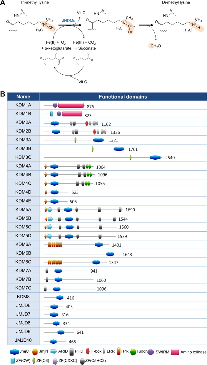
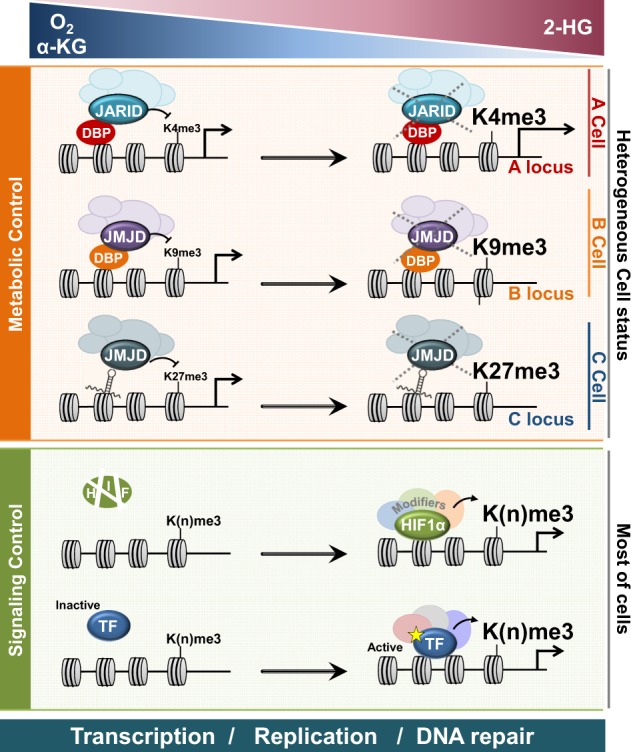
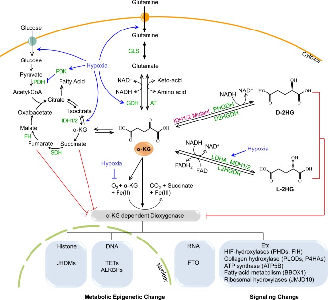
Recent studies on mutations in cancer genomes have distinguished driver mutations from passenger mutations, which occur as byproducts of cancer development. The cancer genome atlas (TCGA) project identified 299 genes and 24 pathways/biological processes that drive tumor progression (Cell 173: 371-385 e318, 2018). Of the 299 driver genes, 12 genes are involved in histones, histone methylation, and demethylation. Among these 12 genes, those encoding the histone demethylases JARID1C/KDM5C and UTX/KDM6A were identified as cancer driver genes. Furthermore, gain-of-function mutations in genes encoding metabolic enzymes, such as isocitrate dehydrogenases (IDH)1/2, drive tumor progression by producing an oncometabolite, D-2-hydroxyglutarate (D-2HG), which is a competitive inhibitor of α-ketoglutarate, O2-dependent dioxygenases such as Jumonji domain-containing histone demethylases, and DNA demethylases. Studies on oncometabolites suggest that histone demethylases mediate metabolic changes in chromatin structure. We have reviewed the most recent findings regarding cancer-specific metabolic reprogramming and the tumor-suppressive roles of JARID1C/KDM5C and UTX/KDM6A. We have also discussed mutations in other isoforms such as the JARID1A, 1B, 1D of KDM5 subfamilies and the JMJD3/KDM6B of KDM6 subfamilies, which play opposing roles in tumor progression as oncogenes or tumor suppressors depending on the cancer cell type. Genes involved in the removal of methyl groups from histones associated with DNA can promote or suppress tumor growth depending on the metabolic status of the cancer cell. Hyunsung Park and colleagues at the University of Seoul, South Korea, review current knowledge of two genes encoding histone demethylases which have been identified by The Cancer Genome Atlas (TCGA) project as cancer driver genes. Because these demethylase enzymes rely on cellular metabolites to function, their effect is influenced by metabolic conditions in the tumor microenvironment such as low oxygen. The mechanisms through which changes in histone methylation affect the expression of genes involved in tumor progression remain unknown. Further understanding of how cancer metabolism affects the modification of histones will help guide the development of more effective cancer treatments.
Overview
The claims and statistics of this paper haven’t been checked yet.
Run the check — free, no account needed. It reads the open-access text and checks the sources it cites.
Integrity signals
Registry
Cites
Statistics
Figures
Verification
Claims the paper makes
The claims and statistics of this paper haven’t been checked yet.
Run the check — free, no account needed. It reads the open-access text and checks the sources it cites.
Claims other papers make about this one
Each is checked against this paper’s own text: does it say what the citing paper says it does?
In the citing paper
“Cancer cells in tumors with IDH1 or IDH2 mutations show oncogenic activity by converting glutamine-derived α-KG to 2-hydroxyglutarate (2-HG), which competitively inhibits α-KG-dependent histone and DNA modification enzymes27.”
From Glutamine reliance in cell metabolism (2020) · cites this paper as [27]
Self-check
Percentages, sums and reported differences re-computed from the paper’s own numbers.
The self-check needs the paper’s full text.
Citations
Cites
An Integrated Genomic Analysis of Human Glioblastoma Multiforme
Parsons, D. W., Jones, S., Zhang, X., Lin, J. C.-H., Leary, R. J., Angenendt, P., Mankoo, P., Carter, H., Siu, I.-M., Gallia, G. L., et al. (2008) · Science · doi:10.1126/science.1164382 · Cited by 5,857
IDH1 and IDH2 Mutations in Gliomas
Yan, H., Parsons, D. W., Jin, G., McLendon, R., Rasheed, B. A., Yuan, W., Kos, I., Batinic-Haberle, I., Jones, S., Riggins, G. J., et al. (2009) · New England Journal of Medicine · doi:10.1056/nejmoa0808710 · Cited by 5,847
Chen, X., Xiu, M., Xing, J., Yu, S., Min, D., & Guo, F. (2017) · Cellular Physiology and Biochemistry · doi:10.1159/000479439 · Cited by 5,760
Mutational landscape and significance across 12 major cancer types
Kandoth, C., McLellan, M. D., Vandin, F., Ye, K., Niu, B., Lu, C., Xie, M., Zhang, Q., McMichael, J. F., Wyczalkowski, M. A., et al. (2013) · Nature · doi:10.1038/nature12634 · Cited by 4,419
Cancer-associated IDH1 mutations produce 2-hydroxyglutarate
Dang, L., White, D. W., Gross, S., Bennett, B. D., Bittinger, M. A., Driggers, E. M., Fantin, V. R., Jang, H. G., Jin, S., Keenan, M. C., et al. (2009) · Nature · doi:10.1038/nature08617 · Cited by 4,154
Histone demethylation mediated by the nuclear amine oxidase homolog LSD1
Shi, Y., Lan, F., Matson, C., Mulligan, P., Whetstine, J. R., Cole, P. A., Casero, R. A., & Shi, Y. (2004) · Cell · doi:10.1016/j.cell.2004.12.012 · Cited by 4,051
Targeting hypoxia in cancer therapy
Wilson, W. R., & Hay, M. P. (2011) · Nature Reviews Cancer · doi:10.1038/nrc3064 · Cited by 3,172
Comprehensive molecular characterization of urothelial bladder carcinoma
Perou, C. M. (2014) · Carolina Digital Repository (University of North Carolina at Chapel Hill) · doi:10.17615/za09-dk57 · Cited by 2,922
Xu, W., Yang, H., Liu, Y., Yang, Y., Wang, P., Kim, S.-H., Ito, S., Yang, C., Wang, P., Xiao, M.-T., et al. (2011) · Cancer Cell · doi:10.1016/j.ccr.2010.12.014 · Cited by 2,875
A Chromatin-Mediated Reversible Drug-Tolerant State in Cancer Cell Subpopulations
Sharma, S. V., Lee, D. Y., Li, B., Quinlan, M. P., Takahashi, F., Maheswaran, S., McDermott, U., Azizian, N., Zou, L., Fischbach, M. A., et al. (2010) · Cell · doi:10.1016/j.cell.2010.02.027 · Cited by 2,770
Cited by
Sorted by citations
Glutamine reliance in cell metabolism
Yoo, H. C., Yu, Y. C., Sung, Y., & Han, J. M. (2020) · Experimental & Molecular Medicine · doi:10.1038/s12276-020-00504-8 · Cited by 1,021
Enhancing the Efficacy of Glutamine Metabolism Inhibitors in Cancer Therapy
Yang, W., Qiu, Y., Stamatatos, O. T., Janowitz, T., & Lukey, M. J. (2021) · Trends in cancer · doi:10.1016/j.trecan.2021.04.003 · Cited by 308
Sunitinib resistance in renal cell carcinoma: From molecular mechanisms to predictive biomarkers
Jin, J., Xie, Y., Zhang, J.-S., Wang, J., Dai, S.-J., He, W., Li, S.-Y., Ashby, C. R., Chen, Z., & He, Q. (2023) · Drug Resistance Updates · doi:10.1016/j.drup.2023.100929 · Cited by 185
Hypoxia-Driven Effects in Cancer: Characterization, Mechanisms, and Therapeutic Implications
Shi, R., Liao, C., & Zhang, Q. H. (2021) · Cells · doi:10.3390/cells10030678 · Cited by 151
“2-HG can also affect the activity of oxygen-dependent enzymes such as JmjC histone demethylases and DNA demethylases [99].”
BackgroundShort-chain fatty acids in cancer pathogenesis
Feitelson, M. A., Arzumanyan, A. L., Medhat, A., & Spector, I. (2023) · Cancer and Metastasis Reviews · doi:10.1007/s10555-023-10117-y · Cited by 151
Immunosuppressive Property of MSCs Mediated by Cell Surface Receptors
Liu, S., Liu, F., Zhou, Y. L., Jin, B., Sun, Q., & Guo, S. (2020) · Frontiers in Immunology · doi:10.3389/fimmu.2020.01076 · Cited by 107
Jiang, Q., Qiu, Y., Kurland, I. J., Drlica, K., Subbian, S., Tyagi, S., & Shi, L. (2022) · mBio · doi:10.1128/mbio.01274-22 · Cited by 98
Role of Metabolism in Bone Development and Homeostasis
Suzuki, A., Minamide, M., Iwaya, C., Ogata, K., & Iwata, J. (2020) · International Journal of Molecular Sciences · doi:10.3390/ijms21238992 · Cited by 92
“Mutations in IDH1 also accelerate the conversion of α-ketoglutarate to D-2-hydroxyglutarate and the accumulation of D-2-hydroxyglutarate, which is known as an oncometabolite competing withα-ketoglutarate in binding to histone and DNA demethylases, leading to chromatin hypermethylation [160,161].”
BackgroundCirculating RNAs in prostate cancer patients
Mugoni, V., Ciani, Y., Nardella, C., & Demichelis, F. (2021) · Cancer Letters · doi:10.1016/j.canlet.2021.10.011 · Cited by 89
KDM6A-ARHGDIB axis blocks metastasis of bladder cancer by inhibiting Rac1
Liu, L., Cui, J., Zhao, Y., Liu, X., Chen, L., Xia, Y., Wang, Y., Chen, S., Sun, S., Shi, B., et al. (2021) · Molecular Cancer · doi:10.1186/s12943-021-01369-9 · Cited by 87
“KDM6A has also been shown to be involved in the establishment of the active histone marks H3K4 methylation and H3K27 acetylation in a demethylase activity-independent manner via recruitment of the COMPASS-like complex and histone acetyltransferase p300 [10, 33].”
BackgroundA noncoding single-nucleotide polymorphism at 8q24 drives IDH1 -mutant glioma formation
Yanchus, C., Drucker, K. L., Kollmeyer, T. M., Tsai, R., Winick‐Ng, W., Liang, M., Malik, A., Pawling, J., Lorenzo, S. B. D., Ali, A., et al. (2022) · Science · doi:10.1126/science.abj2890 · Cited by 77
Vallianatos, C. N., Farrehi, C., Friez, M. J., Burmeister, M., Keegan, C. E., & Iwase, S. (2018) · Frontiers in Molecular Neuroscience · doi:10.3389/fnmol.2018.00104 · Cited by 71
The rapidly changing landscape in mature T‐cell lymphoma (MTCL) biology and management
Marchi, E., & O’Connor, O. A. (2019) · CA A Cancer Journal for Clinicians · doi:10.3322/caac.21589 · Cited by 67
Histone demethylases in the regulation of immunity and inflammation
Qu, L., Yin, T., Zhao, Y., Lv, W., Liu, Z., Chen, C., Liu, K., Shan, S., Zhou, R., Li, X., et al. (2023) · Cell Death Discovery · doi:10.1038/s41420-023-01489-9 · Cited by 64
“Histones were the first proteins identified as lysine methylation substrates, while enzymes mediating methylation and demethylation were called histone methyltransferases (KMTs) and histone demethylases (KDMs) [7, 8].”
BackgroundRegulating Methylation at H3K27: A Trick or Treat for Cancer Cell Plasticity
Das, P., & Taube, J. H. (2020) · Cancers · doi:10.3390/cancers12102792 · Cited by 61
“Since 2-HG inhibits both the Jumanji-C domain containing histone lysine demethylases and the TET family of enzymes [293,294], both pathways for removing methylation may be impacted.”
BackgroundTricarico, R., Nicolas, É., Hall, M. J., & Golemis, E. A. (2020) · Clinical Cancer Research · doi:10.1158/1078-0432.ccr-20-1741 · Cited by 62
Hypoxia and Extracellular Acidification as Drivers of Melanoma Progression and Drug Resistance
Dratkiewicz, E., Simiczyjew, A., Mazurkiewicz, J., Ziętek, M., Matkowski, R., & Nowak, D. (2021) · Cells · doi:10.3390/cells10040862 · Cited by 65
Functions and Interactions of Mammalian KDM5 Demethylases
Pavlenko, E. A., Ruengeler, T., Engel, P., & Poepsel, S. (2022) · Frontiers in Genetics · doi:10.3389/fgene.2022.906662 · Cited by 57
Zinc and its binding proteins: essential roles and therapeutic potential
Kiouri, D. P., Chasapis, C. T., Mavromoustakos, T., Spiliopoulou, C., & Stefanidou, M. E. (2024) · Archives of Toxicology · doi:10.1007/s00204-024-03891-3 · Cited by 56
KDM6 Demethylases and Their Roles in Human Cancers
Hua, C., Chen, J., Li, S., Zhou, J., Fu, J., Sun, W., & Wang, W. (2021) · Frontiers in Oncology · doi:10.3389/fonc.2021.779918 · Cited by 57
“Glucose and glutamine respectively are transported into cells and both are converted to a-KG, which related to 2-hydroxyglutarate (2-HG) (10).”
BackgroundInfluentialLactylation modification in cancer: mechanisms, functions, and therapeutic strategies
Lv, M., Huang, Y., Chen, Y., & Ding, K. (2025) · Experimental Hematology and Oncology · doi:10.1186/s40164-025-00622-x · Cited by 54
“…modifications are involved in various tumor cell processes, including proliferation [4], invasion [5], metastasis [6], and metabolic reprogramming [7], etc. Metabolic alterations are hallmarks of cancer, including increased aerobic glycolysis, elevated glucose uptake, dysregulated glutamine…”
Significance of KDM6A mutation in bladder cancer immune escape
Chen, X., Lin, X., Pang, G., Deng, J., Xie, Q., & Zhang, Z. (2021) · BMC Cancer · doi:10.1186/s12885-021-08372-9 · Cited by 48
“Our present analysis of the somatic mutation landscape data of BC samples from the TCGA dataset (412 cases) and ICGC dataset (101 cases) confirmed that KDM6A was frequently mutated in both cohorts, in line with previous studies [6, 19].”
ResultCui, Z., Li, Y., Lin, Y., Zheng, C., Luo, L., Hu, D., Chen, Y., Xiao, Z., & Sun, Y. (2024) · Frontiers in Endocrinology · doi:10.3389/fendo.2024.1328679 · Cited by 47
Duan, X., Xing, Z., Qiao, L., Qin, S., Zhao, X., Gong, Y., & Li, X. (2024) · Frontiers in Immunology · doi:10.3389/fimmu.2024.1495221 · Cited by 48
Roles of HIF and 2-Oxoglutarate-Dependent Dioxygenases in Controlling Gene Expression in Hypoxia
Frost, J., Frost, M. G., Batie, M., Jiang, H., & Rocha, S. (2021) · Cancers · doi:10.3390/cancers13020350 · Cited by 47
“2-OGDs can be inhibited by metabolites that are accumulated in tumours, including succinate, fumarate and D/L-2-hydroxyglutarate [318,319].”
BackgroundLi, Z., Yu, B., Qi, F., & Li, F. (2021) · Frontiers in Oncology · doi:10.3389/fonc.2021.670218 · Cited by 42
“Advances in recent years, such as the identification of oncogenes and immunotherapy treatments, have provided valuable insight to guide the management of LUAD (6, 7).”
BackgroundA three layered histone epigenetics in breast cancer metastasis
Nandy, D., Rajam, S. M., & Dutta, D. (2020) · Cell & Bioscience · doi:10.1186/s13578-020-00415-1 · Cited by 40
High Risk-Human Papillomavirus in HNSCC: Present and Future Challenges for Epigenetic Therapies
Ghiani, L., & Chiocca, S. (2022) · International Journal of Molecular Sciences · doi:10.3390/ijms23073483 · Cited by 40
“Moreover, HPVmediated downregulation of KDM5C activates the expression of oncogenes such as EGFR and c-MET through the activation of cancer super-enhancers [31,90].”
BackgroundInfluentialThe molecular code of kidney cancer: A path of discovery for gene mutation and precision therapy
Xie, D., Li, G., Zheng, Z., Zhang, X., Wang, S., Jiang, B., Li, X., Wang, X., & Wu, G. (2024) · Molecular Aspects of Medicine · doi:10.1016/j.mam.2024.101335 · Cited by 41
Alterations of Chromatin Regulators in the Pathogenesis of Urinary Bladder Urothelial Carcinoma
Hoffmann, M. J., & Schulz, W. A. (2021) · Cancers · doi:10.3390/cancers13236040 · Cited by 40
Zengin, T., & Önal-Süzek, T. (2021) · Journal of Personalized Medicine · doi:10.3390/jpm11020154 · Cited by 32
“Additionally, EGFR [102], MGA [103], SMARCA4 [104], ATM [105], RBM10 [106] and KDM5C [107] which are lung cancer related genes are mutated only in LUAD but not in LUSC.”
BackgroundInfluentialHypoxia in Lung Cancer Management: A Translational Approach
Ancel, J., Perotin, J., Dewolf, M., Launois, C., Mulette, P., Nawrocki‐Raby, B., Dalstein, V., Gilles, C., Deslée, G., Polette, M., et al. (2021) · Cancers · doi:10.3390/cancers13143421 · Cited by 32
Yang, R., Zhang, G., Meng, Z., Wang, L., Li, Y., Li, H., Yan, S., Wei, X., Wang, S., & Cui, H. (2024) · Neuro-Oncology · doi:10.1093/neuonc/noae222 · Cited by 35
From the (Epi)Genome to Metabolism and Vice Versa; Examples from Hematologic Malignancy
Karagianni, P., Giannouli, S., & Voulgarelis, M. (2021) · International Journal of Molecular Sciences · doi:10.3390/ijms22126321 · Cited by 35
KDM5c Promotes Colon Cancer Cell Proliferation Through the FBXW7-c-Jun Regulatory Axis
Lin, H., Ma, N., Zhao, L., Yang, G., & Cao, B. (2020) · Frontiers in Oncology · doi:10.3389/fonc.2020.535449 · Cited by 30
Liu, C., Kang, N., Guo, Y., & Gong, P. (2021) · Frontiers in Cell and Developmental Biology · doi:10.3389/fcell.2021.710203 · Cited by 29
Qu, L.-H., Fang, Q., Yin, T., Yi, H.-M., Mei, G., Hong, Z.-Z., Qiu, X., Zhou, R., & Dong, H. (2022) · Cancer Immunology Immunotherapy · doi:10.1007/s00262-022-03167-8 · Cited by 29
“However, KDM5C has been found to have a tumor-suppressive role in renal clear cell carcinoma, pancreatic cancer, and HPV-related cancers [62].”
BackgroundThe Epigenetics of Glioma Stem Cells: A Brief Overview
Valor, L. M., & Hervás-Corpión, I. (2020) · Frontiers in Oncology · doi:10.3389/fonc.2020.602378 · Cited by 28
“Additionally, mutations in arginine 132 of the tricarboxylic acid cycle component IDH1 (or in arginine 172 of IDH2), which are associated with longer survival, induce the overproduction of the 2-hydroxybutyrate metabolite that inhibits the a-ketoglutarate-dependent activity of epigenetic enzymes such as JumonjiC histo…”
BackgroundRifayee, S. B. J. S., Chaturvedi, S. S., Warner, C., Wildey, J., White, W., Thompson, M. J., Schofield, C. J., & Christov, C. Z. (2023) · Chemistry - A European Journal · doi:10.1002/chem.202301305 · Cited by 27
Su, L., Bu, J., Yu, J., Jin, M., Meng, G., & Zhu, X. (2024) · Clinical and Translational Medicine · doi:10.1002/ctm2.70066 · Cited by 26
“In addition, histone H4 methylation (H4 M) modification is also a key epigenetic modification and is involved in the development of HCC. 26,27 It also has different patterns under the heterogeneity of HCC. Yu et al. enrolled 2305 HCC samples and performed consensus clustering analysis, then they…”
Fejzo, M. S., Chen, H., Anderson, L. A., McDermott, M. S. J., Karlan, B. Y., Konecny, G. E., & Slamon, D. J. (2020) · Gynecologic Oncology · doi:10.1016/j.ygyno.2020.11.008 · Cited by 25
Classifying Breast Cancer Molecular Subtypes by Using Deep Clustering Approach
Rohani, N., & Eslahchi, C. (2020) · Frontiers in Genetics · doi:10.3389/fgene.2020.553587 · Cited by 26
“Studies on oncometabolite have shown that the KDM 5 C is involved in cancer-related metabolic reprogramming and the tumor suppression (Chang et al., 2019).”
BackgroundCritical roles and clinical perspectives of RNA methylation in cancer
Li, G., Yao, Q., Liu, P., Zhang, H., Liu, Y., Li, S., Shi, Y., Li, Z., & Zhu, W. (2024) · MedComm · doi:10.1002/mco2.559 · Cited by 29
“332,333 Moreover, the accumulation of certain mitochondrial metabolites in the TME has been found to contribute to cancer progression.”
BackgroundDorna, D., & Paluszczak, J. (2022) · Cells · doi:10.3390/cells11061023 · Cited by 25
Chen, D., Cai, B., Zhu, Y., Ma, Y., Yu, X., Xiong, J., Shen, J., Tie, W., Zhang, Y., & Guo, F. (2024) · Clinical Epigenetics · doi:10.1186/s13148-024-01665-3 · Cited by 24
“Previous research has shown that JMJD3 and UTX have oncogenic properties in various malignancies [39, 40].”
BackgroundAntunes, M. A., Braga, C. L., Oliveira, T. B., Kitoko, J. Z., de Castro, L. L., Xisto, D. G., Coelho, M., Rocha, N. N., Silva-Aguiar, R. P., Caruso‐Neves, C., et al. (2021) · Frontiers in Cell and Developmental Biology · doi:10.3389/fcell.2021.661385 · Cited by 24
DNA Hydroxymethylation in Smoking-Associated Cancers
Nia, A. B., Caceres, A., & Tommasi, S. (2022) · International Journal of Molecular Sciences · doi:10.3390/ijms23052657 · Cited by 25
Bajinka, O., Ouédraogo, S. Y., Golubnitschaja, O., Li, N., & Zhan, X. (2024) · The EPMA Journal · doi:10.1007/s13167-024-00357-5 · Cited by 26
Chen, H., Wu, Q., Peng, L., Cao, T., Deng, M.-L., Liu, Y.-W., Huang, J., Hu, Y., Fu, N., Zhou, K.-B., et al. (2021) · Journal of Nanomaterials · doi:10.1155/2021/5164100 · Cited by 22
“IDH mutation changes the active site of the enzyme and endows the enzyme with new catalytic activity, using α-ketoglutar-ate (α-KG) to produce high levels of R-2-hydrox-yglutarate (R-2-HG); the molecular structure of 2-HG and α-KG is very similar [52], and it is speculated that 2-HG may participate in OXPHOS by inhibi…”
BackgroundDevelopment of JmjC-domain-containing histone demethylase (KDM2-7) inhibitors for cancer therapy
Zhang, L., Chen, Y., Li, Z., Lin, C., Zhang, T., & Wang, G. (2023) · Drug Discovery Today · doi:10.1016/j.drudis.2023.103519 · Cited by 22
Wang, M., Yue, S., & Yang, Z. (2023) · Scientific Reports · doi:10.1038/s41598-023-31325-0 · Cited by 23
Chopra, A., Cho, W. C. S., Willmore, W. G., & Biggar, K. K. (2020) · Frontiers in Genetics · doi:10.3389/fgene.2020.579636 · Cited by 23
“The catalytic activity of JmjC KDMs is also specifically tied to the individual affinities for molecular oxygen (Km;O2), in addition to other metabolic factors that are inherent to metabolism in the hypoxic environment (Chang et al., 2019).”
BackgroundMolecular and Metabolic Subtypes in Sporadic and Inherited Clear Cell Renal Cell Carcinoma
Czyzyk-Krzeska, M., Figueroa, J. A. L., Gulati, S., Cunningham, J. T., Meller, J., Shamsaei, B., Vemuri, B., & Plas, D. R. (2021) · Genes · doi:10.3390/genes12030388 · Cited by 23
“Other iron utilizing ketoglutarate-dependent dioxygenases include the enzyme-regulating epigenome, such as TET dioxygenases that remove the methyl group from pyrimidine bases in DNA [100], Jumonji domain containing histone lysine demethylases [101], and AlkB-type dioxygenases that are DNA repair enzymes performing oxi…”
BackgroundEpigenomic and Metabolomic Integration Reveals Dynamic Metabolic Regulation in Bladder Cancer
Loras, A., Segovia, C., & Ruiz-Cerdá, J. L. (2021) · Cancers · doi:10.3390/cancers13112719 · Cited by 23
Structures and nucleic acid-binding preferences of the eukaryotic ARID domain
Korn, S. M., & Schlundt, A. (2022) · Biological Chemistry · doi:10.1515/hsz-2021-0404 · Cited by 21
“While some members of the Arid family actively function as tumor suppressors (Bluemn et al. 2019; Chang et al. 2019; Luchini et al. 2015), overexpression of others provokes opposing effects in tumor growth [reviewed in (Lin et al. 2014)].”
BackgroundChen, L., Xu, X.-Y., Zhong, X.-D., Liu, Y., Zhu, M.-H., Tao, F., Li, C.-Y., She, Q. S., Yang, G.-J., & Chen, J. (2023) · Bioorganic Chemistry · doi:10.1016/j.bioorg.2023.106409 · Cited by 21
Guan, J., Xu, X., Qiu, G., He, C., Lu, X., Wang, K., Liu, X., Li, Y., Ling, Z., Tang, X., et al. (2023) · Journal of Experimental & Clinical Cancer Research · doi:10.1186/s13046-023-02734-w · Cited by 21
“Recent studies on metabolic reprogramming in HNSCC have provided promising potential targets in the treatment of HNSCC [36–38], demonstrating that comprehensively research on metabolic heterogeneity and relevant metabolic mechanisms is significantly demanded.”
Burkardt, D. D., Zachariou, A., Loveday, C., Allen, C. L., Amor, D. J., Ardissone, A., Banka, S., Bourgois, A., Coubes, C., Cytrynbaum, C. S., et al. (2019) · American Journal of Medical Genetics Part A · doi:10.1002/ajmg.a.61321 · Cited by 21
Becker, R., Vergarajauregui, S., Billing, F., Sharkova, M., Lippolis, E., Mamchaoui, K., Ferrazzi, F., & Engel, F. B. (2021) · eLife · doi:10.7554/elife.65672 · Cited by 22
Tian, T., Li, J., Shi, D., Zeng, Y.-P., Yu, B., Li, X., Wei, P., & Zhou, X. (2022) · Cell Death and Disease · doi:10.1038/s41419-022-05208-7 · Cited by 20
The emerging roles of histone demethylases in cancers
Tong, D., Tang, Y., & Zhong, P. (2024) · Cancer and Metastasis Reviews · doi:10.1007/s10555-023-10160-9 · Cited by 20
“Genes encoding the histone demethy-lases KDM5C and KDM6A were identified as cancer-driving genes [271].”
Luo, Y., Liu, L., Li, X., & Shi, Y. (2020) · Biomedicine & Pharmacotherapy · doi:10.1016/j.biopha.2020.110508 · Cited by 20
Role of Chromodomain-Helicase-DNA-Binding Protein 4 (CHD4) in Breast Cancer
Novillo, A., Fernández‐Santander, A., Gaibar, M., Galán, M. Á., Romero‐Lorca, A., Abdellaoui-Soussi, F. E., & Arco, P. G. (2021) · Frontiers in Oncology · doi:10.3389/fonc.2021.633233 · Cited by 21
“Indeed, in the PANCAN study, CHD4 was identified as one of the 12 most important cancer-driving genes involved in chromatin epigenetics (29, 30).”
BackgroundDai, J., Reyimu, A., Sun, A., Duoji, Z., Zhou, W., Song, L., Hu, S., Dai, W., & Xu, X. (2022) · Scientific Reports · doi:10.1038/s41598-022-11760-1 · Cited by 19
Chen, Z., Qi, Y., Shen, J., & Chen, Z. (2023) · Translational Oncology · doi:10.1016/j.tranon.2023.101778 · Cited by 19
Epigenetic modifications in early stage lung cancer: pathogenesis, biomarkers, and early diagnosis
Bi, L., Wang, X., Li, J., Li, W., & Wang, Z. (2025) · MedComm · doi:10.1002/mco2.70080 · Cited by 19
“161 Phosphorylation, that is, phosphorylation modifications can regulate the interaction of histones with other proteins and affect gene expression.”
AEBP1 Promotes Glioblastoma Progression and Activates the Classical NF-κB Pathway
Guo, K., Song, L., Chang, J.-Y., Cao, P., & Liu, Q. (2020) · Behavioural Neurology · doi:10.1155/2020/8890452 · Cited by 18
The Interplay between Dysregulated Metabolism and Epigenetics in Cancer
Bassal, M. A. (2023) · Biomolecules · doi:10.3390/biom13060944 · Cited by 19
Xiang, K., Kunin, M., Larafa, S., Busch, M. A., Dünker, N., Jendrossek, V., & Matschke, J. (2024) · Cell Death Discovery · doi:10.1038/s41420-024-01805-x · Cited by 20
“It has been well established that the aberrant production of 2-hydroxyglutarate (2-HG), succinate and fumarate can induce cancer initiation and progression, linking these oncometabolites to cellular metabolic reprogramming and perturbation of biological processes [8 – 10].”
BackgroundAudu, C. O., Wolf, S., Joshi, A., Moon, J. Y., Melvin, W. J., Sharma, S. B., Davis, F. M., Obi, A. T., Wasikowski, R., Tsoi, L. C., et al. (2024) · JCI Insight · doi:10.1172/jci.insight.172959 · Cited by 18
“Its role in inflammation has only recently been described within the context of neurological disorders, such as glioblastoma multiforme, where it was shown to be important to the hypoxia-inducible factor 1 α (HIF1 α ) inflammation axis (71, 72).”
BackgroundMetabolic heterogeneity in tumor microenvironment – A novel landmark for immunotherapy
Chen, C., Han, P., & Qing, Y. (2024) · Autoimmunity Reviews · doi:10.1016/j.autrev.2024.103579 · Cited by 17
Dynamic Tumor Analysis Using Deep Embedded Clustering (DEC) for Personalized Oncology Treatment
Mahendar, M., Manasa, P., Nimmala, S., Raghavendar, K., Sheikh, A. N. Q., & Kiran, M. A. (2025) · International Conference on Machine Learning and Autonomous Systems (ICMLAS) · doi:10.1109/icmlas64557.2025.10968766 · Cited by 17
“Notwithstanding their utility, these strategies are predominantly linear or unsupervised visualization tools and are deficient in their ability to include clustering into their frameworks [18].”
From Anti-EBV Immune Responses to the EBV Diseasome via Cross-reactivity
Kanduc, D., & Shoenfeld, Y. (2020) · Global Medical Genetics · doi:10.1055/s-0040-1715641 · Cited by 17
Extracellular Vesicles: An Emerging Regenerative Treatment for Oral Disease
He, F., Li, L., Fan, R., Wang, X., Chen, X., & Xu, Y. (2021) · Frontiers in Cell and Developmental Biology · doi:10.3389/fcell.2021.669011 · Cited by 17
Wang, Y., Ye, H., Yang, Y., Li, J., Cen, A., & Zhao, L. (2021) · Journal of Endocrinological Investigation · doi:10.1007/s40618-021-01606-4 · Cited by 16
Luo, S., Wang, W., Feng, J., & Li, R. (2021) · Journal of Immunology Research · doi:10.1155/2021/5975893 · Cited by 16
Epigenetic modifications in esophageal cancer: An evolving biomarker
Liu, W.-J., Zhao, Y., Chen, X., Miao, M.-L., & Zhang, R.-Q. (2023) · Frontiers in Genetics · doi:10.3389/fgene.2022.1087479 · Cited by 16
“In addition, aberrant histone modifications often accompany and interact with DNA methylation, further complicating the mechanisms of gene expression in tumour cells (Chang et al., 2019).”
BackgroundHayakawa, K., Nishitani, K., & Tanaka, S. (2019) · Scientific Reports · doi:10.1038/s41598-019-56341-x · Cited by 16
Li, N., Zhao, Z., Liu, P., Zheng, Y., Cai, S., Sun, Y., & Wang, B. (2020) · Molecular Therapy — Oncolytics · doi:10.1016/j.omto.2020.12.008 · Cited by 16
“In the context of several controversies in oncological research, JMJD3 plays contradictory roles in both tumor suppression and tumorigenesis.(32) JMJD3 was shown to serve as a potential tumor suppressor in different types of cancers.”
BackgroundGASC1 promotes glioma progression by enhancing NOTCH1 signaling
Xiao, Z., Yang, X., Liu, Z., Shao, Z., Song, C., Zhang, K., Wang, X., & Li, Z. (2021) · Molecular Medicine Reports · doi:10.3892/mmr.2021.11949 · Cited by 16
“This finding was in line with previous studies showing that >50% of gliomas exhibit a higher GASC1 expression (30,57,58).”
ResultDeciphering the multifaceted roles and clinical implications of 2-hydroxyglutarate in cancer
Hao, J., Huang, Z., Zhang, S., Song, K., Wang, J., Gao, C., Fang, Z., & Zhang, N. (2024) · Pharmacological Research · doi:10.1016/j.phrs.2024.107437 · Cited by 15
Peripheral T-cell lymphoma: From biology to practice to the future
O’Connor, O. A., Ma, H., Chan, J. Y., Kim, S. J., Yoon, S. E., & Kim, W. S. (2024) · Cancer Treatment Reviews · doi:10.1016/j.ctrv.2024.102793 · Cited by 15
Liu, X., Li, H., Yuan, M., Wan, J., Guo, J., Dong, X., Jin, X., Yang, C., & Wang, G. (2025) · Cancer Letters · doi:10.1016/j.canlet.2025.217972 · Cited by 14
Liu, K.-L., Yin, Y.-W., Lu, B., Niu, Y., Wang, D., Shi, B., Zhang, H., Guo, P., Yang, Z., & Li, W. (2022) · Cancer Cell International · doi:10.1186/s12935-022-02475-4 · Cited by 14
“Studies have shown that abnormal expression of KDM5C is closely related to the occurrence and development of various cancers [18, 19].”
BackgroundHistone methylation and acetylation in cancer: mechanism, progression, and targets
Bu, S., Ye, T., Gao, H., Song, H., & Zhu, Y. (2024) · ONCOLOGIE · doi:10.1515/oncologie-2024-0324 · Cited by 14
KDM6B-Mediated HADHA Demethylation/Lactylation Regulates Cementogenesis
Yang, Z., Wang, H., Xiao, J., Yang, Q., Sun, J., Liu, H., Ma, L., Huang, X., Wang, C., Wang, X., et al. (2024) · Journal of Dental Research · doi:10.1177/00220345241286460 · Cited by 14
“In addition to epigenetic regulation, there exists significant crosstalk between histone modifications and bioenergetic metabolism, which has emerged as a promising therapeutic target (Chang et al. 2019).”
Thonsri, U., Wongkham, S., Wongkham, C., Hino, S., Nakao, M., Roytrakul, S., Koga, T., & Seubwai, W. (2020) · Cancer Science · doi:10.1111/cas.14719 · Cited by 13
Mutation pattern and genotype-phenotype correlations of SETD2 in neurodevelopmental disorders
Chen, M., Quan, Y., Duan, G., Wu, H., Bai, T., Wang, Y., Zhou, S., Ou, J., Shen, Y., Hu, Z., et al. (2021) · European Journal of Medical Genetics · doi:10.1016/j.ejmg.2021.104200 · Cited by 14
Breast cancer: Muscarinic receptors as new targets for tumor therapy
Español, A. J., Salem, A. R., Sánchez, Y., & Sales, M. E. (2021) · World Journal of Clinical Oncology · doi:10.5306/wjco.v12.i6.404 · Cited by 14
Gu, H.-Z., Lin, R., Zheng, F., & ZHANG, Q. (2020) · Journal of Molecular Histology · doi:10.1007/s10735-020-09921-5 · Cited by 14
Fan, M., Shi, H., Yao, H., Wang, W., Zhang, Y., Jiang, C., & Lin, R. (2022) · Stem Cell Research & Therapy · doi:10.1186/s13287-022-02936-7 · Cited by 13
“D-2-hydroxyglutarate (D-2HG), a competitive inhibitor of α-KG, can be used as an inhibitor of histone demethylation [26].”
BackgroundLiu, G., Jia, G., Ren, Y., Yin, C., Xiao, X., Wu, H., Liu, J., & Chen, M. (2025) · Cell Biology and Toxicology · doi:10.1007/s10565-025-10021-x · Cited by 13
“Histone is the first protein identified as a lysine-methylated substrate (Chang et al. 2019; Wu et al. 2022).”
Lysine demethylase 6 (KDM6): A promising therapeutic target in autoimmune disorders and cancer
Nastaranpour, M., Damara, A., Grabbe, S., & Shahneh, F. Z. (2025) · Biomedicine & Pharmacotherapy · doi:10.1016/j.biopha.2025.118254 · Cited by 13
Hoekstra, M., & Biggar, K. K. (2021) · Analytical Biochemistry · doi:10.1016/j.ab.2021.114429 · Cited by 15
IDH1 R132C and ERC2 L309I Mutations Contribute to the Development of Maffucci’s Syndrome
Cheng, P., Chen, K., Zhang, S., Mu, K., Liang, S., & Zhang, Y. (2021) · Frontiers in Endocrinology · doi:10.3389/fendo.2021.763349 · Cited by 13
“The above somatic mutations render IDH1 or IDH2 unable to convert isocitrate to a-ketoglutarate but promote D-2hydroxyglutarate accumulation, the levels of which strongly correlate with tumorigenesis (13, 20, 21).”
BackgroundEvolving therapeutic proteins to precisely kill cancer cells
Zaman, R., Islam, R. A., & Chowdhury, E. H. (2022) · Journal of Controlled Release · doi:10.1016/j.jconrel.2022.09.066 · Cited by 13
Maietta, I., González‐Fernández, Á., & Simón‐Vázquez, R. (2025) · Advanced Drug Delivery Reviews · doi:10.1016/j.addr.2025.115638 · Cited by 13
Edrei, Y., Levy, R., Kaye, D., Marom, A., Radlwimmer, B., & Hellman, A. (2023) · Genome biology · doi:10.1186/s13059-023-03094-6 · Cited by 12
Cui, X., Hou, L., Yan, B., Liu, J., Zhang, C., Sui, P., Tong, S., Luchsinger, L. L., Mendelson, A., Zhou, D., et al. (2025) · Journal of Clinical Investigation · doi:10.1172/jci182125 · Cited by 13
Fazazi, M. R., Ruda, G. F., Brennan, P. E., & Rangachari, M. (2023) · Brain Research Bulletin · doi:10.1016/j.brainresbull.2023.110748 · Cited by 13
“Dysregulation of KDM5C, KDM5D and KDM6A are associated with sex-dependent malignancies such as prostate, breast and pancreatic cancer (Chang et al., 2019; Duan et al., 2022; Varghese et al., 2021).”
Background
Metadata
- Authors
- Chang, S., Yim, S., & Park, H.
- Affiliations
- University of Seoul (KR)
- Corresponding author
- Park, Hyunsung
- Venue
- Experimental & Molecular Medicine
- Published
- 2019-06
- Volume
- 51
- Issue
- 6
- Pages
- 1-17
- ISSN
- 1226-3613 (ISSN-L)
- 2092-6413
- PMC
- PMC6586683
- OpenAlex
- W2950853065
- Cited by
- 228
- References
- 147
- Citations by year
- 2026: 8 · 2025: 31 · 2024: 41 · 2023: 37 · 2022: 32 · 2021: 50 · 2020: 24 · 2019: 4
- FWCI
- 2.46
- Type
- journal article
- Language
- en
- Open access
- gold
- doi.org
- Licence
- Locations
- Experimental & Molecular Medicine (publishedVersion, cc-by)
- PubMed Central (publishedVersion, cc-by)
- PubMed (publishedVersion)
- DOAJ (DOAJ: Directory of Open Access Journals) (submittedVersion, cc-by-sa)
- Funders
- National Research Foundation of Korea (2016R1A2B4012840, 2018R1A4A1025985)
- Topics
- Life Sciences › Biochemistry, Genetics and Molecular Biology › Molecular Biology › Epigenetics and DNA Methylation
- Life Sciences › Biochemistry, Genetics and Molecular Biology › Molecular Biology › Chromatin Remodeling and Cancer
- Life Sciences › Biochemistry, Genetics and Molecular Biology › Cancer Research › Cancer, Hypoxia, and Metabolism
- Keywords
- histone demethylation, histone methylation, cancer driver genes, cancer metabolism, D-2-hydroxyglutarate, oncometabolites, tumor suppressor, JARID1A, JARID1B, chromatin
- MeSH terms
- Tumor Hypoxia, Nucleophosmin, Animals, Histones, Humans, Isocitrate Dehydrogenase, Neoplasms, Gene Expression Regulation, Neoplastic, Histone Demethylases
About these checks
Registry, retraction, Cites and statistics results are computed, not judged, with one exception: a printed reference that no index lists is read once by a model before it counts as a citation error. Claims are extracted by a model and kept only when their quote appears verbatim in the text; each cited source is then checked, with a model’s help, for whether it says what the claim relies on. Those verdicts can be wrong — read the claim’s quote and the cited source before relying on one.
Statistics and arithmetic are recomputed, not judged.
Confidence bands are shown only for fields calibrated on our annotated test set. None is calibrated yet, so no band is shown.
What counts as a claim ›How we measure confidence ›How we check this ›
Are you an author, or do you see a problem with this page? Request a review
