

The DOI matches its Crossref record; 271 of the 272 references it cites were found in a registry or an index.
Recent advances in the development of protein–protein interactions modulators: mechanisms and clinical trials
Open accessAbstract
Protein–protein interactions (PPIs) have pivotal roles in life processes. The studies showed that aberrant PPIs are associated with various diseases, including cancer, infectious diseases, and neurodegenerative diseases. Therefore, targeting PPIs is a direction in treating diseases and an essential strategy for the development of new drugs. In the past few decades, the modulation of PPIs has been recognized as one of the most challenging drug discovery tasks. In recent years, some PPIs modulators have entered clinical studies, some of which been approved for marketing, indicating that the modulators targeting PPIs have broad prospects. Here, we summarize the recent advances in PPIs modulators, including small molecules, peptides, and antibodies, hoping to provide some guidance to the design of novel drugs targeting PPIs in the future.
Overview
The claims and statistics of this paper haven’t been checked yet.
Run the check — free, no account needed. It reads the open-access text and checks the sources it cites.
Integrity signals
Registry
Cites
Statistics
Figures
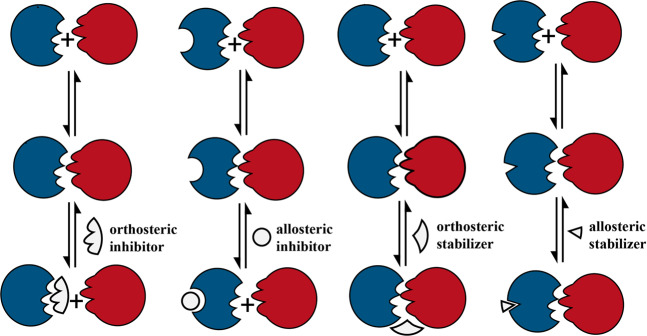
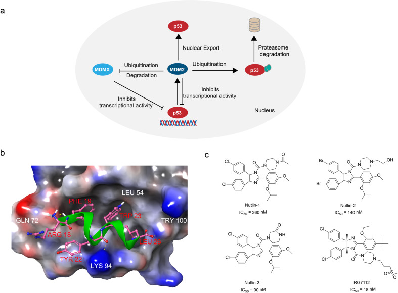
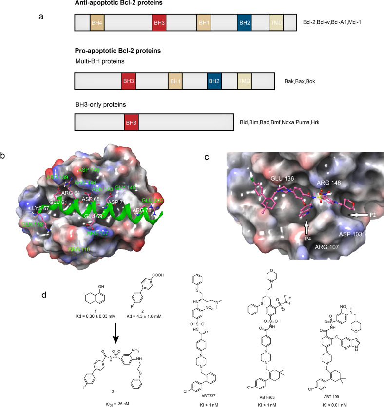
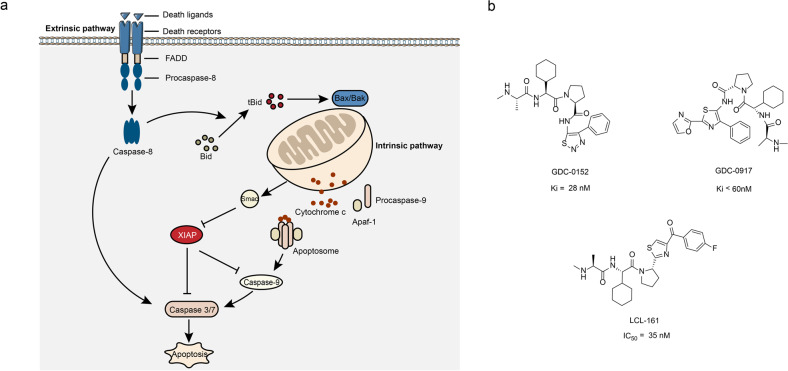
Verification
Claims the paper makes
The claims and statistics of this paper haven’t been checked yet.
Run the check — free, no account needed. It reads the open-access text and checks the sources it cites.
Claims other papers make about this one
Each is checked against this paper’s own text: does it say what the citing paper says it does?
In the citing paper
“However, peptides and proteins have general physicochemical properties, such as instability under physiological conditions, poor oral bioavailability and solubility, which make them less optimal drugs.19”
From Recent advances in targeting the “undruggable” proteins: from drug discovery to clinical trials (2023) · cites this paper as [19]
In the citing paper
“Among them, YAP/TAZ and β-catenin belong to TFs have traditionally been considered “undruggable” targets.”
From Recent advances in targeting the “undruggable” proteins: from drug discovery to clinical trials (2023) · cites this paper as [19]
In the citing paper
“There are several other undruggable proteins can be regulated by PPI modulation, such as YAP/TAZ, β-catenin, XIAP, etc.19”
From Recent advances in targeting the “undruggable” proteins: from drug discovery to clinical trials (2023) · cites this paper as [19]
Self-check
Percentages, sums and reported differences re-computed from the paper’s own numbers.
The self-check needs the paper’s full text.
Citations
Cites
PD-1 and PD-L1 Immune Checkpoint Blockade to Treat Breast Cancer
Hartkopf, A. D., Taran, F., Wallwiener, M., Walter, C. B., Krämer, B., Grischke, E., & Brucker, S. Y. (2016) · Breast Care · doi:10.1159/000453569 · Cited by 30,989
Lipinski, C. A., Lombardo, F., Dominy, B. W., & Feeney, P. J. (2001) · Advanced Drug Delivery Reviews · doi:10.1016/s0169-409x(00)00129-0 · Cited by 18,139
Adverse Renal Effects of Immune Checkpoint Inhibitors: A Narrative Review
Wanchoo, R., Karam, S., Uppal, N. N., Barta, V. S., Deray, G., Devoe, C., Launay‐Vacher, V., Jhaveri, K. D., & on behalf of Cancer and Kidney International Network Workgroup on Immune Checkpoint Inhibitors (2017) · American Journal of Nephrology · doi:10.1159/000455014 · Cited by 15,320
The blockade of immune checkpoints in cancer immunotherapy
Pardoll, D. M. (2012) · Nature Reviews Cancer · doi:10.1038/nrc3239 · Cited by 13,838
Lipinski, C. A., Lombardo, F., Dominy, B. W., & Feeney, P. J. (1997) · Advanced Drug Delivery Reviews · doi:10.1016/s0169-409x(96)00423-1 · Cited by 11,484
Pembrolizumab versus Chemotherapy for PD-L1-Positive Non-Small-Cell Lung Cancer
Reck, M., Rodríguez-Abreu, D., Robinson, A. G., Hui, R., Csőszi, T., Fülöp, A., Gottfried, M., Peled, N., Tafreshi, A., Cuffe, S., et al. (2016) · New England Journal of Medicine · doi:10.1056/nejmoa1606774 · Cited by 10,089
Nivolumab versus docetaxel in advanced nonsquamous non-small-cell lung cancer
Borghaei, H., Paz-Ares, L., Horn, L., Spigel, D. R., Steins, M., Ready, N. E., Chow, L. Q., Vokes, E. E., Felip, E., Holgado, E., et al. (2015) · New England Journal of Medicine · doi:10.1056/nejmoa1507643 · Cited by 9,335
Pembrolizumab for the Treatment of Non–Small-Cell Lung Cancer
Garon, E. B., Rizvi, N. A., Hui, R., Leighl, N., Balmanoukian, A. S., Eder, J. P., Patnaik, A., Aggarwal, C., Gubens, M., Horn, L., et al. (2015) · New England Journal of Medicine · doi:10.1056/nejmoa1501824 · Cited by 6,198
Nivolumab for Recurrent Squamous-Cell Carcinoma of the Head and Neck
Ferris, R. L., Blumenschein, G., Fayette, J., Guigay, J., Colevas, A. D., Licitra, L., Harrington, K., Kasper, S., Vokes, E. E., Even, C., et al. (2016) · New England Journal of Medicine · doi:10.1056/nejmoa1602252 · Cited by 5,101
In Vivo Activation of the p53 Pathway by Small-Molecule Antagonists of MDM2
Vassilev, L. T., Vu, B. T., Graves, B., Carvajal, D., Podlaski, F., Filipovic, Z., Kong, N., Kammlott, U., Lukacs, C., Klein, C., et al. (2004) · Science · doi:10.1126/science.1092472 · Cited by 4,661
Research works printed but not linked: 3
- Ferrari, S., Pellati, F. & Costi, M. P. Disruption of Protein-Protein Interfaces (Springer, Berlin, 2013).
- Chen, L. et al. P53 alpha-helix mimetics antagonize p53/MDM2 interaction and activate p53. Mol. Cancer Therap. 4, 1019–1025 (2005). · DOI
- Li, X., Liu, R. & Fang, H. Bcl-2: research progress from target to launched drug. Acta Pharma. Sin. 53, 509–517 (2018).
Cited by
Sorted by citations
Items 801–899 of 899
Martinez, J. L. T., Loreto, K. R. R., Frio, E. J. L., & Endriga, M. A. (2025) · IEEE International Conference on Bioinformatics and Biomedicine (BIBM) · doi:10.1109/bibm66473.2025.11356373
Santos-Fernandez, M., Fouque, K. J. D., Pita, S. S. D. R., Chapagain, P. P., Leng, F., & Fernandez-Lima, F. A. (2026) · Journal of the American Society for Mass Spectrometry · doi:10.1021/jasms.5c00357
Protein-protein interaction inhibitors in cancer
Hardcastle, I. R., Goujon-Ricci, J., & Thompson, W. I. (2026) · Elsevier eBooks · doi:10.1016/b978-0-443-29808-0.00067-4
Neelima, N., Elrewey, H., Sneha, S., Ruiz, A., Wahajuddin, M., Joshi, G., & Jha, V. (2026) · Bioorganic & Medicinal Chemistry · doi:10.1016/j.bmc.2026.118587
Dynamics of large protein systems by NMR
Vasconcelos, A. A., Bezerra, P. R., de Araújo, G. R., & Almeida, F. C. L. (2026) · Elsevier eBooks · doi:10.1016/b978-0-323-99507-8.00060-3
Wang, Z., Wang, L., Li, Y., You, Z., & Li, Y. (2026) · iScience · doi:10.1016/j.isci.2026.115100
Inspired by ion-specific effects: Repurposing metformin to tackle protein crystallopathy
Zhang, W., Li, S., Wang, Y., Wang, X., Mo, S., Xu, L., Jian, Z., 刘树立, Liu, Z., Yang, Y., et al. (2026) · Nano Today · doi:10.1016/j.nantod.2026.103017
A Pretraining-Based Deep Learning Framework for Protein Mutation Effect Prediction
Zhang, P., Deng, J., Gu, M., & Liu, M. (2025) · International Conference on Algorithms, Computing and Artificial Intelligence (ACAI) · doi:10.1109/acai68217.2025.11406652
Mechanism of AF9 (MLLT3)–Partner Dissociation in Mixed-Lineage Leukemia-Rearranged Leukemia
Sharma, S., Joshi, K., & Saha, A. (2026) · JACS Au · doi:10.1021/jacsau.5c01454
Wei, Z., Wei, L., Hu, Y. A., Chen, P., Chen, Y., Wang, K., Weng, J., Xing, B., Ming, K., Wu, Z., et al. (2026) · Research Square · doi:10.21203/rs.3.rs-8525913/v1
Shanizal, M. R. P. P. (2026) · Research Square · doi:10.21203/rs.3.rs-8841071/v1
Shkedi, A., Grabinska-Rogala, A., Hernandez, J., Dutkiewicz, R., Marszałek, J., & Gestwicki, J. E. (2026) · Chemical Biology & Drug Design · doi:10.1111/cbdd.70281
Chinn, A. J., Quinn, T. R., Boerth, J. A., Kohara, K., & Bommakanti, G. (2026) · Elsevier eBooks · doi:10.1016/b978-0-443-29808-0.00094-7
Structure-Based Virtual Screening of New Skp1 Inhibitor Chemotypes Targeting F-Box Binding Interface
Hussain, M., Lu, Y., Wang, G.-Z., Tariq, M., Zhang, J., Zhou, G., & Liu, J. (2026) · Drug Design Development and Therapy · doi:10.2147/dddt.s578211
Lanthipeptide structure prediction and design with Rosetta
Tydings, C. W., Moretti, R., Meiler, J., & Walker, A. S. (2026) · Methods in enzymology on CD-ROM/Methods in enzymology · doi:10.1016/bs.mie.2026.01.001
TINA: a geometry-aware post-hoc scoring framework for protein-protein interaction
Jiang, L., Li, Y.-R., Su, Q., Cheng, S., Shen, L.-J., Zhang, K., Zhu, K., Zhang, H., Shen, C., Kang, Y., et al. (2026) · Acta Pharmacologica Sinica · doi:10.1038/s41401-026-01937-z
A yeast-based assay to screen protein-protein interaction modulators
Sudevan, A. (2021) · theses.fr (ABES) · doi:10.70675/9ab04dcaz498fz43ebz8762z012f3f86154c
Discovery of SHANK1-PDZ Peptide-Fragment Inhibitors Using a Dynamic Ligation Screening Strategy
Li, Y., Giménez, D., Warriner, S. L., & Wilson, A. J. (2026) · Biochemistry · doi:10.1021/acs.biochem.5c00804
Khodjoyan, S. (2023) · theses.fr (ABES) · doi:10.70675/8a9b378cz83a3z42f8za20bzdd25e08822b5
A nanobody based approach to capture interactomes of dimeric protein complexes in living cells
Sleiman, N. H. (2024) · theses.fr (ABES) · doi:10.70675/633151e1z9a1cz42b0zbcbaz776177601937
Structure-based design of antiviral drugs against respiratory viruses using in silico approaches
Ruano, L. C. (2024) · theses.fr (ABES) · doi:10.70675/434de13dz7ff8z46beza24ezfaf0740f7dea
Nouchikian, L. (2023) · theses.fr (ABES) · doi:10.70675/ae1b34afz3360z4758zb095z89c58db2a12f
Deka, H., Abdelghani, H. T. M., Gangopadhyay, A., Pawar, A. D., Patil, P. C., & Bhowmick, S. (2026) · 3 Biotech · doi:10.1007/s13205-026-04777-0
Nielipinska, D., Orlikowska, M., Nielipinski, M., Sekula, B., Błażewska, K. M., Gendaszewska‐Darmach, E., & Pietrzyk‐Brzezinska, A. J. (2026) · Protein Science · doi:10.1002/pro.70581
Bogár, F., Quintero, M. R., Horváth, J., Násztor, Z., Mero, M., Krekic, S., Veber, A. A., Puškar, L., Dér, A., & Heiner, Z. (2026) · Biomacromolecules · doi:10.1021/acs.biomac.5c02743
Protein–Protein Interaction Stabilizers from MD Simulation-Derived Pharmacophores
Ibrahim, M., & Zacharias, M. (2026) · Journal of Chemical Information and Modeling · doi:10.1021/acs.jcim.6c00290
A systematic evaluation of network subsampling methods for T-cell receptor repertoire network
Wu, H., Le, T., Yang, H., Le, P., Oh, D. Y., & Zhang, L. (2026) · Statistics Innovation · doi:10.48130/stati-0026-0005
Transcription Factors in the Pathogenesis of Schizophrenia
Helaly, A. M., & Al‐Emam, A. (2026) · Life · doi:10.3390/life16050773
Kita, M., & Kigoshi, H. (2026) · Bulletin of the Chemical Society of Japan · doi:10.1093/bulcsj/uoag053
Florentino, B. R., Bonidia, R. P., & de Carvalho, A. C. P. L. F. (2026) · Briefings in Bioinformatics · doi:10.1093/bib/bbag226
Functional flexibility in bacterial hub proteins is driven by proteome expansion
Paradžik, M., Prekpalaj, S., & Paradžik, T. (2026) · Scientific Reports · doi:10.1038/s41598-026-52986-7
Choi, J.-T., Yu, S.-R., Oh, J., Jang, Y.-B., Lee, Y., Cha, H., Won, D., Kim, D., Yu, S., Yu, S., et al. (2026) · bioRxiv (Cold Spring Harbor Laboratory) · doi:10.64898/2026.05.15.725558
Song, X., Tang, K., Gao, J., Liu, X., Jiang, Q., & Ma, L. (2026) · European Journal of Pharmacology · doi:10.1016/j.ejphar.2026.178983
Zerihun, M., Chakraborty, S., Abita, A., Dey, S., Lopez, I., Shlomo, R. W., Sridhar, M. K. C., Davis, L., Ertracht, O., Atar, S., et al. (2026) · Journal of Medicinal Chemistry · doi:10.1021/acs.jmedchem.6c00333
Split-Indigoidine synthetase as optical reporter for benchmarking protein-protein interactionsPreprint
Gonschorek, P., Schelhas, C., Flakowski, M., Schenk, L., Podolski, A., & Bode, H. B. (2026) · bioRxiv (Cold Spring Harbor Laboratory) · doi:10.64898/2026.06.03.729908
Huang, X., Lin, J., & Zhang, Y. (2026) · Neurochemistry International · doi:10.1016/j.neuint.2026.106202
Bahn, Y., Choi, J.-T., Yu, S.-R., Oh, J., Jang, Y.-B., Lee, Y., Cha, H., Won, D., Kim, D., Yu, S., et al. (2026) · Research Square · doi:10.21203/rs.3.rs-9899291/v1
Sequence-based protein binding site prediction using a multi-channel graph neural network
Zhang, Z., Fan, Y., Hang, H., Pang, S., Ma, T., Jiang, M., Zhou, W., & Zhang, Y. (2026) · Neurocomputing · doi:10.1016/j.neucom.2026.134369
Pep2Mol: 3D Molecule Generation Targeting Protein-Protein Interfaces with Diffusion ModelsPreprint
Yue, R., Yang, Z., Seabra, G. D. M., Li, C., & Li, Y. (2026) · bioRxiv (Cold Spring Harbor Laboratory) · doi:10.64898/2026.06.28.734975
Bicyclo[1.1.1]pentane as a Phenyl Isosteric Replacement
Saar, A., Kombo, D. C., Thurairatnam, S., Kothe, M., Merriman, G. H., Tsymbal, A. V., Kane, J. L. J., LaMarche, M. J., Gkeka, P., & Li, Y. (2026) · ACS Medicinal Chemistry Letters · doi:10.1021/acsmedchemlett.6c00246
O’Leary, K. M., Ślężak, T., Le, D. A., & Kossiakoff, A. A. (2026) · Journal of Molecular Biology · doi:10.1016/j.jmb.2026.169953
Lu, S., Li, Y., Tian, Z., Nan, X., Zhou, Q., & Zhang, S. (2026) · Computational and Structural Biotechnology Journal · doi:10.34133/csbj.0200
Self-citationDai, Y., Wu, H., Shen, Y., Yang, F., Liu, X., & Zhuang, T. (2026) · Journal of Enzyme Inhibition and Medicinal Chemistry · doi:10.1080/14756366.2026.2706119
Protein–Protein Interactions: Emerging Drug Targets
Silverman, R. B., Xue, F., & Juncosa, J. I. (2026) · Elsevier eBooks · doi:10.1016/b978-0-443-15862-9.00010-2
Learning Protein–Protein Binding Free Energies from Interface Graphs and Physicochemical Descriptors
Zhao, Q.-J., & Saha, A. (2026) · ACS Physical Chemistry Au · doi:10.1021/acsphyschemau.6c00060
PANCS-Inhibitors: A rapid method to directly select for protein-protein interaction inhibitorsPreprint
Styles, M. J., Xie, V. C., Legault, S., Pixley, J. A., & Dickinson, B. C. (2026) · bioRxiv (Cold Spring Harbor Laboratory) · doi:10.64898/2026.08.05.743105
Protein–protein interaction inhibitors in the human proteome: lessons from 117 targeted interactions
Hyde, E., & Beekman, A. M. (2026) · RSC Medicinal Chemistry · doi:10.1039/d6md00552g
Advances in Small-Molecule Inhibitors for Targeted Cancer Therapy
Husain, A., Singh, N. K., Ahmad, F., Pandey, R. K., & Bala, L. (2026) · Springer eBooks · doi:10.1007/978-981-92-0477-9_6
Synthetic Protein Mimetic Based Therapies for Neurodegeneration
Stillman, N. H. (2025) · Digital Commons - DU (University of Denver) · doi:10.56902/etdcrp.2025.171
Untangling the Complexity of Binding Across Biomolecular Interfaces with Interaction Network Models
Sládek, V., & Fedorov, D. G. (2026) · Methods in molecular biology · doi:10.1007/978-1-0716-5420-0_9
Protein–Protein Interactions with the Fragment Molecular Orbital Method
Woods, D. C., Monteleone, S., & Labouille, S. (2026) · Methods in molecular biology · doi:10.1007/978-1-0716-5420-0_5
Chemocarrier-Enabled Peptide Inhibition of MYC-Cofactor Interactions in Plants
Liu, R., Uehara, Y., Mino, K., Idei, A., Kimoto, K., Fujiwara, D., Yoshida, M., Ueda, M., & Takaoka, Y. (2026) · ACS Chemical Biology · doi:10.1021/acschembio.6c00523
Targeting Aberrant Protein-Protein Interactions in Cancer and Neurodegenerative Diseases
Ball, T. D. (2025) · Digital Commons - DU (University of Denver) · doi:10.56902/etdcrp.2025.222
HighMorph: De Novo Cyclic Peptide Sequence Design via Protein–Protein Interaction Recapitulation
Lan, M., Zhang, C., Wang, W., Haomeng, H., Lin, H., Cao, S., Guo, J., & Duan, H. (2026) · Journal of Medicinal Chemistry · doi:10.1021/acs.jmedchem.6c01926
Artificial Synthetic Protein Mimetics As Pathology-Targeted Therapeutics
Dohoney, R. A. (2026) · Digital Commons - DU (University of Denver) · doi:10.56902/etdcrp.2026.62
SNOOPPI: a sequence-normalized database of on- and off-target protein–protein interactions
Vincoff, S., & Chatterjee, P. (2026) · Bioinformatics · doi:10.1093/bioinformatics/btag736
Roberts, S. K., Clarke, D., Galdadas, I., & Martin-Fernandez, M. L. (2026) · Philosophical Transactions of the Royal Society B Biological Sciences · doi:10.1098/rstb.2024.0513
Cellular Regenerative and Gene Therapy for Male Sexual Dysfunction: A Reality?
Truong, M. N., Alipour, H., & Taher, A. (2026) · Springer eBooks · doi:10.1007/978-3-032-29112-7_17
Discovery of Cbl-b Targeting Cyclic Peptide Ligands by Phage Display
Yang, X.-Q., 卫明, Zhai, W., Huang, R., Li, Z.-H., Li, K., Bai, L., & Lei, X. (2026) · Organic Letters · doi:10.1021/acs.orglett.6c03654
Macrocyclization of native peptides through (thio)urea crosslinking of two amines
Liu, J., Chen, P., Tang, M., Chen, Q., Liao, Y., Liu, Y., Ullah, S., Zhao, J., Long, Y., Chen, G., et al. (2026) · Nature Communications · doi:10.1038/s41467-026-77301-w
Tian, Z., Fu, L., Yan, X., Chen, Y., Yang, W., Chen, Y., Liu, H., Cai, Y., & Dai, S. (2026) · European Journal of Medicinal Chemistry · doi:10.1016/j.ejmech.2026.119296
Synthetic Targeted Peptides in Therapy
Shiryaeva, O. S., Smirnov, E. A., Znamensky, V. A., Murashko, E. A., Kolobov, A. A., Бондаренко, А. Б., Korolev, D. V., Galagudza, M. M., & Cheburkin, Y. V. (2026) · Nanobiotechnology Reports · doi:10.1134/s2635167626602366
Hossain, M. M., Singdha, T. R., Sakib, S. I., Adhikari, J., Choudhury, P., Miya, M. S., Sarker, A. K., Das, M., & Shibly, A. Z. (2026) · Bioinformatics and Biology Insights · doi:10.1177/11779322261489770
Grignard Reagents Unlock Three-Dimensional Nitrogen Heterocycles via Single-Carbon-Atom Insertion
Koronatov, A. N., Sakharov, P. А., Kaushansky, A., Fridman, N., Schreiner, P. R., & Gandelman, M. (2026) · Journal of the American Chemical Society · doi:10.1021/jacs.6c08636
Conformationally restrained de novo peptides to modulate Skp1–Skp2 interaction of the SCF E3 ligase
Tolani, S., Tripathi, N. M., Antil, H., Ghosh, S., Mitra, D., Banerjee, A., Kumar, S., Bandyopadhyay, A., & Kumar, A. (2026) · RSC Medicinal Chemistry · doi:10.1039/d6md00660d
C-RAF Sequence-Encoded Cooperativity Hotspots for 14-3-3/C-RAF Molecular Glues
Pennings, M. A. M., Lauffer, G.-A. A. M., Konstantinidou, M., de Vries, R. M. J. M., Ottmann, C., Arkin, M. R., & Brunsveld, L. (2026) · Journal of Medicinal Chemistry · doi:10.1021/acs.jmedchem.6c02049
Targeting the IL-17A Cytokine Interface: Structural Insights and Small-Molecule Modulators
Kim, G. U., Lee, A., Lee, S., Jeon, B., Park, H., Kang, T., & Choo, H. (2026) · Journal of Medicinal Chemistry · doi:10.1021/acs.jmedchem.6c01296
Protein–Protein Interaction-Guided Peptides Interfere with Early Zika Virus Entry-Associated Events
Rodríguez-Salazar, C. A., Recalde-Reyes, D. P., Linares-Jiménez, S., Lopez-Cardoso, C., & Giraldo, M. I. (2026) · Viruses · doi:10.3390/v18101066
Progress and challenges in traceless ligand-directed labelling chemistry
Hopkinson, T., Cox, L. R., & Wilson, A. J. (2026) · Communications Chemistry · doi:10.1038/s42004-026-02222-0
Chen, S., Solazzo, R., Fouché, M., ROTH, H. ‐., Dittrich, B., & Riniker, S. (2026) · Journal of the American Chemical Society · doi:10.1021/jacs.6c12127
Kim, J., Ha, T., Park, S., Kang, D., Lee, S., Lee, S., Oh, S., Chung, M., Chi, S., Kim, S. J., et al. (2026) · Biosensors and Bioelectronics · doi:10.1016/j.bios.2026.119295
Mao, S., Meng, S., & Xiang, Y. (2026) · iScience · doi:10.1016/j.isci.2026.117693
Development of a Lactam-Bridged Peptide as Interleukin-11 Antagonist for Renal Fibrosis Treatment
Yang, X., Zhao, N., Zheng, R., Li, J., Wu, Y., Ma, Z., Pang, N., Meng, X., Liu, R., Xiong, X., et al. (2026) · ACS Medicinal Chemistry Letters · doi:10.1021/acsmedchemlett.6c00034
Protein interactions with small molecules, peptides and proteins share residue cluster classes
Castillo, J. M. H., Macias, E. H. V., & del Rio, G. (2026) · Journal of Molecular Graphics and Modelling · doi:10.1016/j.jmgm.2026.109614
Discovery of Small Peptides as Dual Inhibitors of IL-4 and IL-13 Signaling
Mei, C., Atawuchugi, P., Iqbal, T., Stolyar, M., Penner, M., Acquah, F. A., & Fetse, J. (2026) · ACS Medicinal Chemistry Letters · doi:10.1021/acsmedchemlett.6c00403
Bhandare, V. V., Khatal, S., Scheele, J., Agarwal, A., Sarode, K., Chatterjee, S., Kondhalkar, U., Joshi, P., Ahluwalia, A., & Yadav, R. (2026) · Research Square · doi:10.21203/rs.3.rs-11295356/v1
Guo, R., Norford, K., Xu, L., Wei, M., Wang, W., Halene, S., & Pyle, A. M. (2026) · Nature Communications · doi:10.1038/s41467-026-77982-3
Jahangiri-Manesh, A., Amjadi, N., Nikkhah, M., & Hosseinkhani, S. (2026) · ACS Applied Nano Materials · doi:10.1021/acsanm.6c03247
BindingDB (2023) · UC San Diego · doi:10.7270/q2hh6q46
BindingDB Entry 50023549: Classifying covalent protein binders by their targeted binding site.
BindingDB (2026) · UC San Diego · doi:10.7270/q2xk8mgm
BindingDB (2026) · UC San Diego · doi:10.7270/q2th8sjz
A BromoTag-Based Toolkit for Rapid Modulation of Chemically Induced Proximity
Colombo, G. M., DaWon, K., Hassan, M. M., & Corsello, S. M. (2026) · ACS Chemical Biology · doi:10.1021/acschembio.6c00616
Metadata
- Authors
- Lu, H., Zhou, Q., He, J., Jiang, Z., Peng, C., Tong, R., & Shi, J.
- Affiliations
- University of Electronic Science and Technology of China (CN)
- Sichuan Academy of Medical Sciences & Sichuan Provincial People's Hospital (CN)
- Sichuan University (CN)
- University of Miami (US)
- Chengdu University of Traditional Chinese Medicine (CN)
- Corresponding author
- Peng, Cheng
- Tong, Rongsheng
- Shi, Jianyou
- Venue
- Signal Transduction and Targeted Therapy
- Published
- 2020-09-23
- Volume
- 5
- Issue
- 1
- Pages
- 213-213
- ISSN
- 2059-3635 (ISSN-L)
- PMC
- PMC7511340
- OpenAlex
- W3087769169
- Cited by
- 891
- References
- 272
- Citations by year
- 2026: 125 · 2025: 195 · 2024: 192 · 2023: 159 · 2022: 141 · 2021: 75 · 2020: 1
- FWCI
- 9.07
- Type
- journal article
- Language
- en
- Open access
- gold
- doi.org
- Locations
- Signal Transduction and Targeted Therapy (publishedVersion, cc-by)
- PubMed Central (publishedVersion, cc-by)
- PubMed (publishedVersion)
- Funders
- Chengdu Science and Technology Bureau (No. 2015-HM01-00463-SF)
- State Administration of Traditional Chinese Medicine of the People’ Republic of China (JDZX2015210)
- the Open Research Fund of Chengdu University of Traditional Chinese Medicine Key Laboratory of Systematic Research of Distinctive Chinese Medicine Resources in Southwest China
- the Key Research and Development Program of Science and Technology Department of Sichuan Provinc
- Topics
- Life Sciences › Biochemistry, Genetics and Molecular Biology › Molecular Biology › Protein Degradation and Inhibitors
- Physical Sciences › Chemistry › Organic Chemistry › Chemical Synthesis and Analysis
- Life Sciences › Pharmacology, Toxicology and Pharmaceutics › Drug Discovery › Computational Drug Discovery Methods
- Keywords
- protein-protein interaction, PPI inhibitors, drug discovery, small molecules, peptides, immunoglobulins, cancer, infectious diseases, neurodegenerative diseases, clinical trials
- MeSH terms
- Humans, Infections, Neoplasms, Drug Delivery Systems, Neurodegenerative Diseases, Drug Discovery, Protein Interaction Maps
About these checks
Registry, retraction, Cites and statistics results are computed, not judged, with one exception: a printed reference that no index lists is read once by a model before it counts as a citation error. Claims are extracted by a model and kept only when their quote appears verbatim in the text; each cited source is then checked, with a model’s help, for whether it says what the claim relies on. Those verdicts can be wrong — read the claim’s quote and the cited source before relying on one.
Statistics and arithmetic are recomputed, not judged.
Confidence bands are shown only for fields calibrated on our annotated test set. None is calibrated yet, so no band is shown.
What counts as a claim ›How we measure confidence ›How we check this ›
Are you an author, or do you see a problem with this page? Request a review
