

The DOI matches its Crossref record; 24 of the 24 references it cites were found in a registry or an index.
SARS-CoV-2 triggers inflammatory responses and cell death through caspase-8 activation
Open accessAbstract
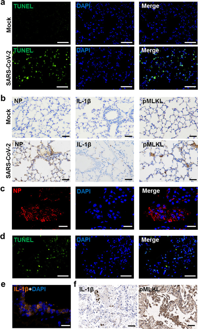
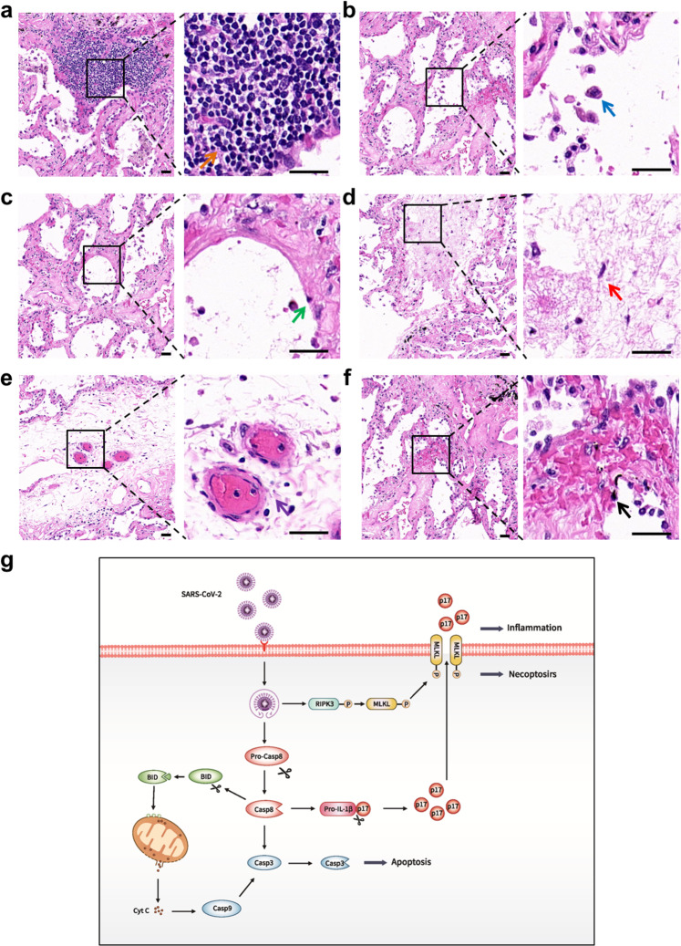
Severe acute respiratory syndrome coronavirus 2 (SARS-CoV-2) infection can lead to respiratory illness and multi-organ failure in critically ill patients. Although the virus-induced lung damage and inflammatory cytokine storm are believed to be directly associated with coronavirus disease 2019 (COVID-19) clinical manifestations, the underlying mechanisms of virus-triggered inflammatory responses are currently unknown. Here we report that SARS-CoV-2 infection activates caspase-8 to trigger cell apoptosis and inflammatory cytokine processing in the lung epithelial cells. The processed inflammatory cytokines are released through the virus-induced necroptosis pathway. Virus-induced apoptosis, necroptosis, and inflammation activation were also observed in the lung sections of SARS-CoV-2-infected HFH4-hACE2 transgenic mouse model, a valid model for studying SARS-CoV-2 pathogenesis. Furthermore, analysis of the postmortem lung sections of fatal COVID-19 patients revealed not only apoptosis and necroptosis but also massive inflammatory cell infiltration, necrotic cell debris, and pulmonary interstitial fibrosis, typical of immune pathogenesis in the lung. The SARS-CoV-2 infection triggered a dual mode of cell death pathways and caspase-8-dependent inflammatory responses may lead to the lung damage in the COVID-19 patients. These discoveries might assist the development of therapeutic strategies to treat COVID-19.
Overview
The claims and statistics of this paper haven’t been checked yet.
Run the check — free, no account needed. It reads the open-access text and checks the sources it cites.
Integrity signals
Registry
Cites
Statistics
Figures
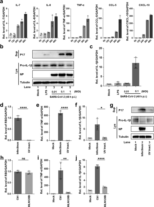
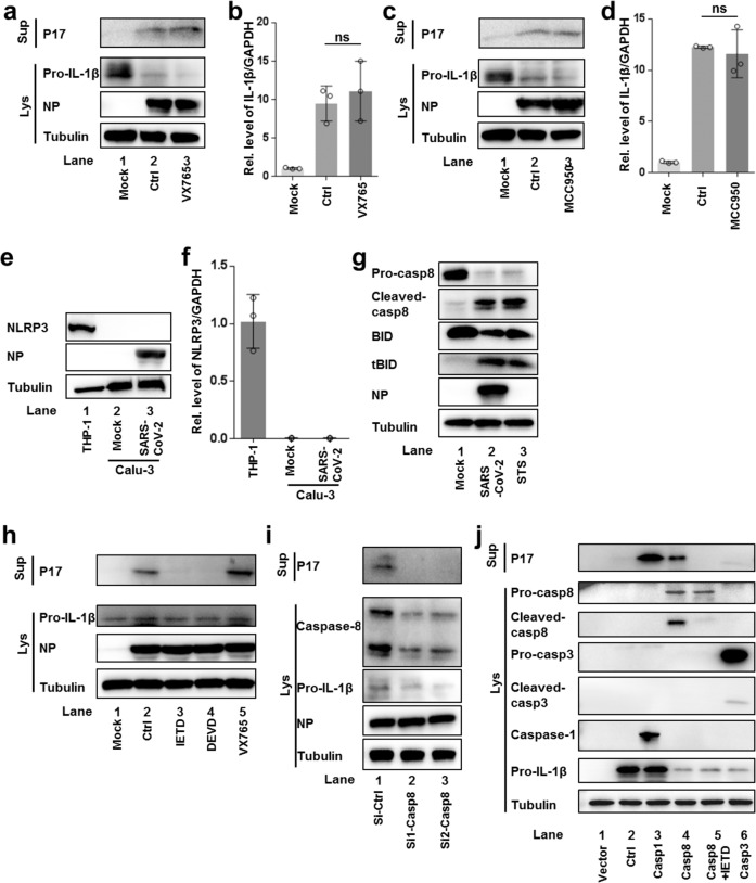
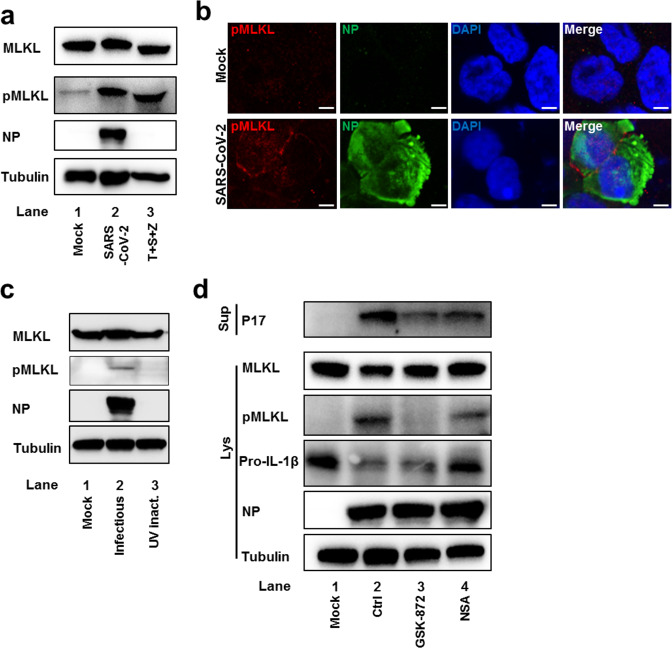
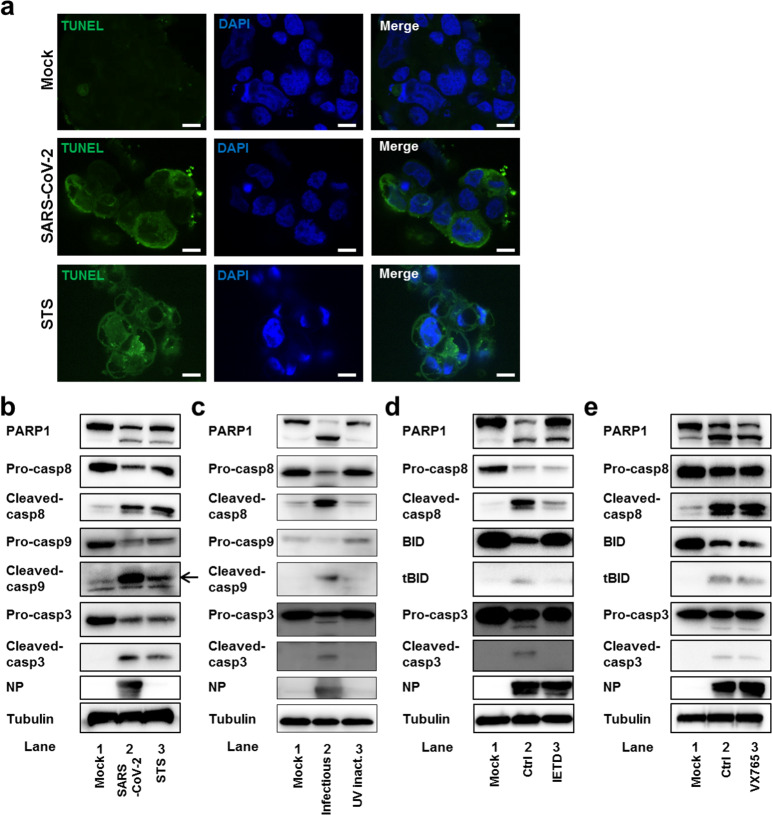
Verification
Claims the paper makes
The claims and statistics of this paper haven’t been checked yet.
Run the check — free, no account needed. It reads the open-access text and checks the sources it cites.
Claims other papers make about this one
Each is checked against this paper’s own text: does it say what the citing paper says it does?
In the citing paper
“Increased TNF and caspase 8 concentrations with minimal evidence of caspase 3–dependent apoptosis suggested a role for inflammatory-driven cell death pathways (e.g., necroptosis), as previously reported (39).”
From Lung epithelial and endothelial damage, loss of tissue repair, inhibition of fibrinolysis, and cellular senescence in fatal COVID-19 (2021) · cites this paper as [39]
Self-check
Percentages, sums and reported differences re-computed from the paper’s own numbers.
The self-check needs the paper’s full text.
Citations
Cites
A pneumonia outbreak associated with a new coronavirus of probable bat origin
Zhou, P., Yang, X.-L., Wang, X.-G., Hu, B., Zhang, L., Zhang, W., Si, H.-R., Zhu, Y., Li, B., Huang, C.-L., et al. (2020) · Nature · doi:10.1038/s41586-020-2012-7 · Cited by 21,969
Self-citationGorbalenya, A. E., Baker, S. C., Baric, R. S., de Groot, R. J., Drosten, C., Gulyaeva, A. A., Haagmans, B. L., Lauber, C., Leontovich, A. M., Neuman, B. W., et al. (2020) · Nature Microbiology · doi:10.1038/s41564-020-0695-z · Cited by 8,745
Ruan, Q., Yang, K., Wang, W., Jiang, L., & Song, J. (2020) · Intensive Care Medicine · doi:10.1007/s00134-020-05991-x · Cited by 4,898
Clinical and immunological features of severe and moderate coronavirus disease 2019
Chen, G., Wu, D., Guo, W., Cao, Y., Huang, D., Wang, H., Wang, T., Zhang, X., Chen, H., Yu, H., et al. (2020) · Journal of Clinical Investigation · doi:10.1172/jci137244 · Cited by 4,750
Self-citationThe trinity of COVID-19: immunity, inflammation and intervention
Tay, M. Z., Poh, C. M., Rénia, L., MacAry, P. A., & Ng, L. F. P. (2020) · Nature Reviews Immunology · doi:10.1038/s41577-020-0311-8 · Cited by 4,360
Necroptosis and its role in inflammation
Pasparakis, M., & Vandenabeele, P. (2015) · Nature · doi:10.1038/nature14191 · Cited by 2,148
Cytokine release syndrome in severe COVID-19
Moore, J. B., & June, C. H. (2020) · Science · doi:10.1126/science.abb8925 · Cited by 1,912
COVID-19 cytokine storm: the interplay between inflammation and coagulation
José, R. J. P., & Manuel, A. R. (2020) · The Lancet Respiratory Medicine · doi:10.1016/s2213-2600(20)30216-2 · Cited by 1,254
Caspase-8 is the molecular switch for apoptosis, necroptosis and pyroptosis
Fritsch, M., Günther, S. D., Schwarzer, R., Albert, M., Schorn, F., Werthenbach, J. P., Schiffmann, L. M., Stair, N., Stocks, H., Seeger, J. M., et al. (2019) · Nature · doi:10.1038/s41586-019-1770-6 · Cited by 1,122
Caspase‐8: regulating life and death
Tummers, B., & Green, D. R. (2017) · Immunological Reviews · doi:10.1111/imr.12541 · Cited by 779
Cited by
Sorted by citations
Items 201–300 of 365
Yang, Y., Shen, X.-R., Zhang, Y., Jiang, R.-D., Wang, X., Guan, Z., Li, Q., Yao, Y., Gong, Q.-C., Geng, R., et al. (2023) · mBio · doi:10.1128/mbio.03285-22 · Cited by 9
Self-citationGuo, Y., Zhou, J., Wang, Y., Wu, X., Mou, Y., & Song, X. (2023) · Immunological Reviews · doi:10.1111/imr.13282 · Cited by 9
Soriano‐Correa, C., Vichi-Ramírez, M. M., Herrera-Valencia, E. E., & Barrientos‐Salcedo, C. (2023) · Journal of Molecular Graphics and Modelling · doi:10.1016/j.jmgm.2023.108472 · Cited by 9
Bagato, O., Balkema‐Buschmann, A., Tödt, D., Weber, S., Gömer, A., Qu, B., Miskey, C., Ivics, Z., Mettenleiter, T. C., Finke, S., et al. (2023) · Microbiology Spectrum · doi:10.1128/spectrum.02469-23 · Cited by 7
Li, Y., Yu, Q., Huang, R., Chen, H., Ren, H., Ma, L., He, Y., & Li, W. (2023) · mBio · doi:10.1128/mbio.03359-22 · Cited by 8
Intestinal IL-1β Plays a Role in Protecting against SARS-CoV-2 Infection
Lücke, J., Heinrich, F., Malsy, J., Meins, N., Schnell, J., Böttcher, M., Nawrocki, M., Zhang, T., Bertram, F., Sabihi, M., et al. (2023) · The Journal of Immunology · doi:10.4049/jimmunol.2200844 · Cited by 8
Wu, J., Chen, G., Fan, C., Shen, F., Yang, Y., Pang, W., Zhao, Z., Guan, H., Wu, H., Lu, Y., et al. (2023) · Advanced Functional Materials · doi:10.1002/adfm.202306010 · Cited by 8
Fraga‐Silva, T. F. D. C., Cipriano, U. G., Fumagalli, M. J., Corrêa, G. F., Fuzo, C. A., dos-Santos, D., Mestriner, F., Becari, C., Teixeira‐Carvalho, A., Coelho-dos-Reis, J. G. A., et al. (2023) · iScience · doi:10.1016/j.isci.2023.108366 · Cited by 9
Cheng, L., Rui, Y., Wang, Y., Chen, S., Su, J., & Yu, X. (2024) · Journal of Biomedical Science · doi:10.1186/s12929-024-01062-1 · Cited by 9
Zulbaran‐Rojas, A., Bara, R. O., Lee, M., Bargas-Ochoa, M., Phan, T., Pacheco, M., Flores‐Camargo, A., Kazmi, S. M., Rouzi, M. D., Modi, D., et al. (2024) · Scientific Reports · doi:10.1038/s41598-024-78651-5 · Cited by 9
Craddock, V. D., Cook, C. M., & Dhillon, N. K. (2022) · Extracellular Vesicles and Circulating Nucleic Acids · doi:10.20517/evcna.2022.19 · Cited by 8
Yi, J., Miao, J., Zuo, Q., Owusu, F., Dong, Q., Lin, P., Wang, Q., Gao, R., Kong, X., & Yang, L. (2022) · Frontiers in Immunology · doi:10.3389/fimmu.2022.978619 · Cited by 9
Feng, L., Li, Z., Xiong, Y., Yan, T., Fu, C., Zeng, Q., & Wang, H. (2023) · Journal of Oncology · doi:10.1155/2023/4067418 · Cited by 8
Acat, M., Gülhan, P. Y., Eröz, R., Özkan, A. E., Koca, O., & Çınar, C. (2023) · Molecular Biology Reports · doi:10.1007/s11033-023-08244-4 · Cited by 8
Pyroptosis targeting via mitochondria: An educated guess to innovate COVID‐19 therapies
Singh, A., Strobbe, D., & Campanella, M. (2021) · British Journal of Pharmacology · doi:10.1111/bph.15670 · Cited by 7
SARS-CoV-2 recombinant spike protein induces cell apoptosis in rat taste buds
Yamamoto, T., Koyama, Y., Ujita, T., Sawada, E., Kishimoto, N., & Seo, K. (2022) · Journal of Dental Sciences · doi:10.1016/j.jds.2022.08.016 · Cited by 7
Association between expression of ZBP1, AIM2, and MDA5 genes and severity of COVID-19
Arefinia, N., Ramezani, A., Farokhnia, M., Zadeh, A. M. A., Yaghobi, R., & Sarvari, J. (2022) · EXCLI journal · doi:10.17179/excli2022-5141 · Cited by 7
ZBP1 inflames the SARS-CoV-2-infected lung
Yin, C., & Balachandran, S. (2023) · Cell Research · doi:10.1038/s41422-023-00784-5 · Cited by 7
Targeting necroptosis in fibrosis
Hassanein, E. H. M., Ibrahim, I. M., El‐Maksoud, M. S. A., El‐Aziz, M. K. A., Abd‐alhameed, E. K., & Althagafy, H. S. (2023) · Molecular Biology Reports · doi:10.1007/s11033-023-08857-9 · Cited by 9
Cappelletti, G., Colombrita, C., Limanaqi, F., Invernizzi, S., Garziano, M., Vanetti, C., Moscheni, C., Santangelo, S., Zecchini, S., Trabattoni, D., et al. (2023) · Frontiers in Cellular Neuroscience · doi:10.3389/fncel.2023.1285836 · Cited by 7
High fusion and cytopathy of SARS-CoV-2 variant B.1.640.1
Bolland, W., Michel, V., Planas, D., Hubert, M., Guivel‐Benhassine, F., Porrot, F., Staropoli, I., N’Debi, M., Rodriguez, C., Fourati, S., et al. (2023) · Journal of Virology · doi:10.1128/jvi.01351-23 · Cited by 8
Yuan, Z., Yang, X., Hu, Z., Gao, Y., Wang, M., Xie, L., Zhu, H., Chen, C., Lu, H., & Bai, Y. (2024) · Phytotherapy Research · doi:10.1002/ptr.8073 · Cited by 7
Martiáñez–Vendrell, X., Bloeme, J., Hutchinson, R., Guy, C., Bowie, A., & Kikkert, M. (2024) · Viruses · doi:10.3390/v16060898 · Cited by 7
Investigation of CYP2E1 and Caspase-3 Gene Expressions in COVID-19 patients
Uzunçakmak, S. K., Dirican, E., Naldan, M. E., Can, F. K., & Halıcı, Z. (2022) · Gene Reports · doi:10.1016/j.genrep.2022.101497 · Cited by 7
Qu, H., Zong, Q., Wang, H., Wu, S., Cai, D., & Bao, W. (2022) · Frontiers in Immunology · doi:10.3389/fimmu.2022.881289 · Cited by 7
Okada, Y., Numata, T., Sabirov, R. Z., Kashio, M., Merzlyak, P. G., & Sato-Numata, K. (2023) · Frontiers in Cell and Developmental Biology · doi:10.3389/fcell.2023.1246955 · Cited by 7
COVID-19 coagulopathy and antiphospholipid syndrome
Serrano, M., Espinosa, G., Cervera, R., & Serrano, A. (2021) · Revista Colombiana de Reumatología · doi:10.1016/j.rcreu.2021.02.013 · Cited by 7
Evangelou, K., Veroutis, D., Foukas, P. G., Paschalaki, K. E., Lаgopati, N., Dimitriou, M., Papaspyropoulos, A., Hazapis, O. G., Polyzou, A., Havaki, S., et al. (2021) · bioRxiv (Cold Spring Harbor Laboratory) · doi:10.1101/2021.01.02.424917 · Cited by 7
Lustig, G., Ganga, Y., Rodel, H. E., Tegally, H., Jackson, L., Cele, S., Khan, K., Jule, Z., Reedoy, K., Karim, F., et al. (2022) · medRxiv · doi:10.1101/2022.11.23.22282673 · Cited by 9
Soares, V. C., Dias, S. D. S. G., Santos, J. C., & Bozza, P. T. (2024) · Journal of Leukocyte Biology · doi:10.1093/jleuko/qiae170 · Cited by 6
Qi, Q., Li, Y., Ding, M., Huang, C., Omar, S. M., Shi, Y., Liu, P., Cai, G., Zheng, Z., Guo, X., et al. (2024) · International Journal of Molecular Sciences · doi:10.3390/ijms25158194 · Cited by 6
Bakshi, A., Gangopadhyay, K., Basak, S., De, R. K., Sengupta, S., & Dasgupta, A. (2025) · Journal of the American Medical Informatics Association · doi:10.1093/jamia/ocaf035 · Cited by 6
mTOR-Mediated Cell Death and Infection
Li, S., Wang, Q., & Su, B. (2021) · Infectious Microbes & Diseases · doi:10.1097/im9.0000000000000063 · Cited by 6
Self-citationTong, H., Chen, H., & Williams, C. (2022) · Viruses · doi:10.3390/v14040837 · Cited by 6
Elevated Levels of Cytotoxicity, Cytokines, and Anti-SARS-CoV-2 Antibodies in Mild Cases of COVID-19
Fernández‐Rojas, M. A., Ávila, G., Romero‐Valdovinos, M., Plett-Torres, T., Salazar, A. M., Sordo, M., Chávez-Vargas, M., Ángeles, C. J. C., Cruz-Rivera, M. Y., Santiago-Olivares, C., et al. (2023) · Viral Immunology · doi:10.1089/vim.2023.0012 · Cited by 6
The severity of COVID-19 upon hospital admission is associated with plasma omega-3 fatty acids
Fernandes, L. P., Murai, I. H., Fernandes, A. L., Sales, L. P., Rogero, M. M., Gualano, B., Barroso, L. P., Milne, G. L., Pereira, R. M. R., & Castro, I. A. (2024) · Scientific Reports · doi:10.1038/s41598-024-60815-y · Cited by 6
Proteomics approaches for the identification of protease substrates during virus infection
Martiáñez–Vendrell, X., & Kikkert, M. (2021) · Advances in virus research · doi:10.1016/bs.aivir.2021.03.003 · Cited by 6
Suzuki, S., Imamura, M., Mouri, M., Tsuchida, T., Tomita, H., Matsuoka, S., Takita, M., Kakinuma, K., Kawasaki, T., Sakurai, K., et al. (2022) · Respiratory Investigation · doi:10.1016/j.resinv.2022.06.007 · Cited by 7
SARS-CoV-2 immune complex triggers human monocyte necroptosis
Santos, L. D., Antunes, K. H., Cassão, G., Gonçalves, J. I., Abbadi, B. L., Bizarro, C. V., Basso, L. A., Machado, P., de Souza, A. P. D., & Porto, B. N. (2023) · International Immunopharmacology · doi:10.1016/j.intimp.2023.109954 · Cited by 6
Çetin, Z. (2021) · Stem Cell Reviews and Reports · doi:10.1007/s12015-021-10166-2 · Cited by 6
Sheerin, D., Phan, T. K., Eriksson, E. M., & Coussens, A. K. (2023) · medRxiv · doi:10.1101/2023.05.24.23290499 · Cited by 7
Grant, C., Duffin, E., O’Connell, F., Nadarajan, P., Bergin, C. J., Keane, J., & O’Sullivan, M. P. (2025) · Immunology · doi:10.1111/imm.13922 · Cited by 5
Bader, S. M., Scherer, L., Bhandari, R., Motyer, A., Cooney, J. P., Mackiewicz, L., Dayton, M., Sheerin, D., Romero, D. V. L., Schaefer, J., et al. (2025) · Nature Communications · doi:10.1038/s41467-025-65098-z · Cited by 8
The BCG vaccine and SARS-CoV-2: Could there be a beneficial relationship?
Peña‐Bates, C., Lascuraín, R., Ortiz-Navarrete, V. F., & Chávez‐Galán, L. (2024) · Heliyon · doi:10.1016/j.heliyon.2024.e38085 · Cited by 5
Hsieh, L.-C., Hsieh, S.-L., Ping, T.-N., Huang, Y.-C., Lin, S.-J., Chi, H.-Y., & Wu, C. (2024) · Pharmaceutical Biology · doi:10.1080/13880209.2024.2433994 · Cited by 5
Cappelletti, G., Brambilla, L., Strizzi, S., Limanaqi, F., Melzi, V., Rizzuti, M., Nizzardo, M., Saulle, I., Trabattoni, D., Corti, S. P., et al. (2025) · The FASEB Journal · doi:10.1096/fj.202401604rrr · Cited by 5
Tan, T. H., Patton, E., Munro, C. A., Corzo-León, D., Porter, A. J., & Palliyil, S. (2021) · Viruses · doi:10.3390/v13112201 · Cited by 5
Intestinal Shedding of SARS-CoV-2 in Children: No Evidence for Infectious Potential
Nogueira, F., Obrová, K., Haas, M., Tucek, E., Kosulin, K., Fortschegger, M., Fürhacker, P., Walter, C., Größlinger, L., Peter, S., et al. (2022) · Microorganisms · doi:10.3390/microorganisms11010033 · Cited by 5
Das, R., Sinnarasan, V. S. P., Paul, D., & Venkatesan, A. (2023) · Biochemical Genetics · doi:10.1007/s10528-023-10458-x · Cited by 5
Wang, E., Yang, Q., Xu, X., Zou, Q., Long, Y., Ma, G., Deng, Z., Zhao, J., Li, M., & Zeng, J. (2024) · Journal of Medical Virology · doi:10.1002/jmv.29357 · Cited by 5
Li, H., Zhao, X., Zhang, X., Zheng, H., Wang, Y., Hou, J., Li, J., Zhao, Y., Peng, S., Li, Y., et al. (2024) · APOPTOSIS · doi:10.1007/s10495-024-01940-x · Cited by 5
Self-citationCao, J., Zhu, S.-T., Huang, X., Wang, X.-Y., Wu, M., Li, X., Liu, F., Chen, L., Zheng, Y., & Wang, J. (2024) · Virologica Sinica · doi:10.1016/j.virs.2024.03.001 · Cited by 8
Yin, Y., Zeng, Z., Wei, S., Shen, Z., Cong, Z., & Zhu, X. D. (2024) · International Immunopharmacology · doi:10.1016/j.intimp.2024.112670 · Cited by 5
Bispo, E. C. I., Argañaraz, E. R., Neves, F. D. A. R., Carvalho, J. L., & Saldanha‐Araújo, F. (2024) · Scientific Reports · doi:10.1038/s41598-024-75776-5 · Cited by 6
Pazopanib Is a Potential Treatment for Coronavirus-Induced Lung Injuries
Luan, Y., Yuan, Q., Wang, Q., Compton, S. R., Wu, D., & Tang, W. (2022) · The Journal of Immunology · doi:10.4049/jimmunol.2100968 · Cited by 5
Xia, S., Jiao, F., Chen, J., Wang, L., Lu, T., Wang, Q., Xu, W., Wang, X., Sun, F., Zhu, Y., et al. (2025) · Cell Reports Medicine · doi:10.1016/j.xcrm.2025.102277 · Cited by 4
Self-citationROS-Mediated Necroptosis Promotes Coxsackievirus B3 Replication and Myocardial Injury
Huang, J., Di, Y., Song, Q., Cheng, Z., Wu, H., Wu, M., He, M., Zhang, G., Wang, F., & Tong, L. (2025) · Microorganisms · doi:10.3390/microorganisms13102389 · Cited by 4
Zhou, Y., Zhang, C., Zhang, Y., Li, F., & Shen, J. (2024) · Biosafety and Health · doi:10.1016/j.bsheal.2024.03.003 · Cited by 4
Self-citationSun, Z., Hu, Y., Qu, J., Zhao, Q., Gao, H., & Peng, Z. (2024) · Frontiers in Genetics · doi:10.3389/fgene.2024.1389630 · Cited by 4
Chatterjee, D., Kurup, D., & Smeyne, R. J. (2025) · npj Parkinson s Disease · doi:10.1038/s41531-025-00925-0 · Cited by 4
Saksena, N. K., Bonam, S. R., & Saksena, M. M. (2021) · Exploration of Immunology · doi:10.37349/ei.2021.00007 · Cited by 4
Kapuy, O., & Korcsmaros, T. D. (2022) · PLoS ONE · doi:10.1371/journal.pone.0266337 · Cited by 4
COVID-19 in Children: Molecular Profile and Pathological Features
Nasyrov, R. A., Иванов, Д. О., Krasnogorskaya, O. L., Тимченко, В. Н., Fedotova, E. P., Chepelev, A. S., Galichina, V. A., Sidorova, N. A., & Аничков, Н. М. (2023) · International Journal of Molecular Sciences · doi:10.3390/ijms242316750 · Cited by 4
Liu, J., Chen, Y., Nie, L., Xiao, L., Huang, W., & Li, R. (2022) · Frontiers in Pharmacology · doi:10.3389/fphar.2022.955482 · Cited by 4
Oseghale, O., Quinn, K. M., Coward‐Smith, M., Liong, F., Miles, M. A., Brooks, R. D., Vlahos, R., O’Leary, J. J., Brooks, D. A., Liong, S., et al. (2024) · American Journal of Physiology-Heart and Circulatory Physiology · doi:10.1152/ajpheart.00646.2023 · Cited by 3
Pradhan, A. J., & Atilla‐Gokcumen, G. E. (2023) · Molecular Omics · doi:10.1039/d2mo00318j · Cited by 3
Anti‐inflammatory effects of sodium‐glucose cotransporter‐2 inhibitors in COVID‐19
Yaribeygi, H., Maleki, M., Atkin, S. L., Kesharwani, P., Jamialahmadi, T., & Sahebkar, A. (2023) · IUBMB Life · doi:10.1002/iub.2719 · Cited by 4
Rationale for combined therapies in severe-to-critical COVID-19 patients
Gonzaga, A., Andreu, E., Blasco, L., Meseguer, R., Al‐Akioui‐Sanz, K., Soria-Juan, B., Sanjuan-Gimenez, J. C., Ferreras, C., Tejedo, J. R., López‐Lluch, G., et al. (2023) · Frontiers in Immunology · doi:10.3389/fimmu.2023.1232472 · Cited by 3
Suryawanshi, P. V., Patil‐Takbhate, B., Athavale, P. V., MIRZA, S. B., Tripathy, A. S., Kanitkar, S. A., Shivnitwar, S. K., Barthwal, M. S., Dole, S. S., Chavan, H., et al. (2024) · Immunity Inflammation and Disease · doi:10.1002/iid3.1238 · Cited by 3
Programmed Cell Death in SARS-CoV-2 Infection: A Short Review
Deshpande, R., & Zou, C. (2021) · Journal of Respiration · doi:10.3390/jor1040021 · Cited by 3
Dysregulated Innate Immune and Inflammatory Responses in SARS-CoV-2 Infection and COVID-19 Severity
Ghosh, P., Nagaraja, S., Basavaraju, S., & Kesavardhana, S. (2021) · Critical Reviews in Immunology · doi:10.1615/critrevimmunol.2021039716 · Cited by 3
The Role of Inflammasomes in COVID-19: Potential Therapeutic Targets
Sun, C., Zhao, H., Han, Y., Wang, Y., & Sun, X. (2022) · Journal of Interferon & Cytokine Research · doi:10.1089/jir.2022.0061 · Cited by 3
Minu Treeza, M., Augustine, S., Mathew, A. A., Kanthlal, S. K., & Panonnummal, R. (2023) · Advanced Pharmaceutical Bulletin · doi:10.34172/apb.2023.069 · Cited by 3
Yadav, B., Prasad, N., Kushwaha, R. S., Patel, M. R., BHADAURIA, D. S., & Kaul, A. (2023) · Indian Journal of Transplantation · doi:10.4103/ijot.ijot_37_23 · Cited by 3
Wang, L., Mao, Z., & Shao, F. (2023) · IET Systems Biology · doi:10.1049/syb2.12079 · Cited by 3
In vitro comparison of viral replication and cytopathology induced by SARS-CoV-2 variants
Phadke, K. S., Higdon, N. B. A., & Bellaire, B. (2024) · Access Microbiology · doi:10.1099/acmi.0.000716.v3
Wang, Y., Pang, X., Li, R., Chen, J., Chen, W., Zhu, H., Long, T., Li, J., Zheng, L., Deng, Y., et al. (2025) · Research · doi:10.34133/research.0594 · Cited by 2
COVID-19-Related Pulmonary Embolism: Incidence, Characteristics, and Risk Factors
Bukhari, Z. M., Alqarni, M. S., Abukhodair, A. W., Alzahrani, A. S., Alzahrani, A., Alsrhani, H., Alasadi, F., Alotaibi, A. F., & Althobaiti, M. (2021) · Cureus · doi:10.7759/cureus.19738 · Cited by 3
Ong, J., & Dan, Y. Y. (2021) · Digestive Diseases and Sciences · doi:10.1007/s10620-020-06767-5 · Cited by 3
Kelleni, M. T. (2021) · doi:10.31219/osf.io/2rf98 · Cited by 3
Senigagliesi, F., Scialla, S., & Marasco, M. L. (2024) · International Journal of Osteopathic Medicine · doi:10.1016/j.ijosm.2024.100737 · Cited by 2
Iketani, T., Miyazaki, K., Iwata‐Yoshikawa, N., Sakai, Y., Shiwa, N., Ozono, S., Asanuma, H., Hasegawa, H., Suzuki, T., & Nagata, N. (2024) · Microbiology and Immunology · doi:10.1111/1348-0421.13184 · Cited by 2
Soluble tissue factor generated by necroptosis-triggered shedding is responsible for thrombosis
Wan, P., Choksi, S., Park, Y.-J., Chen, X., Yan, J., Foroutannejad, S., Liu, Z., Chen, J., Lake, R., Liu, Z.-G., et al. (2025) · Cell Research · doi:10.1038/s41422-025-01167-8 · Cited by 2
Self-citationLi, Q., Ma, X., Shang, X., Nian, X., Mao, R., Luo, Z., Zhu, Z., Ru, Y., Yang, F., Cao, W., et al. (2026) · PLoS Pathogens · doi:10.1371/journal.ppat.1013874 · Cited by 2
Self-citationMorus alba L. alleviates influenza viral pneumonia. Evidences of its mechanism of action
Jing, J., Sun, Y., Shui, Y., Wang, J., Ye, W., Chen, R., Wu, L., Xing, L., Huang, R., Zhou, T., et al. (2025) · Phytomedicine · doi:10.1016/j.phymed.2025.156574 · Cited by 2
Wijerathne, S. V. T., Pandit, R., Ezeuko, C. C., & Matthews, Q. L. (2025) · Veterinary Sciences · doi:10.3390/vetsci12050477 · Cited by 2
Singh, A. A., Song, M., & Kim, G. (2025) · Current Issues in Molecular Biology · doi:10.3390/cimb47060401 · Cited by 2
Regulation of Apoptosis and PANoptosis by Coronavirus: An Overview
Yuan, L., Wang, B., & Huang, Y. (2025) · Journal of Medical Virology · doi:10.1002/jmv.70672 · Cited by 2
Immunoediting in SARS-CoV-2: Mutual relationship between the virus and the host
Kheshtchin, N., Bakhshi, P., Arab, S., & Nourizadeh, M. (2022) · International Immunopharmacology · doi:10.1016/j.intimp.2022.108531 · Cited by 2
Heidarvand, M., Hosseini, R., Kazemi, M., Andalib, A., Sami, R., Eskandari, N., & Ghezelbash, B. (2023) · Advanced Biomedical Research · doi:10.4103/abr.abr_232_22 · Cited by 2
Bonaffini, P. A., Stanco, F., Dulcetta, L., Poli, G., Brambilla, P., Marra, P., Valle, C., Lorini, F. L., Mazzoleni, M., Sonzogni, B., et al. (2023) · Tomography · doi:10.3390/tomography9060171 · Cited by 1
Urinary Mitochondrial DNA Induces an Inflammatory Response in Peripheral Blood Mononuclear Cells
Yadav, B., Prasad, N., Kushwaha, R. S., Singh, A., Yadav, D., BHADAURIA, D. S., & Kaul, A. (2024) · Indian Journal of Transplantation · doi:10.4103/ijot.ijot_94_23 · Cited by 1
Understanding the Impact of SARS-CoV-2 on Lung Endothelial Cells: Brief Mechanisms Unveiled
Shah, F. H., Bang, J. Y., Nam, Y. S., Hwang, I. S., Kim, D. H., Ki, M., Salman, S., & Lee, H. (2024) · Cell Biochemistry and Biophysics · doi:10.1007/s12013-024-01529-w · Cited by 1
Cinnamon: health benefits and its potential role in prevention and treatment of COVID-19
Aswani, B. S., Sajeev, A., Hegde, M., & Kunnumakkara, A. B. (2025) · Elsevier eBooks · doi:10.1016/b978-0-443-21820-0.00017-9 · Cited by 1
SARS-CoV-2 causes gastric damage: structural and ultrastructural evaluation
Yilmaz, E. S., Onen, E. A., Mutlu, H. S., Solakoğlu, S., & Demirci, E. (2025) · Journal of Molecular Histology · doi:10.1007/s10735-025-10477-5 · Cited by 1
The antimicrobial peptide nisin promotes host cell survival during SARS-CoV-2 infection
English, B. C., Radaic, A., Barrios-Medina, R., Lin, K., Kamarajan, P., Sil, A., & Kapila, Y. L. (2025) · Virology Journal · doi:10.1186/s12985-025-02921-5 · Cited by 1
Kundu, S., Riegler, A. N., Chelappan, R., Mishra, D. K., Dave, T. V., Leal, S. M., & Roy, S. (2025) · Medical Mycology · doi:10.1093/mmy/myaf104 · Cited by 1
CASPASE-3 expression and activity in PBMCs associate with SARS-CoV-2 infection and clinical features
de Carvalho, V. M. F., Silva, B. D. O., de Oliveira, P. S. S., da Costa, T. B., da Rosa, M. M., Rêgo, M. J. B. D. M., Pereira, M. C., & Pitta, M. G. D. R. (2025) · Clinical Immunology Communications · doi:10.1016/j.clicom.2025.09.004 · Cited by 1
Wang, S., Wang, H., Li, F., Zhao, Y., Pei, Z., Lu, W., Zhao, J., & Lu, S. (2026) · Foods · doi:10.3390/foods15020261 · Cited by 1
COVID-19 Subclinical Infection and Immunity
Bartekwa, J. W., Abene, E., Luka, P. D., Yilgwan, C. S., & Shehu, N. Y. (2021) · Nigerian Journal of Medicine · doi:10.4103/njm.njm_85_21 · Cited by 1
Metadata
- Authors
- Li, S., Zhang, Y., Guan, Z., Li, H., Ye, M., Chen, X., Shen, J., Zhou, Y., Shi, Z.-L., Zhou, P., & Peng, K.
- Affiliations
- Chinese Academy of Sciences (CN)
- Wuhan No.1 Hospital (CN)
- Children's Hospital of Fudan University (CN)
- Huazhong University of Science and Technology (CN)
- Corresponding author
- Zhou, Peng
- Peng, Ke
- Venue
- Signal Transduction and Targeted Therapy
- Published
- 2020-10-09
- Volume
- 5
- Issue
- 1
- Pages
- 235-235
- ISSN
- 2059-3635 (ISSN-L)
- PMC
- PMC7545816
- OpenAlex
- W3092576617
- Cited by
- 365
- References
- 24
- Citations by year
- 2026: 12 · 2025: 34 · 2024: 54 · 2023: 74 · 2022: 81 · 2021: 108 · 2020: 2
- FWCI
- 8.68
- Type
- journal article
- Language
- en
- Open access
- gold
- doi.org
- Locations
- Signal Transduction and Targeted Therapy (publishedVersion, cc-by)
- PubMed Central (publishedVersion, cc-by)
- PubMed (publishedVersion)
- Funders
- National Natural Science Foundation of China (31770188)
- Topics
- Health Sciences › Medicine › Infectious Diseases › COVID-19 Clinical Research Studies
- Health Sciences › Medicine › Infectious Diseases › SARS-CoV-2 and COVID-19 Research
- Life Sciences › Biochemistry, Genetics and Molecular Biology › Molecular Biology › Cell death mechanisms and regulation
- Keywords
- SARS-CoV-2, COVID-19 pandemic, caspase-8, apoptosis, necroptosis, inflammatory cytokines, cytokine processing, lung epithelial cells, lung injury, interstitial pulmonary fibrosis, inflammatory cell infiltration, cell death pathways
- MeSH terms
- Betacoronavirus, Necroptosis, Cytokine Release Syndrome, COVID-19, SARS-CoV-2, Animals, Disease Models, Animal, Epithelial Cells, Gene Expression Regulation, Humans, Lung, Mice, Transgenic, Pneumonia, Viral, Pulmonary Fibrosis, Tumor Necrosis Factor-alpha, Interleukin-7, Interleukin-8, Apoptosis, Coronavirus Infections, Chemokine CCL5, Cell Line, Tumor, Mice, Caspase 8, Interleukin-1beta, Chemokine CXCL10, Pandemics
About these checks
Registry, retraction, Cites and statistics results are computed, not judged, with one exception: a printed reference that no index lists is read once by a model before it counts as a citation error. Claims are extracted by a model and kept only when their quote appears verbatim in the text; each cited source is then checked, with a model’s help, for whether it says what the claim relies on. Those verdicts can be wrong — read the claim’s quote and the cited source before relying on one.
Statistics and arithmetic are recomputed, not judged.
Confidence bands are shown only for fields calibrated on our annotated test set. None is calibrated yet, so no band is shown.
What counts as a claim ›How we measure confidence ›How we check this ›
Are you an author, or do you see a problem with this page? Request a review
