

The DOI matches its Crossref record; 521 of the 522 references it cites were found in a registry or an index.
Regulating tumor suppressor genes: post-translational modifications
Open accessAbstract
Tumor suppressor genes cooperate with each other in tumors. Three important tumor suppressor proteins, retinoblastoma (Rb), p53, phosphatase, and tensin homolog deleted on chromosome ten (PTEN) are functionally associated and they regulated by post-translational modification (PTMs) as well. PTMs include phosphorylation, SUMOylation, acetylation, and other novel modifications becoming growing appreciated. Because most of PTMs are reversible, normal cells use them as a switch to control the state of cells being the resting or proliferating, and PTMs also involve in cell survival and cell cycle, which may lead to abnormal proliferation and tumorigenesis. Although a lot of studies focus on the importance of each kind of PTM, further discoveries shows that tumor suppressor genes (TSGs) form a complex “network” by the interaction of modification. Recently, there are several promising strategies for TSGs for they change more frequently than carcinogenic genes in cancers. We here review the necessity, characteristics, and mechanisms of each kind of post-translational modification on Rb, p53, PTEN, and its influence on the precise and selective function. We also discuss the current antitumoral therapies of Rb, p53 and PTEN as predictive, prognostic, and therapeutic target in cancer.
Overview
The claims and statistics of this paper haven’t been checked yet.
Run the check — free, no account needed. It reads the open-access text and checks the sources it cites.
Integrity signals
Registry
Cites
Statistics
Figures

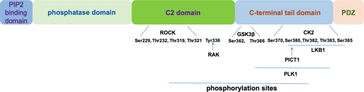
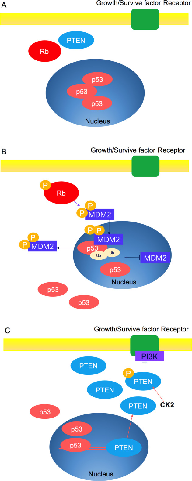
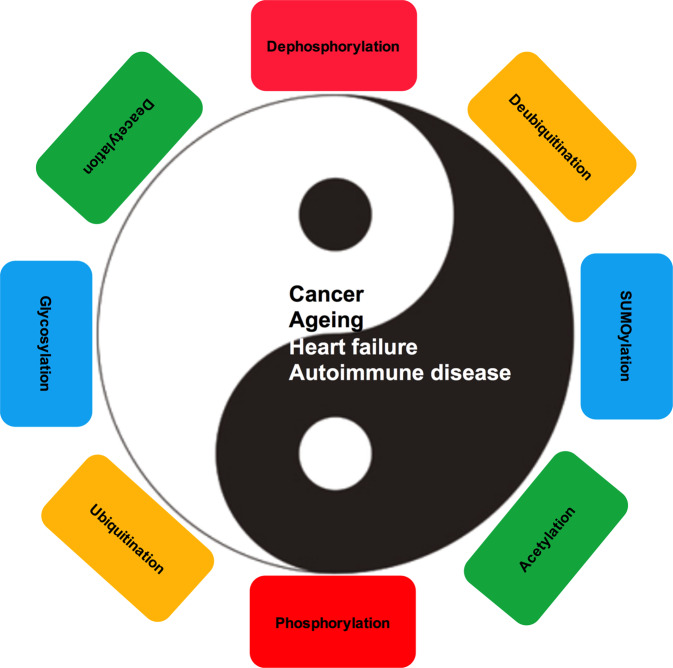
Verification
Claims the paper makes
The claims and statistics of this paper haven’t been checked yet.
Run the check — free, no account needed. It reads the open-access text and checks the sources it cites.
Claims other papers make about this one
Each is checked against this paper’s own text: does it say what the citing paper says it does?
The cited source says what the claim attributes to it.
In the citing paper
“PTMs are known to function as molecular switches by affecting protein-protein interactions (Chen, Liu et al., 2020, Nishi, Hashimoto et al., 2011).”
From SHARPIN S146 phosphorylation mediates ARP2/3 interaction, cancer cell invasion and metastasis (2022) · cites this paper as Chen, Liu et al., 2020
Self-check
Percentages, sums and reported differences re-computed from the paper’s own numbers.
The self-check needs the paper’s full text.
Citations
Cites
1 retracted
Song, P., Wu, Y., Xu, J., Xie, Z., Dong, Y., Zhang, M., & Zou, M. (2007) · Circulation · doi:10.1161/circulationaha.107.716498 · Cited by 144
RetractedHershko, A., & Ciechanover, A. (1998) · Annual Review of Biochemistry · doi:10.1146/annurev.biochem.67.1.425 · Cited by 8,840
p53 Mutations in Human Cancers
Hollstein, M., Sidransky, D., Vogelstein, B., & Harris, C. C. (1991) · Science · doi:10.1126/science.1905840 · Cited by 8,018
Comprehensive genomic characterization defines human glioblastoma genes and core pathways
The Cancer Genome Atlas Research Network (2008) · Nature · doi:10.1038/nature07385 · Cited by 7,735
p53, the cellular gatekeeper for growth and division
Levine, A. J. (1997) · Cell · doi:10.1016/s0092-8674(00)81871-1 · Cited by 7,693
Mutation and cancer: statistical study of retinoblastoma
Knudson, A. G. (1971) · Proceedings of the National Academy of Sciences · doi:10.1073/pnas.68.4.820 · Cited by 7,612
Nowell, P. C. (1976) · Science · doi:10.1126/science.959840 · Cited by 6,471
AKT/PKB signaling: navigating downstream
Manning, B. D., & Cantley, L. C. (2007) · Cell · doi:10.1016/j.cell.2007.06.009 · Cited by 6,115
An Integrated Genomic Analysis of Human Glioblastoma Multiforme
Parsons, D. W., Jones, S., Zhang, X., Lin, J. C.-H., Leary, R. J., Angenendt, P., Mankoo, P., Carter, H., Siu, I.-M., Gallia, G. L., et al. (2008) · Science · doi:10.1126/science.1164382 · Cited by 5,857
The retinoblastoma protein and cell cycle control
Weinberg, R. A. (1995) · Cell · doi:10.1016/0092-8674(95)90385-2 · Cited by 4,862
Cited by
Sorted by citations
Items 101–200 of 452
Ning, S., Liu, C., Wang, K., Cai, Y., Ning, Z., Li, M.-Y., & Zeng, L. (2023) · Journal of Translational Medicine · doi:10.1186/s12967-023-04236-x · Cited by 23
The Burden of Post-Translational Modification (PTM)—Disrupting Mutations in the Tumor Matrisome
Holstein, E., Dittmann, A., Kääriäinen, A., Pesola, V., Koivunen, J. T., Pihlajaniemi, T. A., Naba, A., & Izzi, V. (2021) · Cancers · doi:10.3390/cancers13051081 · Cited by 24
LncRNA‐Mediated TPI1 and PKM2 Promote Self‐Renewal and Chemoresistance in GBM
Yang, C., Li, Z., Tian, K., Meng, X., Wang, X., Song, D., Wang, X., Xu, T., Sun, P., Zhong, J., et al. (2024) · Advanced Science · doi:10.1002/advs.202402600 · Cited by 22
p53: from understanding its structure to advances in therapeutic targeting
Wang, W., Liu, X., Liu, H., Abolhassani, H., Yan, H., Zhang, H., & Wang, X. (2026) · Signal Transduction and Targeted Therapy · doi:10.1038/s41392-025-02549-5 · Cited by 29
Yuan, H., Lu, Y., Chan, Y.-T., Zhang, C., Wang, N., & Feng, Y. (2021) · Cancers · doi:10.3390/cancers13225700 · Cited by 22
Zhang, N., Zhu, H., Huang, W. C., Wen, X., Xie, X., Jiang, X., Peng, C., Han, B., & He, G. (2022) · Experimental Hematology and Oncology · doi:10.1186/s40164-022-00321-x · Cited by 22
Lv, J., Wu, Q., Li, K., Bai, K., Yu, H., Zhuang, J., Sun, H., Yang, H., Yang, X. F., & Lü, Q. (2023) · International Journal of Biological Sciences · doi:10.7150/ijbs.87182 · Cited by 22
Feng, R., Cheng, D., Chen, X., Yang, L., & Wu, H. (2023) · Biochemical and Biophysical Research Communications · doi:10.1016/j.bbrc.2023.149325 · Cited by 21
Li, K., Lin, H., Liu, A., Qiu, C., Rao, Z., Wang, Z., Chen, S., She, X., Zhu, S., Liu, L., et al. (2024) · Cancer Letters · doi:10.1016/j.canlet.2024.216842 · Cited by 21
Post-translational modifications in tumor-associated carbonic anhydrases
Fiore, A. D., Supuran, C. T., Scaloni, A., & Simone, G. D. (2021) · Amino Acids · doi:10.1007/s00726-021-03063-y · Cited by 21
Zhang, L., Hou, N., Chen, B., Kan, C., Han, F., Zhang, J., & Sun, X. (2022) · Frontiers in Pharmacology · doi:10.3389/fphar.2022.908772 · Cited by 21
Zhang, R., Ai, J., Wang, J., sun, C., Lu, H., He, A., Li, M., Liao, Y. A., Lei, J., Zhou, F., et al. (2022) · Journal of Translational Medicine · doi:10.1186/s12967-022-03519-z · Cited by 21
Global landscape of 2-hydroxyisobutyrylation in human pancreatic cancer
Lü, Y., Li, X., Zhao, K., Qiu, P., Deng, Z., Yao, W., & Wang, J. (2022) · Frontiers in Oncology · doi:10.3389/fonc.2022.1001807 · Cited by 21
N-linked glycosylation is essential for anti-tumor activities of KIAA1324 in gastric cancer
Yun, R., Hong, E., Kim, J., Park, B., Kim, S. J., Lee, B., Song, Y. S., Kim, S., Park, S., & Kang, J. M. (2023) · Cell Death and Disease · doi:10.1038/s41419-023-06083-6 · Cited by 21
Emerging trends in post-translational modification: Shedding light on Glioblastoma multiforme
Kumari, S., Gupta, R. V., Ambasta, R. K., & Kumar, P. (2023) · Biochimica et Biophysica Acta (BBA) - Reviews on Cancer · doi:10.1016/j.bbcan.2023.188999 · Cited by 22
Interrogating epigenetic mechanisms with chemically customized chromatin
Hananya, N., Koren, S., & Muir, T. W. (2023) · Nature Reviews Genetics · doi:10.1038/s41576-023-00664-z · Cited by 20
Icaritin activates p53 and inhibits aerobic glycolysis in liver cancer cells
Zhou, X., Wu, D., Mi, T., Li, R., Guo, T., & Li, W. (2024) · Chemico-Biological Interactions · doi:10.1016/j.cbi.2024.110926 · Cited by 21
Lipid nanoparticle-mediated targeted mRNA delivery and its application in cancer therapy
Sui, Y., Hou, X., Zhang, J., Hong, X., Wang, H., Xiao, Y., & Zeng, X. (2025) · Journal of Materials Chemistry B · doi:10.1039/d5tb01556a · Cited by 25
SETDB1-mediated CD147-K71 di-methylation promotes cell apoptosis in non-small cell lung cancer
Shi, M.-Y., Wang, Y., Shi, Y. Q., Tian, R.-F., Chen, X., Zhang, H., Wang, K., Chen, Z., & Chen, R. (2023) · Genes & Diseases · doi:10.1016/j.gendis.2023.02.015 · Cited by 20
Clinical trial designs for evaluating and exploiting cancer evolution
Garces, A. I., Porta, N., Graham, T. A., & Banerji, U. (2023) · Cancer Treatment Reviews · doi:10.1016/j.ctrv.2023.102583 · Cited by 19
Acyl post-translational modification of proteins by metabolites in cancer cells
Wang, X., Guo, Y., Fu, Y., Zhang, C., Chen, W., Tang, X. T., Yu, Y., Chen, Y., Ding, G., & Zhang, J. (2025) · Cell Death Discovery · doi:10.1038/s41420-025-02535-4 · Cited by 20
Qi, L., Yi, J., Ji, Y., Jiang, H., Sun, J., Shen, Y., & Sun, H. (2026) · Biochemical Pharmacology · doi:10.1016/j.bcp.2026.117999 · Cited by 19
Hou, W., Liu, G., Ren, X., Liu, X., He, L., & Huang, H. (2022) · Oxidative Medicine and Cellular Longevity · doi:10.1155/2022/4592170 · Cited by 19
Luo, W., He, M., Luo, Q., & Li, Y. (2023) · Frontiers in Cardiovascular Medicine · doi:10.3389/fcvm.2022.1066822 · Cited by 20
Weh, K. M., Howard, C. L., Zhang, Y., Tripp, B. A., Clarke, J., Howell, A. B., Rubenstein, J. H., Abrams, J. A., Westerhoff, M. A., & Kresty, L. A. (2024) · JCI Insight · doi:10.1172/jci.insight.168112 · Cited by 19
Ferroptosis in Cancer: Epigenetic Control and Therapeutic Opportunities
Tranchese, R. V., Battista, S., Cerchia, L., & Fedele, M. (2024) · Biomolecules · doi:10.3390/biom14111443 · Cited by 20
Targeted protein modification as a paradigm shift in drug discovery
Amirian, R., Azadi Badrbani, M., Izadi, Z., Samadian, H., Bahrami, G., Sarvari, S., Abdolmaleki, S., Nabavi, S. M., Derakhshankhah, H., & Jaymand, M. (2023) · European Journal of Medicinal Chemistry · doi:10.1016/j.ejmech.2023.115765 · Cited by 18
Parkin as a Molecular Bridge Linking Alzheimer’s and Parkinson’s Diseases?
Checler, F., & da Costa, C. A. (2022) · Biomolecules · doi:10.3390/biom12040559 · Cited by 19
Emerging Role of Ubiquitin-Specific Protease 19 in Oncogenesis and Cancer Development
Rossi, F. A., & Rossi, M. (2022) · Frontiers in Cell and Developmental Biology · doi:10.3389/fcell.2022.889166 · Cited by 20
QSER1 preserves the suppressive status of the pro-apoptotic genes to prevent apoptosis
Zhao, X., Fang, K., Liu, X., Yao, R., Wang, M., Li, F., Hao, S., He, J., Wang, Y., Fan, M., et al. (2022) · Cell Death and Differentiation · doi:10.1038/s41418-022-01085-x · Cited by 18
Kaleem, M., Azmi, L., Shahzad, N., Taha, M., Kumar, S., Mujtaba, M. A., Hazazi, A. Y., & Kayali, A. (2025) · Naunyn-Schmiedeberg s Archives of Pharmacology · doi:10.1007/s00210-025-04217-5 · Cited by 21
Shahhosseini, A., a-Flin, E. B., Derakhshan, S., Aminishakib, P., & Goudarzi, A. (2023) · Biomedicine · doi:10.37796/2211-8039.1391 · Cited by 17
Posttranslational regulation of liver kinase B1 in human cancer
Hu, L., Liu, M., Tang, B., Li, Q., Pan, B., Xu, C. S., & Lin, H. (2023) · Journal of Biological Chemistry · doi:10.1016/j.jbc.2023.104570 · Cited by 18
Kabil, M. F., Badary, O. A., Bier, F. F., Mousa, S. A., & El-sherbiny, I. M. (2023) · Journal of Liposome Research · doi:10.1080/08982104.2023.2204372 · Cited by 17
Post‐translational modifications in diabetic cardiomyopathy
Li, Z., Chen, J., Huang, H., Zhan, Q., Wang, F., Chen, Z., Lu, X., & Sun, G. (2024) · Journal of Cellular and Molecular Medicine · doi:10.1111/jcmm.18158 · Cited by 17
Wang, Y., Sandrine, I. K., Ma, L., Chen, K., Chen, X., Yu, Y., Wang, S., Xiao, L., Li, C., Liu, Y., et al. (2024) · Cell Death and Disease · doi:10.1038/s41419-024-06897-y · Cited by 17
Zhang, Q., Zhu, F., Yin, T., Shi, D., & Zhang, J. (2024) · Scientific Reports · doi:10.1038/s41598-024-69233-6 · Cited by 17
FGFR2-mediated phosphorylation of PTEN at tyrosine 240 contributes to the radioresistance of glioma
Yan, Y., Li, Z., Zeng, S., Wang, X., Gong, Z., & Xu, Z. (2019) · Journal of Cell Communication and Signaling · doi:10.1007/s12079-019-00518-6 · Cited by 17
Therapeutic Targeting PLK1 by ON-01910.Na Is Effective in Local Treatment of Retinoblastoma
Ma, H., Nie, C., Chen, Y., Li, J., Xie, Y., Tang, Z., Gao, Y., Ai, S., Mao, Y., Sun, Q., et al. (2021) · Oncology Research Featuring Preclinical and Clinical Cancer Therapeutics · doi:10.3727/096504021x16130322409507 · Cited by 17
Uddin, M. A., Akhter, M. S., Kubra, K., & Barabutis, N. (2021) · Tissue Barriers · doi:10.1080/21688370.2021.1929787 · Cited by 17
Aberrant Energy Metabolism in Tumors and Potential Therapeutic Targets
Fan, S., Guo, J., Nie, H., Xiong, H., & Xia, Y. (2024) · Genes Chromosomes and Cancer · doi:10.1002/gcc.70008 · Cited by 20
The role of oxidative post-translational modifications in type 1 diabetes pathogenesis
Alhamar, G., Vinci, C., Franzese, V., Tramontana, F., Goux, N. L., Ludvigsson, J., Nissim, A., & Strollo, R. (2025) · Frontiers in Immunology · doi:10.3389/fimmu.2025.1537405 · Cited by 16
The current advances of lncRNAs in breast cancer immunobiology research
Fonseca-Montaño, M. A., Vázquez-Santillán, K., & Hidalgo‐Miranda, A. (2023) · Frontiers in Immunology · doi:10.3389/fimmu.2023.1194300 · Cited by 18
Rani, N., Sahu, M., Ambasta, R. K., & Kumar, P. (2023) · Ageing Research Reviews · doi:10.1016/j.arr.2023.102174 · Cited by 16
Enhanced innate responses in microglia derived from retinoblastoma patient‐specific iPSCs
Xu, J. M., Yu, S., Sun, S., Li, Y., Zhang, X., Jin, K., & Jin, Z. (2024) · Glia · doi:10.1002/glia.24507 · Cited by 16
Exploring the landscape of post-translational modification in drug discovery
Cao, Y., Yu, T., Zhu, Z., Zhang, Y., Sun, S., Li, N., Gu, C., & Yang, Y. (2024) · Pharmacology & Therapeutics · doi:10.1016/j.pharmthera.2024.108749 · Cited by 17
Improved prediction of post-translational modification crosstalk within proteins using DeepPCT
Huang, Y., & Liu, R. (2024) · Bioinformatics · doi:10.1093/bioinformatics/btae675 · Cited by 16
The hallmarks of oncogenic signaling: From pathways to resistance in cancer therapy
Tufail, M., Gong, K., Ijaz, B., Patel, H. H., Lui, W., Wang, X., & Li, J. (2026) · Drug Resistance Updates · doi:10.1016/j.drup.2026.101355 · Cited by 16
Tang, Q., Chen, Z., Xie, J., Mo, C., Lu, J., Zhang, Q., Wang, Z., Wu, W., & Wang, H. (2022) · Frontiers in Immunology · doi:10.3389/fimmu.2022.915709 · Cited by 15
Chen, J., Wang, D., Wu, G., Xiong, F., Liu, W., Wang, Q., Kuai, Y., Huang, W., Qi, Y., Wang, B., et al. (2024) · Journal of Experimental & Clinical Cancer Research · doi:10.1186/s13046-024-03186-6 · Cited by 15
Insights on post-translational modifications in fatty liver and fibrosis progression
Raju, C. N., & Sankaranarayanan, K. (2025) · Biochimica et Biophysica Acta (BBA) - Molecular Basis of Disease · doi:10.1016/j.bbadis.2025.167659 · Cited by 15
Identification of biomarkers and pathways of mitochondria in sepsis patientsPreprint
Yuan, G. (2021) · bioRxiv (Cold Spring Harbor Laboratory) · doi:10.1101/2021.03.29.437586 · Cited by 15
Zhang, Q., Zhao, R.-M., Lei, Y., Tian, X., Hu, Y., Zhang, L., & Zhang, J. (2025) · Biomacromolecules · doi:10.1021/acs.biomac.4c01731 · Cited by 14
Liu, X., Li, H., Yuan, M., Wan, J., Guo, J., Dong, X., Jin, X., Yang, C., & Wang, G. (2025) · Cancer Letters · doi:10.1016/j.canlet.2025.217972 · Cited by 14
Pan cancer research reveals the role of PTGDS in tumor suppression and immune regulation
Li, J., Qu, Z., Zhu, D., Lu, Y., Lu, J., Wu, Z., Zhang, L., Zhang, P., Xia, Y., & Pei, S. (2025) · npj Precision Oncology · doi:10.1038/s41698-025-01097-z · Cited by 14
Behl, T., Kumar, A., vishakha, V., Sehgal, A., Singh, S., Sharma, N., Yadav, S., Rashid, S., Ali, N., Ahmed, A. S., et al. (2023) · International Journal of Biological Macromolecules · doi:10.1016/j.ijbiomac.2023.126595 · Cited by 14
Liang, F., Wang, M., Li, J., & Guo, J. (2024) · Cancer Cell International · doi:10.1186/s12935-024-03568-y · Cited by 14
Huang, J., Tan, X., Liu, Y., Jiang, K., & Luo, J. (2023) · Molecular Carcinogenesis · doi:10.1002/mc.23518 · Cited by 14
Rhythms in Remodeling: Posttranslational Regulation of Bone by the Circadian Clock
Yuan, G. (2025) · Biomedicines · doi:10.3390/biomedicines13030705 · Cited by 13
Han, Y., Li, S., Oyang, L., Cui, S., Zhang, W., Yang, W., Peng, M., Tan, S., Xia, L., Lin, J., et al. (2025) · Cellular Oncology · doi:10.1007/s13402-025-01086-1 · Cited by 14
USP5-Mediated PD-L1 deubiquitination regulates immunotherapy efficacy in melanoma
Xie, J., Zhang, P., Liu, Y., Wu, D., Ou, X., Wang, M., Yan, W., Liu, J.-L., Tang, J., Ni, T., et al. (2025) · Journal of Translational Medicine · doi:10.1186/s12967-025-06812-9 · Cited by 15
Zhang, T., Wang, B., Gu, B., Su, F., Xiang, L., Liu, L., Li, X., Wang, X., Gao, L., & Chen, H. (2022) · Journal of Oncology · doi:10.1155/2022/3938652 · Cited by 13
Wei, N., Chao-yang, G., Zhou, W., Ze-yuan, L., Shi, Y., Shun-bai, Z., Kai, Z., Yan-chao, M., & Zhang, H.-H. (2022) · Frontiers in Pharmacology · doi:10.3389/fphar.2022.904448 · Cited by 13
Gene signature and prognostic value of ubiquitination-related genes in endometrial cancer
Wang, Z., Cheng, S., Liu, Y., Zhao, R., Zhang, J., Zhou, X., Shu, W., Feng, D., & Wang, H. (2023) · World Journal of Surgical Oncology · doi:10.1186/s12957-022-02875-w · Cited by 13
Zhang, R., Wang, X., Ying, X., Huang, Y., Zhai, S., Shi, M., Tang, X., Liu, J., Shi, Y., Li, F., et al. (2023) · International Journal of Medical Sciences · doi:10.7150/ijms.87833 · Cited by 13
Zhang, G., Zhang, C., Cai, M., Luo, C., Zhu, F., & Liang, Z. T. (2024) · International Journal of Biological Macromolecules · doi:10.1016/j.ijbiomac.2024.131180 · Cited by 14
Chen, J., Wang, T., Zhang, D., Wang, H., Huang, Z., Yang, Z., Li, J., Hu, T., Wang, X., & Li, X. (2024) · Proceedings of the National Academy of Sciences · doi:10.1073/pnas.2402022121 · Cited by 13
The overview of lactylation in the placenta of preeclampsia
Feng, Q., Yang, P., Lyu, J., Liu, X., Zhong, S., Liang, Y., Liu, P., Huang, L.-T., Fan, S., & Zhang, X. (2025) · Placenta · doi:10.1016/j.placenta.2025.01.003 · Cited by 13
Review: Protein O-GlcNAcylation regulates DNA damage response: A novel target for cancer therapy
Zhu, Z., Li, S., Yin, X., Sun, K., Song, J., Ren, W., Gao, L., & Zhi, K. (2024) · International Journal of Biological Macromolecules · doi:10.1016/j.ijbiomac.2024.130351 · Cited by 13
Hu, Y., & Luo, M. (2024) · Aging · doi:10.18632/aging.205661 · Cited by 12
Antitumor Activity of Bioactive Compounds from Rapana venosa against Human Breast Cell Lines
Petrova, M., Vlahova, Z., Schröder, M., Todorova, J., Tzintzarov, A., Gospodinov, A. G., Velkova, L. G., Kaynarov, D., Dolashki, A., Dolashka, P. A., et al. (2023) · Pharmaceuticals · doi:10.3390/ph16020181 · Cited by 12
Phosphoproteomics data-driven signalling network inference: Does it work?
Sriraja, L., Werhli, A. V., & Petsalaki, E. (2022) · Computational and Structural Biotechnology Journal · doi:10.1016/j.csbj.2022.12.010 · Cited by 11
Biglari-Zadeh, G., Sargazi, S., Mohammadi, M., Ghasemi, M., Majidpour, M., Saravani, R., & Mirinejad, S. (2023) · Biochemical Genetics · doi:10.1007/s10528-023-10349-1 · Cited by 11
ANP32B suppresses B‐cell acute lymphoblastic leukemia through activation of PU .1 in mice
Yang, Q., Liu, H., Yang, S., Wei, Y., Zhu, X., Zhi, Z., Zhu, D., Chen, G., & Yu, Y. (2023) · Cancer Science · doi:10.1111/cas.15822 · Cited by 10
Qi, Q., Yang, S., Li, J., Li, P., & Du, L. (2023) · Antioxidants and Redox Signaling · doi:10.1089/ars.2023.0371 · Cited by 12
Exploring novel protein-based biomarkers for advancing breast cancer diagnosis: A review
Vellan, C. J., Islam, T., Silva, S. D., Taib, N. A. M., Prasanna, G., & Jayapalan, J. J. (2024) · Clinical Biochemistry · doi:10.1016/j.clinbiochem.2024.110776 · Cited by 13
Verdugo‐Sivianes, E. M., & Carnero, A. (2021) · Cancers · doi:10.3390/cancers13092226 · Cited by 14
Mitochondrial Proteins as Source of Cancer Neoantigens
Prota, G., Lechuga‐Vieco, A. V., & Libero, G. D. (2022) · International Journal of Molecular Sciences · doi:10.3390/ijms23052627 · Cited by 12
Enzymatic nucleosome acetylation selectively affects activity of histone methyltransferases in vitro
Trush, V. V., Feller, C., Li, A. S. M., Allali‐Hassani, A., Szewczyk, M. M., Chau, I., Eram, M. S., Jiang, B., Luu, R., Zhang, F., et al. (2022) · Biochimica et Biophysica Acta (BBA) - Gene Regulatory Mechanisms · doi:10.1016/j.bbagrm.2022.194845 · Cited by 11
Molfetta, R., Petillo, S., Cippitelli, M., & Paolini, R. (2023) · Frontiers in Cell and Developmental Biology · doi:10.3389/fcell.2023.1213114 · Cited by 12
Source, co-occurrence, and prognostic value of PTEN mutations or loss in colorectal cancer
Serebriiskii, I. G., Pavlov, V. A., Andrianov, G. V., Litwin, S., Basickes, S., Newberg, J. Y., Frampton, G. M., Meyer, J. E., & Golemis, E. A. (2023) · npj Genomic Medicine · doi:10.1038/s41525-023-00384-7 · Cited by 11
Yang, Y., Zhao, C.-J., Jin, Z.-F., Zheng, J., & Ma, L. (2023) · Frontiers in Oncology · doi:10.3389/fonc.2023.1120828 · Cited by 11
Molecular chaperones: Guardians of tumor suppressor stability and function
Heritz, J. A., Backe, S. J., & Mollapour, M. (2024) · Oncotarget · doi:10.18632/oncotarget.28653 · Cited by 11
Liquid-liquid phase separation in human diseases: Functions, mechanisms and treatments
Yue, T., Zhang, F., Wei, Y., & Wang, Z. (2024) · Nano Today · doi:10.1016/j.nantod.2024.102521 · Cited by 11
Effiong, M. E., Bella-Omunagbe, M., Afolabi, I. S., & Chinedu, S. N. (2025) · Bioinformatics and Biology Insights · doi:10.1177/11779322251316864 · Cited by 10
Zhao, S., Liu, X., Luo, R., Jian, Z., Xu, C., Hou, Y., Liu, X., & Zhang, P. (2025) · Cell Death and Differentiation · doi:10.1038/s41418-025-01462-2 · Cited by 10
SET8 inhibition preserves PTEN to attenuate kidney cell apoptosis in cisplatin nephrotoxicity
Yang, X., Guan, Y. A., Bayliss, G. P., Zhao, T. C., & Zhuang, S. (2025) · Cell Death and Disease · doi:10.1038/s41419-025-07526-y · Cited by 10
A review on cullin neddylation and strategies to identify its inhibitors for cancer therapy
Bano, I., Malhi, M., Zhao, M., Giurgiulescu, L., Sajjad, H., & Kieliszek, M. (2022) · 3 Biotech · doi:10.1007/s13205-022-03162-x · Cited by 10
Mechanisms of carcinogenic activity triggered by lysine-specific demethylase 1A
Yang, C., Li, D., Zang, S., Zhang, L., Zhong, Z., & Zhou, Y. (2022) · Frontiers in Pharmacology · doi:10.3389/fphar.2022.955218 · Cited by 10
Mass spectrometry analysis of phosphotyrosine‐containing proteins
Li, J., & Zhan, X. (2023) · Mass Spectrometry Reviews · doi:10.1002/mas.21836 · Cited by 10
Circular RNA Controls Tumor Occurrence and Development via Cell Cycle Regulation
Liu, F., Qu, R., Yang, L., Shi, G., Hao, S., & Hu, C. (2022) · OncoTargets and Therapy · doi:10.2147/ott.s371629 · Cited by 9
Tanim, M. T. H., Nath, S. D., Khan, S. F., Khan, A., & Sajib, A. A. (2024) · Cancer Treatment and Research Communications · doi:10.1016/j.ctarc.2024.100808 · Cited by 9
Guo, Y., Wen, H., Chen, Z., Jiao, M., Zhang, Y., Ge, D., Liu, R., & Gu, J. (2024) · Briefings in Bioinformatics · doi:10.1093/bib/bbae415 · Cited by 9
Valtrate alleviates inflammation by targeting USP9X to enhance NLRP3 degradation
Su, K., Ye, N., Lin, L., Wu, J., Zhao, M., Jiang, X., Zhang, R., Cai, X., Zhang, X., Peng, J., et al. (2025) · Phytomedicine · doi:10.1016/j.phymed.2025.156835 · Cited by 9
Self-citationMETTL21A promotes hepatocellular carcinoma progression via methylating and stabilizing BAG3
Zhan, P., Cheng, Y., Wu, Y., Lü, J., Wen, J. A., Chi, X., Luo, C., Peng, Y., Chen, X., Wang, F., et al. (2025) · npj Precision Oncology · doi:10.1038/s41698-025-01021-5 · Cited by 9
Wan, Q., Yang, Z., Huang, L., Xia, Y., Long, L., Xu, Z., & Li, J. (2026) · Molecular Biomedicine · doi:10.1186/s43556-026-00411-w · Cited by 10
Zhao, Y., Wu, D., Fu, Z., Liu, W., Yao, Y., & Liang, Y. J. (2024) · BMC Complementary Medicine and Therapies · doi:10.1186/s12906-023-04306-z · Cited by 9
(In)Distinctive Role of Long Non-Coding RNAs in Common and Rare Ovarian Cancers
Sabol, M., Calleja‐Agius, J., Fiore, R. D., Suleiman, S., Özcan, S., Ward, M. P., & Ozretić, P. (2021) · Cancers · doi:10.3390/cancers13205040 · Cited by 9
Cui, Y., Chen, Z., Pan, B., Chen, T., Ding, H., Li, Q., Wan, L., Luo, G., Sun, L., Ding, C., et al. (2022) · Frontiers in Cell and Developmental Biology · doi:10.3389/fcell.2022.979262 · Cited by 9
Zhang, W., Yang, Y., Lin, L., He, J., Dong, J., Yan, B., Cai, W., Chen, Y., Yin, L., Tang, D., et al. (2022) · Journal of Translational Medicine · doi:10.1186/s12967-022-03645-8 · Cited by 9
Metadata
- Authors
- Chen, L., Liu, S., & Tao, Y.
- Affiliations
- Central South University (CN)
- Corresponding author
- Tao, Yongguang
- Venue
- Signal Transduction and Targeted Therapy
- Published
- 2020-06-10
- Volume
- 5
- Issue
- 1
- Pages
- 90-90
- ISSN
- 2059-3635 (ISSN-L)
- PMC
- PMC7293209
- OpenAlex
- W3035758066
- Cited by
- 449
- References
- 522
- Citations by year
- 2026: 52 · 2025: 89 · 2024: 110 · 2023: 87 · 2022: 65 · 2021: 38 · 2020: 3
- FWCI
- 5.23
- Type
- journal article
- Language
- en
- Open access
- gold
- doi.org
- Locations
- Signal Transduction and Targeted Therapy (publishedVersion, cc-by)
- PubMed Central (publishedVersion, cc-by)
- PubMed (publishedVersion)
- Funders
- National Natural Science Foundation of China (81672787, 111 Project, 81672991, 81874139)
- Higher Education Discipline Innovation Project
- Overseas Expertise Introduction Project for Discipline Innovation (“111” Project)
- Topics
- Health Sciences › Medicine › Oncology › Cancer-related Molecular Pathways
- Life Sciences › Biochemistry, Genetics and Molecular Biology › Molecular Biology › PI3K/AKT/mTOR signaling in cancer
- Life Sciences › Biochemistry, Genetics and Molecular Biology › Molecular Biology › Cancer-related gene regulation
- Keywords
- tumor suppressor, post-translational modification, retinoblastoma protein, p53, PTEN, phosphorylation, SUMOylation, acetylation, tumor suppressor network, cell cycle regulation, carcinogenesis, cancer therapy, therapeutic target
- MeSH terms
- Humans, Neoplasms, Protein Processing, Post-Translational, Genes, Tumor Suppressor, Tumor Suppressor Protein p53, Retinoblastoma Protein, PTEN Phosphohydrolase, Carcinogenesis
About these checks
Registry, retraction, Cites and statistics results are computed, not judged, with one exception: a printed reference that no index lists is read once by a model before it counts as a citation error. Claims are extracted by a model and kept only when their quote appears verbatim in the text; each cited source is then checked, with a model’s help, for whether it says what the claim relies on. Those verdicts can be wrong — read the claim’s quote and the cited source before relying on one.
Statistics and arithmetic are recomputed, not judged.
Confidence bands are shown only for fields calibrated on our annotated test set. None is calibrated yet, so no band is shown.
What counts as a claim ›How we measure confidence ›How we check this ›
Are you an author, or do you see a problem with this page? Request a review
