

The DOI matches its Crossref record; 3 of the 3 references it cites were found in a registry or an index.
50 years of scanning electron microscopy of bone—a comprehensive overview of the important discoveries made and insights gained into bone material properties in health, disease, and taphonomy
Abstract
Bone is an architecturally complex system that constantly undergoes structural and functional optimisation through renewal and repair. The scanning electron microscope (SEM) is among the most frequently used instruments for examining bone. It offers the key advantage of very high spatial resolution coupled with a large depth of field and wide field of view. Interactions between incident electrons and atoms on the sample surface generate backscattered electrons, secondary electrons, and various other signals including X-rays that relay compositional and topographical information. Through selective removal or preservation of specific tissue components (organic, inorganic, cellular, vascular), their individual contribution(s) to the overall functional competence can be elucidated. With few restrictions on sample geometry and a variety of applicable sample-processing routes, a given sample may be conveniently adapted for multiple analytical methods. While a conventional SEM operates at high vacuum conditions that demand clean, dry, and electrically conductive samples, non-conductive materials (e.g., bone) can be imaged without significant modification from the natural state using an environmental scanning electron microscope. This review highlights important insights gained into bone microstructure and pathophysiology, bone response to implanted biomaterials, elemental analysis, SEM in paleoarchaeology, 3D imaging using focused ion beam techniques, correlative microscopy and in situ experiments. The capacity to image seamlessly across multiple length scales within the meso-micro-nano-continuum, the SEM lends itself to many unique and diverse applications, which attest to the versatility and user-friendly nature of this instrument for studying bone. Significant technological developments are anticipated for analysing bone using the SEM.
Overview
The claims and statistics of this paper haven’t been checked yet.
Run the check — free, no account needed. It reads the open-access text and checks the sources it cites.
Integrity signals
Registry
Cites
Statistics
Figures
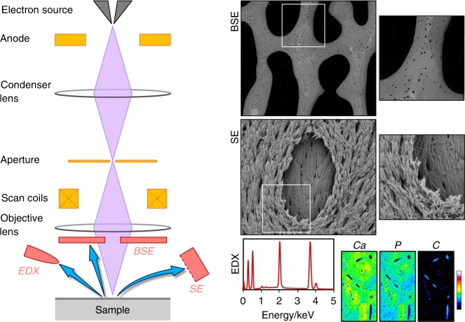
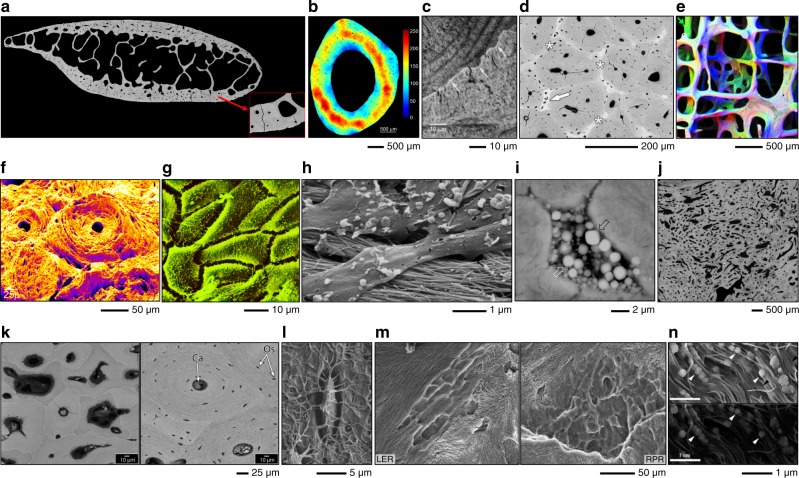
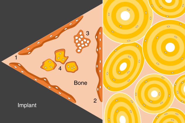

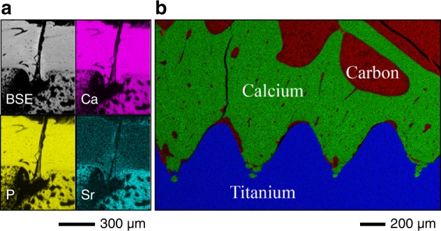

Verification
Claims the paper makes
The claims and statistics of this paper haven’t been checked yet.
Run the check — free, no account needed. It reads the open-access text and checks the sources it cites.
Claims other papers make about this one
Each is checked against this paper’s own text: does it say what the citing paper says it does?
In the citing paper
“The model must be trained to accurately identify key structural components, including the layered bone matrix (lamellae), honeycomb-like network (cancellous bone), osteoblasts, osteoclasts, osteocytes, mineralized bone matrix, and canaliculi.82”
From SEMTWIST Quantification of Biofilm Infection in Human Chronic Wound Using Scanning Electron Microscopy and Machine Learning (2025) · cites this paper as [82]
Self-check
Percentages, sums and reported differences re-computed from the paper’s own numbers.
The self-check needs the paper’s full text.
Citations
Cites
Feng, J. Q., Ward, L. M., Liu, S., Lu, Y., Xie, Y., Yuan, B., Yu, X., Rauch, F., Davis, S. I., Zhang, S., et al. (2006) · Nature Genetics · doi:10.1038/ng1905 · Cited by 1,199
Mahamid, J., Sharir, A., Gur, D., Zelzer, E., Addadi, L., & Weiner, S. (2011) · Journal of Structural Biology · doi:10.1016/j.jsb.2011.03.014 · Cited by 272
Fungal ferromanganese mineralisation in cretaceous dinosaur bones from the gobi desert, Mongolia
Owocki, K., Kremer, B., Wrzosek, B., Królikowska, A., & Kaźmierczak, J. (2016) · PLOS ONE · doi:10.1371/journal.pone.0146293 · Cited by 25
Research works printed but not linked: 258
- Boyde, A. & Jones, S. J. Scanning electron microscopy of cementum and sharpey fibre bone. Z. Zellforsch. Mikros. Anat. 92, 536–548 (1968). · DOI
- Boyde, A. & Hobdell, M. H. Scanning electron microscopy of lamellar bone. Z. Zellforsch. Mikros. Anat. 93, 213–231 (1968). · DOI
- Georgiadis, M., Müller, R. & Schneider, P. Techniques to assess bone ultrastructure organization: orientation and arrangement of mineralized collagen fibrils. J. R. Soc. Interface 13, 20160088 (2016). pii. · DOI
- Palmquist, A. A multiscale analytical approach to evaluate osseointegration. J. Mater. Sci. Mater. Med. 29, 60 (2018). · DOI
- Shah, F. A., Thomsen, P. & Palmquist, A. Osseointegration and current interpretations of the bone-implant interface. Acta Biomater. 84, 1–15 (2019). · DOI
- Goldstein, J. I. et al., Scanning Electron Microscopy and X-ray Microanalysis, 1–550 (Springer-Verlag, New York, 2018).
- Bereshiem, A. C., Pfeiffer, S. K., Grynpas, M. D. & Alblas, A. Use of backscattered scanning electron microscopy to quantify the bone tissues of midthoracic human ribs. Am. J. Phys. Anthrpol. 168, 262–278 (2019). · DOI
- Granke, M., Makowski, A. J., Uppuganti, S. & Nyman, J. S. Prevalent role of porosity and osteonal area over mineralization heterogeneity in the fracture toughness of human cortical bone. J. Biomech. 49, 2748–2755 (2016). · DOI
- Zebaze, R. M. et al. Intracortical remodelling and porosity in the distal radius and post-mortem femurs of women: a cross-sectional study. Lancet 375, 1729–1736 (2010). · DOI
- Shah, F. A., Stoica, A., Cardemil, C. & Palmquist, A. Multiscale characterization of cortical bone composition, microstructure, and nanomechanical properties in experimentally induced osteoporosis. J. Biomed. Mater. Res A 106(4), 997–1007 (2018). · DOI
- Misof, B. M. et al. Intravenous treatment with ibandronate normalizes bone matrix mineralization and reduces cortical porosity after two years in male osteoporosis: a paired biopsy study. J. Bone Miner. Res. 29, 440–449 (2014). · DOI
- Bach-Gansmo, F. L. et al. Osteocyte lacunar properties in rat cortical bone: differences between lamellar and central bone. J. Struct. Biol. 191, 59–67 (2015). · DOI
- Reid, S. A. & Boyde, A. Changes in the mineral density distribution in human bone with age: Image analysis using backscattered electrons in the SEM. J. Bone Miner. Res. 2, 13–22 (1987). · DOI
- Bloebaum, R. D., Skedros, J. G., Vajda, E. G., Bachus, K. N. & Constantz, B. R. Determining mineral content variations in bone using backscattered electron imaging. Bone 20, 485–490 (1997). · DOI
- Hoerth, R. M. et al. Correlations between nanostructure and micromechanical properties of healing bone. J. Mech. Behav. Biomed. Mater. 77, 258–266 (2018). · DOI
- Cui, F. Z., Zhang, Y., Wen, H. B. & Zhu, X. D. Microstructural evolution in external callus of human long bone. Mater. Sci. Eng. C 11, 27–33 (2000). · DOI
- Banse, X. et al. Irreversible perforations in vertebral trabeculae? J. Bone Miner. Res. 18, 1247–1253 (2003). · DOI
- Banse, X., Devogelaer, J. P., Holmyard, D. & Grynpas, M. Vertebral cancellous bone turn-over: Microcallus and bridges in backscatter electron microscopy. Micron 36, 710–714 (2005). · DOI
- Blaskovic, M. et al. Bone healing following different types of osteotomy: scanning electron microscopy (SEM) and three-dimensional SEM analyses. Microsc. Microanal. 22, 1170–1178 (2016). · DOI
- Skedros, J. G., Holmes, J. L., Vajda, E. G. & Bloebaum, R. D. Cement lines of secondary osteons in human bone are not mineral-deficient: new data in a historical perspective. Anat. Rec. A 286A, 781–803 (2005). · DOI
- Milovanovic, P. et al. Bone tissue aging affects mineralization of cement lines. Bone 110, 187–193 (2018). · DOI
- Bach-Gansmo, F. L., Irvine, S. C., Bruel, A., Thomsen, J. S. & Birkedal, H. Calcified cartilage islands in rat cortical bone. Calcif. Tissue Int. 92, 330–338 (2013). · DOI
- Shipov, A. et al. Unremodeled endochondral bone is a major architectural component of the cortical bone of the rat (Rattus norvegicus). J. Struct. Biol. 183, 132–140 (2013). · DOI
- Kingsmill, V. J. & Boyde, A. Mineralisation density of human mandibular bone: quantitative backscattered electron image analysis. J. Anat. 192, 245–256 (1998). · DOI
- Trivellato, A. E. et al. Osteopetrosis complicated by osteomyelitis of the maxilla and mandible: light and electron microscopic findings. Head. Neck Pathol. 3, 320–326 (2009). · DOI
- Boyde, A. et al. A distinctive patchy osteomalacia characterises Phospho1-deficient mice. J. Anat. 231, 298–308 (2017). · DOI
- Shabestari, M. et al. Presence of pyrophosphate in bone from an atypical femoral fracture site: A case report. Bone Rep. 6, 81–86 (2017). · DOI
- Regelsberger, J. et al. Changes to the cell, tissue and architecture levels in cranial suture synostosis reveal a problem of timing in bone development. Eur. Cell Mater. 24, 441–458 (2012). · DOI
- Launey, M. E., Chen, P. Y., McKittrick, J. & Ritchie, R. O. Mechanistic aspects of the fracture toughness of elk antler bone. Acta Biomater. 6, 1505–1514 (2010). · DOI
- Kierdorf, U., Flohr, S., Gomez, S., Landete-Castillejos, T. & Kierdorf, H. The structure of pedicle and hard antler bone in the European roe deer (Capreolus capreolus): a light microscope and backscattered electron imaging study. J. Anat. 223, 364–384 (2013). · DOI
- Boyde, A. Improved digital SEM of cancellous bone: scanning direction of detection, through focus for in-focus and sample orientation. J. Anat. 202, 183–194 (2003). · DOI
- Marks, S. C. Jr, Cielinski, M. J. & Sundquist, K. T. Bone surface morphology reflects local skeletal metabolism. Microsc Res Tech. 33, 121–127 (1996). · DOI
- Jones, S. J., Boyde, A. & Pawley, J. B. Osteoblasts and collagen orientation. Cell Tissue Res 159, 73–80 (1975). · DOI
- Reid, S. A. Micromorphological characterisation of normal human bone surfaces as a function of age. Scanning Microsc 1, 579–597 (1987).
- Mosekilde, L. Consequences of the remodelling process for vertebral trabecular bone structure: a scanning electron microscopy study (uncoupling of unloaded structures). Bone Miner. 10, 13–35 (1990). · DOI
- Jayasinghe, J. A. P., Jones, S. J. & Boyde, A. Scanning electron microscopy of human lumbar vertebral trabecular bone surfaces. Virchows Arch. A 422, 25–34 (1993). · DOI
- Pazzaglia, U. E., Congiu, T., Raspanti, M., Ranchetti, F. & Quacci, D. Anatomy of the Intracortical Canal system: scanning electron microscopy study in Rabbit Femur. Clin. Orthop. Relat. Res 467, 2446–2456 (2009). · DOI
- Corondan, G. & Haworth, W. L. A fractographic study of human long bone. J. Biomech. 19, 207–218 (1986). · DOI
- Tomlin, J. L., Lawes, T. J., Blunn, G. W., Goodship, A. E. & Muir, P. Fractographic examination of racing greyhound central (navicular) tarsal bone failure surfaces using scanning electron microscopy. Calcif. Tissue Int 67, 260–266 (2000). · DOI
- Weiner, S., Arad, T. & Traub, W. Crystal organization in rat bone lamellae. FEBS Lett. 285, 49–54 (1991). · DOI
- Braidotti, P., Bemporad, E., D'Alessio, T., Sciuto, S. A. & Stagni, L. Tensile experiments and SEM fractography on bovine subchondral bone. J. Biomech. 33, 1153–1157 (2000). · DOI
- Zhang, W., Tekalur, S. A., Baumann, M. & McCabe, L. R. The effects of damage accumulation on the tensile strength and toughness of compact bovine bone. J. Biomech. 46, 964–972 (2013). · DOI
- Wise, L. M., Wang, Z. & Grynpas, M. D. The use of fractography to supplement analysis of bone mechanical properties in different strains of mice. Bone 41, 620–630 (2007). · DOI
- Braidotti, P., Branca, F. P. & Stagni, L. Scanning electron microscopy of human cortical bone failure surfaces. J. Biomech. 30, 155–162 (1997). · DOI
- Vashishth, D., Tanner, K. E. & Bonfield, W. Contribution, development and morphology of microcracking in cortical bone during crack propagation. J. Biomech. 33, 1169–1174 (2000). · DOI · Paper page
- Piekarski, K. Fracture of Bone. J. Appl. Phys. 41, 215–223 (1970). · DOI · Paper page
- Hiller, L. P. et al. Osteon pullout in the equine third metacarpal bone: effects of ex vivo fatigue. J. Orthop. Res. 21, 481–488 (2003). · DOI
- Li, S., Abdel-Wahab, A. & Silberschmidt, V. V. Analysis of fracture processes in cortical bone tissue. Eng. Fract. Mech. 110, 448–458 (2013). · DOI
- Fantner, G. E. et al. Sacrificial bonds and hidden length dissipate energy as mineralized fibrils separate during bone fracture. Nat. Mater. 4, 612–616 (2005). · DOI · Paper page
- Nalla, R. K., Kinney, J. H. & Ritchie, R. O. Mechanistic fracture criteria for the failure of human cortical bone. Nat. Mater. 2, 164–168 (2003). · DOI
- Tang, T. et al. Shear deformation and fracture of human cortical bone. Bone 71, 25–35 (2015). · DOI
- Utku, F. S., Klein, E., Saybasili, H., Yucesoy, C. A. & Weiner, S. Probing the role of water in lamellar bone by dehydration in the environmental scanning electron microscope. J. Struct. Biol. 162, 361–367 (2008). · DOI
- Jimenez-Palomar, I., Shipov, A., Shahar, R. & Barber, A. H. Influence of SEM vacuum on bone micromechanics using in situ AFM. J. Mech. Behav. Biomed. Mater. 5, 149–155 (2012). · DOI
- Moffat, K. L. et al. Characterization of the structure–function relationship at the ligament-to-bone interface. Proc. Natl Acad. Sci. USA 105, 7947 (2008). · DOI
- Cury, D. P., Dias, F. J., Miglino, M. A. & Watanabe, I. S. Structural and ultrastructural characteristics of bone–tendon junction of the calcaneal tendon of adult and elderly wistar rats. PLoS ONE 11, e0153568 (2016). · DOI
- Boyde, A. Scanning electron microscopy of bone. Methods Mol. Biol. 816, 365–400 (2012). · DOI
- Jones, S. J. Secretory territories and rate of matrix production of osteoblasts. Calcif. Tissue Res. 14, 309–315 (1974). · DOI
- Menton, D. N., Simmons, D. J., Chang, S. L. & Orr, B. Y. From bone lining cell to osteocyte—an SEM study. Anat. Rec. 209, 29–39 (1984). · DOI
- Pazzaglia, U. E., Congiu, T., Sibilia, V. & Quacci, D. Osteoblast-osteocyte transformation. A SEM densitometric analysis of endosteal apposition in rabbit femur. J. Anat. 224, 132–141 (2014). · DOI
- Shah, F. A., Zanghellini, E., Matic, A., Thomsen, P. & Palmquist, A. The orientation of nanoscale apatite platelets in relation to osteoblastic-osteocyte lacunae on trabecular bone surface. Calcif. Tissue Int 98, 193–205 (2016). · DOI
- Pazzaglia, U. E., Congiu, T., Marchese, M. & Dell'Orbo, C. The shape modulation of osteoblast-osteocyte transformation and its correlation with the fibrillar organization in secondary osteons: a SEM study employing the graded osmic maceration technique. Cell Tissue Res. 340, 533–540 (2010). · DOI
- Qing, H. et al. Demonstration of osteocytic perilacunar/canalicular remodeling in mice during lactation. J. Bone Miner. Res. 27, 1018–1029 (2012). · DOI · Paper page
- Kaya, S. et al. Lactation-induced changes in the volume of osteocyte lacunar-canalicular space alter mechanical properties in cortical bone tissue. J. Bone Miner. Res. 32, 688–697 (2017). · DOI
- Skedros, J. G. Osteocyte lacuna population densities in sheep, elk and horse calcanei. Cells Tissues Organs 181, 23–37 (2005). · DOI
- Busse, B. et al. Decrease in the osteocyte lacunar density accompanied by hypermineralized lacunar occlusion reveals failure and delay of remodeling in aged human bone. Aging cell 9, 1065–1075 (2010). · DOI
- Boyde, A. The real response of bone to exercise. J. Anat. 203, 173–189 (2003). · DOI
- Jones, S. J., Glorieux, F. H., Travers, R. & Boyde, A. The microscopic structure of bone in normal children and patients with osteogenesis imperfecta: a survey using backscattered electron imaging. Calcif. Tissue Int. 64, 8–17 (1999). · DOI
- Carpentier, V. T. et al. Increased proportion of hypermineralized osteocyte lacunae in osteoporotic and osteoarthritic human trabecular bone: implications for bone remodeling. Bone 50, 688–694 (2012). · DOI
- Shah, F. A. et al. Micrometer-sized magnesium whitlockite crystals in micropetrosis of bisphosphonate-exposed human alveolar bone. Nano Lett. 17, 6210–6216 (2017). · DOI
- Rolvien, T. et al. Early bone tissue aging in human auditory ossicles is accompanied by excessive hypermineralization, osteocyte death and micropetrosis. Sci. Rep. 8, 1920 (2018). · DOI
- Bell, L. S., Kayser, M. & Jones, C. The mineralized osteocyte: a living fossil. Am. J. Phys. Anthrpol. 137, 449–456 (2008). · DOI
- N. Fratzl-Zelman et al. Melorheostotic bone lesions caused by somatic mutations in MAP2K1 have deteriorated microarchitecture and periosteal reaction. J. Bone Miner. Res. https://doi.org/10.1002/jbmr.3656 (2019). · DOI
- Shah, F. A., Jergéus, E., Chiba, A. & Palmquist, A. Osseointegration of 3D printed microalloyed CoCr implants—addition of 0.04% Zr to CoCr does not alter bone material properties. J. Biomed. Mater. Res. A 106, 1655–1663 (2018). · DOI
- Shah, F. A., Snis, A., Matic, A., Thomsen, P. & Palmquist, A. 3D printed Ti6Al4V implant surface promotes bone maturation and retains a higher density of less aged osteocytes at the bone-implant interface. Acta Biomater. 30, 357–367 (2016). · DOI
- Shah, F. A. et al. Long-term osseointegration of 3D printed CoCr constructs with an interconnected open-pore architecture prepared by electron beam melting. Acta Biomater. 36, 296–309 (2016). · DOI
- Shah, F. A., Thomsen, P. & Palmquist, A. A review of the impact of implant biomaterials on osteocytes. J. Dent. Res. 97, 977–986 (2018). · DOI
- Atkins, A. et al. Remodeling in bone without osteocytes: billfish challenge bone structure-function paradigms. Proc. Natl Acad. Sci. USA 111, 16047–16052 (2014). · DOI
- Kubek, D. J., Gattone, V. H. & Allen, M. R. Methodological assessment of acid-etching for visualizing the osteocyte lacunar-canalicular networks using scanning electron microscopy. Microsc. Res. Tech. 73, 182–186 (2010). · DOI
- Rangiani, A. et al. Protective roles of DMP1 in high phosphate homeostasis. PLoS ONE 7, e42329 (2012). · DOI
- Zuo, G. L. et al. Activation of HIFa pathway in mature osteoblasts disrupts the integrity of the osteocyte/canalicular network. PLoS ONE 10, e0121266 (2015). · DOI
- Zhang, D. et al. Retention of osteocytic micromorphology by sclerostin antibody in a concurrent ovariectomy and functional disuse model. Ann. N. Y. Acad. Sci. 1442, 91–103 (2019). · DOI
- Shah, F. A. et al. Laser-modified surface enhances osseointegration and biomechanical anchorage of commercially pure titanium implants for bone-anchored hearing systems. PLoS ONE 11, e0157504 (2016). · DOI
- Gentzsch, C., Delling, G. & Kaiser, E. Microstructural classification of resorption lacunae and perforations in human proximal femora. Calcif. Tissue Int. 72, 698–709 (2003). · DOI
- Gentzsch, C., Pueschel, K., Deuretzbacher, G., Delling, G. & Kaiser, E. First inventory of resorption lacunae on rods and plates of trabecular bone as observed by scanning electron microscopy. Calcif. Tissue Int. 76, 154–162 (2005). · DOI
- Ren, S., Takano, H. & Abe, K. Two types of bone resorption lacunae in the mouse parietal bones as revealed by scanning electron microscopy and histochemistry. Arch. Histol. Cytol. 68, 103–113 (2005). · DOI
- Fuller, K., Thong, J. T., Breton, B. C. & Chambers, T. J. Automated three-dimensional characterization of osteoclastic resorption lacunae by stereoscopic scanning electron microscopy. J. Bone Miner. Res. 9, 17–23 (1994). · DOI
- Roschger, P., Fratzl, P., Eschberger, J. & Klaushofer, K. Validation of quantitative backscattered electron imaging for the measurement of mineral density distribution in human bone biopsies. Bone 23, 319–326 (1998). · DOI
- Roschger, P., Paschalis, E. P., Fratzl, P. & Klaushofer, K. Bone mineralization density distribution in health and disease. Bone 42, 456–466 (2008). · DOI
- Pritchard, J. M. et al. Bone mineralization is elevated and less heterogeneous in adults with type 2 diabetes and osteoarthritis compared to controls with osteoarthritis alone. Bone 54, 76–82 (2013). · DOI
- Busse, B. et al. Increased calcium content and inhomogeneity of mineralization render bone toughness in osteoporosis: mineralization, morphology and biomechanics of human single trabeculae. Bone 45, 1034–1043 (2009). · DOI
- Fratzl-Zelman, N. et al. Mineral particle size in children with osteogenesis imperfecta type I is not increased independently of specific collagen mutations. Bone 60, 122–128 (2014). · DOI
- Fratzl-Zelman, N. et al. Unique micro- and nano-scale mineralization pattern of human osteogenesis imperfecta type VI bone. Bone 73, 233–241 (2015). · DOI
- Roschger, P. et al. Alendronate increases degree and uniformity of mineralization in cancellous bone and decreases the porosity in cortical bone of osteoporotic women. Bone 29, 185–191 (2001). · DOI
- Koehne, T. et al. Trends in trabecular architecture and bone mineral density distribution in 152 individuals aged 30–90 years. Bone 66, 31–38 (2014). · DOI
- Stewart, T. L. et al. Association of COLIA1 Sp1 alleles with defective bone nodule formation in vitro and abnormal bone mineralization in vivo. Calcif. Tissue Int. 77, 113–118 (2005). · DOI
- Mahamid, J. et al. Mapping amorphous calcium phosphate transformation into crystalline mineral from the cell to the bone in zebrafish fin rays. Proc. Natl Acad. Sci. USA 107, 6316–6321 (2010). · DOI · Paper page
- Kerschnitzki, M. et al. Bone mineralization pathways during the rapid growth of embryonic chicken long bones. J. Struct. Biol. 195, 82–92 (2016). · DOI
- Karazisis, D. et al. The role of well-defined nanotopography of titanium implants on osseointegration: cellular and molecular events in vivo. Int. J. Nanomed. 11, 1367–1382 (2016).
- Davies, J. E., Ajami, E., Moineddin, R. & Mendes, V. C. The roles of different scale ranges of surface implant topography on the stability of the bone/implant interface. Biomaterials 34, 3535–3546 (2013). · DOI
- Davies, J. E., Mendes, V. C., Ko, J. C. & Ajami, E. Topographic scale-range synergy at the functional bone/implant interface. Biomaterials 35, 25–35 (2014). · DOI
- Traini, T., Degidi, M., Strocchi, R., Caputi, S. & Piattelli, A. Collagen fiber orientation near dental implants in human bone: do their organization reflect differences in loading? J. Biomed. Mater. Res. B 74B, 538–546 (2005). · DOI
- Davies, J. E. Understanding peri-implant endosseous healing. J. Dent. Educ. 67, 932–949 (2003). · DOI
- Hoerth, R. M. et al. Mechanical and structural properties of bone in non-critical and critical healing in rat. Acta Biomater. 10, 4009–4019 (2014). · DOI
- Pobloth, A.-M. et al. Mechanobiologically optimized 3D titanium-mesh scaffolds enhance bone regeneration in critical segmental defects in sheep. Sci. Transl. Med. 10, eaam8828 (2018). · DOI
- Kerschnitzki, M. et al. Poorly ordered bone as an endogenous scaffold for the deposition of highly oriented lamellar tissue in rapidly growing ovine bone. Cells Tissues Organs 194, 119–123 (2011). · DOI
- Franchi, M. et al. Early detachment of titanium particles from various different surfaces of endosseous dental implants. Biomaterials 25, 2239–2246 (2004). · DOI
- Franchi, M. et al. Biological fixation of endosseous implants. Micron 36, 665–671 (2005). · DOI
- Shah, F. A. & Palmquist, A. Evidence that osteocytes in autogenous bone fragments can repair disrupted canalicular networks and connect with osteocytes in de novo formed bone on the fragment surface. Calcif. Tissue Int. 101, 321–327 (2017). · DOI
- Sumner, D. R., Bryan, J. M., Urban, R. M. & Kuszak, J. R. Measuring the volume fraction of bone ingrowth: a comparison of three techniques. J. Orthop. Res. 8, 448–452 (1990). · DOI
- Shah, F. A., Nilson, B., Brånemark, R., Thomsen, P. & Palmquist, A. The bone–implant interface—nanoscale analysis of clinically retrieved dental implants. Nanomedicine 10, 1729–1737 (2014). · DOI
- Stenlund, P. et al. Osseointegration enhancement by Zr doping of Co–Cr–Mo implants fabricated by electron beam melting. Addit. Manuf. 6, 6–15 (2015). · DOI
- Omar, O. M. et al. The correlation between gene expression of proinflammatory markers and bone formation during osseointegration with titanium implants. Biomaterials 32, 374–386 (2011). · DOI
- Palmquist, A., Emanuelsson, L., Brånemark, R. & Thomsen, P. Biomechanical, histological and ultrastructural analyses of laser micro- and nano-structured titanium implant after 6 months in rabbit. J. Biomed. Mater. Res B 97, 289–298 (2011). · DOI
- Palmquist, A. et al. Biomechanical, histological, and ultrastructural analyses of laser micro- and nano-structured titanium alloy implants: a study in rabbit. J. Biomed. Mater. Res. A 92, 1476–1486 (2010). · DOI
- Karlsson, J. et al. Ex vivo alendronate localization at the mesoporous titania implant/bone interface. J. Mater. Sci. Mater. Med. 26, 11 (2015). · DOI
- Braem, A. et al. Peri- and intra-implant bone response to microporous Ti coatings with surface modification. Acta Biomater. 10, 986–995 (2014). · DOI
- Stenlund, P. et al. Bone response to a novel Ti–Ta–Nb–Zr alloy. Acta Biomater. 20, 165–175 (2015). · DOI
- Palmquist, A., Windahl, S. H., Norlindh, B., Brånemark, R. & Thomsen, P. Retrieved bone-anchored percutaneous amputation prosthesis showing maintained osseointegration after 11 years—a case report. Acta Orthop. 85, 442–445 (2014). · DOI
- Bloebaum, R. D., Koller, K. E., Willie, B. M. & Hofmann, A. A. Does using autograft bone chips achieve consistent bone ingrowth in primary TKA? Clin. Orthop. Relat. Res. 470, 1869–1878 (2012). · DOI
- Bobyn, J. D., Stackpool, G. J., Hacking, S. A., Tanzer, M. & Krygier, J. J. Characteristics of bone ingrowth and interface mechanics of a new porous tantalum biomaterial. J. Bone Jt. Surg. Br. 81, 907–914 (1999). · DOI
- Hofmann, A. A., Bloebaum, R. D. & Bachus, K. N. Progression of human bone ingrowth into porous-coated implants. Rate bone ingrowth in humans. Acta Orthop. Scand. 68, 161–166 (1997). · DOI
- Palmquist, A., Snis, A., Emanuelsson, L., Browne, M. & Thomsen, P. Long-term biocompatibility and osseointegration of electron beam melted, free-form-fabricated solid and porous titanium alloy: experimental studies in sheep. J. Biomater. Appl. 27, 1003–1016 (2013). · DOI
- Willie, B. M. et al. Osseointegration into a novel titanium foam implant in the distal femur of a rabbit. J. Biomed. Mater. Res B 92, 479–488 (2010). · DOI
- Willie, B. M. et al. Cancellous bone osseointegration is enhanced by in vivo loading. Tissue Eng. Part C 16, 1399–1406 (2010). · DOI
- Itälä, A. I., Ylänen, H. O., Ekholm, C., Karlsson, K. H. & Aro, H. T. Pore diameter of more than 100 microm is not requisite for bone ingrowth in rabbits. J. Biomed. Mater. Res. 58, 679–683 (2001). · DOI
- Mendes, V. C., Moineddin, R. & Davies, J. E. The effect of discrete calcium phosphate nanocrystals on bone-bonding to titanium surfaces. Biomaterials 28, 4748–4755 (2007). · DOI
- Omar, O. et al. In vivo gene expression in response to anodically oxidized versus machined titanium implants. J. Biomed. Mater. Res. A 92, 1552–1566 (2010). · DOI
- Luklinska, Z. B. & Bonfield, W. Morphology and ultrastructure of the interface between hydroxyapatite–polyhydroxybutyrate composite implant and bone. J. Mater. Sci. Mater. Med. 8, 379–383 (1997). · DOI
- Ma, R. et al. Osseointegration of nanohydroxyapatite- or nano-calcium silicate-incorporated polyetheretherketone bioactive composites in vivo. Int. J. Nanomed. 11, 6023–6033 (2016). · DOI
- Merolli, A., Tranquilli Leali, P. & De Santis, E. A back-scattered electron microscopy (BSEM) study of the tight apposition between bone and hydroxyapatite coating. J. Orthop. Trauma 1, 11–16 (2000). · DOI
- Stadelmann, V. A., Gauthier, O., Terrier, A., Bouler, J. M. & Pioletti, D. P. Implants delivering bisphosphonate locally increase periprosthetic bone density in an osteoporotic sheep model. A pilot study. Eur. Cells Mater. 16, 10–16 (2008). · DOI
- Peter, B. et al. Calcium phosphate drug delivery system: influence of local zoledronate release on bone implant osteointegration. Bone 36, 52–60 (2005). · DOI
- Peter, B. et al. Local delivery of bisphosphonate from coated orthopedic implants increases implants mechanical stability in osteoporotic rats. J. Biomed. Mater. Res. A 76A, 133–143 (2006). · DOI
- Stadelmann, V. A., Terrier, A., Gauthier, O., Bouler, J. M. & Pioletti, D. P. Prediction of bone density around orthopedic implants delivering bisphosphonate. J. Biomech. 42, 1206–1211 (2009). · DOI
- Verron, E. et al. In vivo bone augmentation in an osteoporotic environment using bisphosphonate-loaded calcium deficient apatite. Biomaterials 31, 7776–7784 (2010). · DOI
- Boyde, A., Corsi, A., Quarto, R., Cancedda, R. & Bianco, P. Osteoconduction in large macroporous hydroxyapatite ceramic implants: evidence for a complementary integration and disintegration mechanism. Bone 24, 579–589 (1999). · DOI
- Elgali, I. et al. Molecular and structural patterns of bone regeneration in surgically created defects containing bone substitutes. Biomaterials 35, 3229–3242 (2014). · DOI
- Dias, A. G. et al. In vivo performance of biodegradable calcium phosphate glass ceramics using the rabbit model: histological and SEM observation. J. Biomater. Appl. 20, 253–266 (2006). · DOI
- Le Nihouannen, D. et al. Ectopic bone formation by microporous calcium phosphate ceramic particles in sheep muscles. Bone 36, 1086–1093 (2005). · DOI
- Le Nihouannen, D. et al. Bone tissue formation in sheep muscles induced by a biphasic calcium phosphate ceramic and fibrin glue composite. J. Mater. Sci. Mater. Med. 19, 667–675 (2008). · DOI
- Le Nihouannen, D. et al. Hybrid composites of calcium phosphate granules, fibrin glue, and bone marrow for skeletal repair. J. Biomed. Mater. Res. A 81, 399–408 (2007). · DOI
- Bellucci, D., Anesi, A., Salvatori, R., Chiarini, L. & Cannillo, V. A comparative in vivo evaluation of bioactive glasses and bioactive glass-based composites for bone tissue repair. Mater. Sci. Eng. C 79, 286–295 (2017). · DOI
- Freeman, C. O. et al. Crystallization modifies osteoconductivity in an apatite-mullite glass-ceramic. J. Mater. Sci. Mater. Med. 14, 985–990 (2003). · DOI
- Goodridge, R. D., Wood, D. J., Ohtsuki, C. & Dalgarno, K. W. Biological evaluation of an apatite-mullite glass-ceramic produced via selective laser sintering. Acta Biomater. 3, 221–231 (2007). · DOI
- Liu, X., Rahaman, M. N. & Fu, Q. Bone regeneration in strong porous bioactive glass (13-93) scaffolds with an oriented microstructure implanted in rat calvarial defects. Acta Biomater. 9, 4889–4898 (2013). · DOI
- Liu, X., Rahaman, M. N., Liu, Y., Bal, B. S. & Bonewald, L. F. Enhanced bone regeneration in rat calvarial defects implanted with surface-modified and BMP-loaded bioactive glass (13-93) scaffolds. Acta Biomater. 9, 7506–7517 (2013). · DOI
- Müller-Mai, C., Voigt, C., de Almeida Reis, S. R., Herbst, H. & Gross, U. M. Substitution of natural coral by cortical bone and bone marrow in the rat femur. J. Mater. Sci. Mater. Med. 7, 479–488 (1996). · DOI
- Liao, H., Mutvei, H., Sjöström, M., Hammarström, L. & Li, J. Tissue responses to natural aragonite (Margaritifera shell) implants in vivo. Biomaterials 21, 457–468 (2000). · DOI
- Pascaretti-Grizon, F. et al. The interface between nacre and bone after implantation in the sheep: a nanotomographic and Raman study. J. Raman Spectrosc. 45, 558–564 (2014). · DOI
- Cipitria, A. et al. Porous scaffold architecture guides tissue formation. J. Bone Miner. Res. 27, 1275–1288 (2012). · DOI
- Paris, M. et al. Scaffold curvature-mediated novel biomineralization process originates a continuous soft tissue-to-bone interface. Acta Biomater. 60, 64–80 (2017). · DOI
- Woodruff, M. A., Lange, C., Chen, F., Fratzl, P. & Hutmacher, D. W. Nano- to macroscale remodeling of functional tissue-engineered bone. Adv. Healthc. Mater. 2, 546–551 (2013). · DOI
- Cipitria, A. et al. BMP delivery complements the guiding effect of scaffold architecture without altering bone microstructure in critical-sized long bone defects: a multiscale analysis. Acta Biomater. 23, 282–294 (2015). · DOI
- Reichert, J. C. et al. A tissue engineering solution for segmental defect regeneration in load-bearing long bones. Sci. Transl. Med. 4, 141ra193 (2012). · DOI
- Shah, F. A., Stenlund, P., Martinelli, A., Thomsen, P. & Palmquist, A. Direct communication between osteocytes and acid-etched titanium implants with a sub-micron topography. J. Mater. Sci. Mater. Med. 27, 167 (2016). · DOI
- Du, Z., Ivanovski, S., Hamlet, S. M., Feng, J. Q. & Xiao, Y. The ultrastructural relationship between osteocytes and dental implants following osseointegration. Clin. Implant Dent. Relat. Res. 18, 270–280 (2016). · DOI
- Shah, F. A., Wang, X., Thomsen, P., Grandfield, K. & Palmquist, A. High-resolution visualization of the osteocyte lacuno-canalicular network juxtaposed to the surface of nanotextured titanium implants in human. ACS Biomater. Sci. Eng. 1, 305–313 (2015). · DOI
- Gorustovich, A. A. Imaging resin-cast osteocyte lacuno-canalicular system at bone-bioactive glass interface by scanning electron microscopy. Microsc. Microanal. 16, 132–136 (2010). · DOI
- Åkesson, K., Grynpas, M. D., Hancock, R. G., Odselius, R. & Obrant, K. J. Energy-dispersive X-ray microanalysis of the bone mineral content in human trabecular bone: a comparison with ICPES and neutron activation analysis. Calcif. Tissue Int. 55, 236–239 (1994). · DOI
- Obrant, K. J. & Odselius, R. Electron microprobe investigation of calcium and phosphorus concentration in human bone trabeculae—both normal and in posttraumatic osteopenia. Calcif. Tissue Int. 37, 117–120 (1985). · DOI
- Wierzchos, J. et al. Advances in the ultrastructural study of the implant–bone interface by backscattered electron imaging. Micron 39, 1363–1370 (2008). · DOI
- Gupta, H. S. et al. Two different correlations between nanoindentation modulus and mineral content in the bone–cartilage interface. J. Struct. Biol. 149, 138–148 (2005). · DOI
- Milovanovic, P. et al. The formation of calcified nanospherites during micropetrosis represents a unique mineralization mechanism in aged human bone. Small 13, 1602215 (2017). · DOI
- Brüel, A. et al. Strontium is incorporated into the fracture callus but does not influence the mechanical strength of healing rat fractures. Calcif. Tissue Int. 88, 142–152 (2011). · DOI
- Ballo, A. M. et al. Bone tissue reactions to biomimetic ion-substituted apatite surfaces on titanium implants. J. R. Soc. Interface 9, 1615–1624 (2012). · DOI
- Ballo, A. M. et al. Osseointegration of fiber-reinforced composite implants: histological and ultrastructural observations. Dent. Mater. 30, e384–e395 (2014). · DOI
- Thomas, A. et al. Direct observation of bone coherence with dental implants. J. Eur. Ceram. Soc. 32, 2607–2612 (2012). · DOI
- Chen, P.-Y. & McKittrick, J. Compressive mechanical properties of demineralized and deproteinized cancellous bone. J. Mech. Behav. Biomed. Mater. 4, 961–973 (2011). · DOI
- Congiu, T., Pazzaglia, U. E., Basso, P. & Quacci, D. Chemical etching in processing cortical bone specimens for scanning electron microscopy. Microsc. Res. Tech. 77, 653–660 (2014). · DOI
- Chen, P.-Y., Toroian, D., Price, P. A. & McKittrick, J. Minerals form a continuum phase in mature cancellous bone. Calcif. Tissue Int. 88, 351–361 (2011). · DOI
- Yamamoto, T. et al. Structure and formation of the twisted plywood pattern of collagen fibrils in rat lamellar bone. J. Electron Microsc. 61, 113–121 (2012).
- Ardizzoni, A. Osteocyte lacunar size-lamellar thickness relationships in human secondary osteons. Bone 28, 215–219 (2001). · DOI
- Pazzaglia, U. E., Congiu, T., Marchese, M., Spagnuolo, F. & Quacci, D. Morphometry and patterns of lamellar bone in human Haversian systems. Anat. Rec. 295, 1421–1429 (2012). · DOI
- Congiu, T. & Pazzaglia, U. E. The sealed osteons of cortical diaphyseal bone. Early observations revisited with scanning electron microscopy. Anat. Rec. 294, 193–198 (2011). · DOI
- Mahamid, J., Sharir, A., Addadi, L. & Weiner, S. Amorphous calcium phosphate is a major component of the forming fin bones of zebrafish: Indications for an amorphous precursor phase. Proc. Natl Acad. Sci. USA 105, 12748–12753 (2008). · DOI · Paper page
- Midura, R. J. et al. Calcospherulites isolated from the mineralization front of bone induce the mineralization of type I collagen. Bone 41, 1005–1016 (2007). · DOI
- Midura, R. J., Vasanji, A., Su, X., Midura, S. B. & Gorski, J. P. Isolation of calcospherulites from the mineralization front of bone. Cells Tissues Organs 189, 75–79 (2009). · DOI
- Pazzaglia, U. E. et al. The application of heat-deproteinization to the morphological study of cortical bone: a contribution to the knowledge of the osteonal structure. Microsc. Res. Tech. 79, 691–699 (2016). · DOI
- Holden, J. L., Phakey, P. P. & Clement, J. G. Scanning electron microscope observations of heat-treated human bone. Forensic Sci. Int. 74, 29–45 (1995). · DOI
- Ejiri, S. & Ozawa, H. Scanning electron microscopic observations of rat tibia using the HCl-collagenase method. Arch. Histol. Jpn. 45, 399–404 (1982). · DOI
- Abe, K., Hashizume, H. & Ushiki, T. An EDTA-KOH method to expose bone cells for scanning electron microscopy. J. Electron Microsc. 41, 113–115 (1992).
- Pazzaglia, U. E. & Congiu, T. The cast imaging of the osteon lacunar-canalicular system and the implications with functional models of intracanalicular flow. J. Anat. 222, 193–202 (2013). · DOI
- Bohner, M. et al. Characterization and distribution of mechanically competent mineralized tissue in micropores of β-tricalcium phosphate bone substitutes. Mater. Today 20, 106–115 (2017). · DOI
- Du, Z., Xiao, Y., Hashimi, S., Hamlet, S. M. & Ivanovski, S. The effects of implant topography on osseointegration under estrogen deficiency induced osteoporotic conditions: Histomorphometric, transcriptional and ultrastructural analysis. Acta Biomater. 42, 351–363 (2016). · DOI
- Lin, T. C., Su, C. Y. & Chang, C. S. Stereomorphologic observation of bone tissue response to hydroxyapatite using SEM with the EDTA-KOH method. J. Biomed. Mater. Res. 36, 91–97 (1997). · DOI
- Gung, Y. W., Cheng, C. K. & Su, C. Y. A stereomorphologic study of bone matrix apposition in HA-implanted cavities observed with SEM, being prepared by a microvascular cast and freeze-fracture method. Med. Eng. Phys. 25, 565–571 (2003). · DOI
- Chang, C. S., Su, C. Y. & Lin, T. C. Scanning electron microscopy observation of vascularization around hydroxyapatite using vascular corrosion casts. J. Biomed. Mater. Res. 48, 411–416 (1999). · DOI
- Martinez-Maza, C., Rosas, A., García-Vargas, S., Estalrrich, A. & de la Rasilla, M. Bone remodelling in Neanderthal mandibles from the El Sidrón site (Asturias, Spain). Biol. Lett. 7, 593–596 (2011). · DOI
- Mansilla, J. et al. On porosity of archeological bones II. Textural characterization of Mesoamerican human bones. Palaeogeogr. Palaeoclim. Palaeoecol. 414, 493–499 (2014). · DOI
- Bosch, P. et al. On porosity of archeological bones I—textural characterization of pathological Spanish medieval human bones. Palaeogeogr. Palaeoclim. Palaeoecol. 414, 486–492 (2014). · DOI
- Brickley, M. & Ives, R. Skeletal manifestations of infantile scurvy. Am. J. Phys. Anthropol. 129, 163–172 (2006). · DOI
- Mays, S., Brickley, M. & Ives, R. Skeletal evidence for hyperparathyroidism in a 19th century child with rickets. Int. J. Osteoarchaeol. 17, 73–81 (2007). · DOI
- Brickley, M., Mays, S. & Ives, R. An investigation of skeletal indicators of vitamin D deficiency in adults: effective markers for interpreting past living conditions and pollution levels in 18th and 19th century Birmingham, England. Am. J. Phys. Anthropol. 132, 67–79 (2007). · DOI
- Bell, L. S. in Forensic Microscopy for Skeletal Tissues, Vol. 915, Ch. 911, 173–190 (ed. Bell, L. S.) (Humana Press Totowa, NJ, 2012). · DOI
- Turner-Walker, G. & Jans, M. Reconstructing taphonomic histories using histological analysis. Palaeogeogr. Palaeoclim. Palaeoecol. 266, 227–235 (2008). · DOI
- Fernández-Jalvo, Y. et al. Early bone diagenesis in temperate environments: Part I: Surface features and histology. Palaeogeogr. Palaeoclim. Palaeoecol. 288, 62–81 (2010). · DOI
- Keenan, S. W. & Engel, A. S. Reconstructing diagenetic conditions of bone at the Gray Fossil Site, Tennessee, USA. Palaeogeogr. Palaeoclim. Palaeoecol. 471, 48–57 (2017). · DOI
- Dal Sasso, G., Maritan, L., Usai, D., Angelini, I. & Artioli, G. Bone diagenesis at the micro-scale: Bone alteration patterns during multiple burial phases at Al Khiday (Khartoum, Sudan) between the Early Holocene and the II century AD. Palaeogeogr. Palaeoclim. Palaeoecol. 416, 30–42 (2014). · DOI
- Hollund, H. I., Blank, M. & Sjögren, K. G. Dead and buried? Variation in post-mortem histories revealed through histotaphonomic characterisation of human bone from megalithic graves in Sweden. PLoS One 13, e0204662 (2018). · DOI
- Tjelldén, A. K. E., Kristiansen, S. M., Birkedal, H. & Jans, M. M. E. The pattern of human bone dissolution—a histological study of Iron Age warriors from a Danish wetland site. Int J. Osteoarchaeol. 28, 407–418 (2018). · DOI
200 of 258 shown
Cited by
Sorted by citations
Singh, S., Muniz De Oliveira, F., Wang, C., Kumar, M., Xuan, Y., DeMazumder, D., Sen, C. K., & Roy, S. (2025) · Advances in Wound Care · doi:10.1089/wound.2024.0291 · Cited by 6
Microscopic characteristics of peri- and postmortem fracture surfaces
Skinner, J., Langley, N., Fahrenholtz, S., Shanavas, Y., Waletzki, B., Brown, R., Herrick, J., Shyamsunder, L., Goguen, P., & Rajan, S. (2024) · Forensic Science International · doi:10.1016/j.forsciint.2024.112223 · Cited by 4
Metadata
- Authors
- Shah, F. A., Ruscsák, K., & Palmquist, A.
- Venue
- Bone Research
- Published
- 2019-05-22
- Volume
- 7
- Issue
- 1
- ISSN
- 2095-6231
- PMC
- PMC6531483
- Cited by
- 115
- References
- 3
- Language
- en
About these checks
Registry, retraction, Cites and statistics results are computed, not judged, with one exception: a printed reference that no index lists is read once by a model before it counts as a citation error. Claims are extracted by a model and kept only when their quote appears verbatim in the text; each cited source is then checked, with a model’s help, for whether it says what the claim relies on. Those verdicts can be wrong — read the claim’s quote and the cited source before relying on one.
Statistics and arithmetic are recomputed, not judged.
Confidence bands are shown only for fields calibrated on our annotated test set. None is calibrated yet, so no band is shown.
What counts as a claim ›How we measure confidence ›How we check this ›
Are you an author, or do you see a problem with this page? Request a review
How to cite
Cite CoChat for the checks on this page, and the paper for its own findings.
- This page
- CoChat. (2026). Citation check: 50 years of scanning electron microscopy of bone—a comprehensive overview of the important discoveries made and insights gained into bone material properties in health, disease, and taphonomy. https://app.cochat.ai/papers/10.1038/s41413-019-0053-z
- The paper
- Shah, F. A., Ruscsák, K., & Palmquist, A. (2019). 50 years of scanning electron microscopy of bone—a comprehensive overview of the important discoveries made and insights gained into bone material properties in health, disease, and taphonomy. Bone Research. https://doi.org/10.1038/s41413-019-0053-z
