

The DOI matches its Crossref record; 7 of the 7 references it cites were found in a registry or an index.
miR-182-5p and miR-378a-3p regulate ferroptosis in I/R-induced renal injury
Abstract
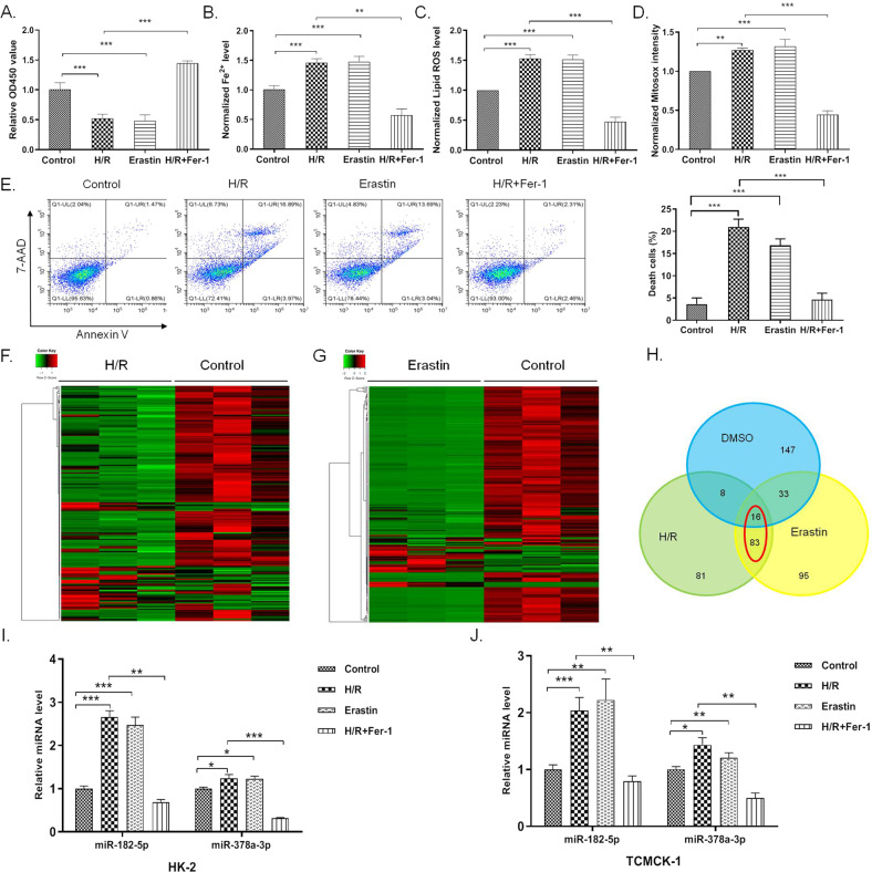
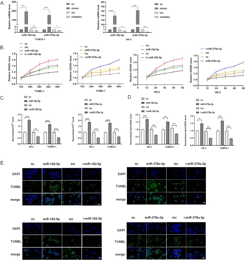
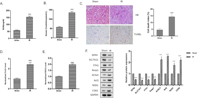
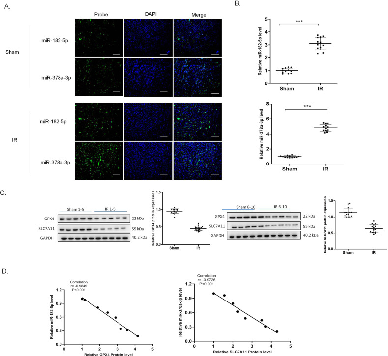
Renal tubular cell death is the key factor of the pathogenesis of ischemia/reperfusion (I/R) kidney injury. Ferroptosis is a type of regulated cell death (RCD) found in various diseases. However, the underlying molecular mechanisms related to ferroptosis in renal I/R injury remain unclear. In the present study, we investigated the regulatory role of microRNAs on ferroptosis in I/R-induced renal injury. We established the I/R-induced renal injury model in rats, and H/R induced HK-2 cells injury in vitro. CCK-8 was used to measure cell viability. Fe 2+ and ROS levels were assayed to evaluate the activation of ferroptosis. We performed RNA sequencing to profile the miRNAs expression in H/R-induced injury and ferroptosis. Western blot analysis was used to detect the protein expression. qRT-PCR was used to detect the mRNA and miRNA levels in cells and tissues. We further used luciferase reporter assay to verify the direct targeting effect of miRNA. We found that ischemia/reperfusion-induced ferroptosis in rat’s kidney. We identified that miR-182-5p and miR-378a-3p were upregulated in the ferroptosis and H/R-induced injury, and correlates reversely with glutathione peroxidases 4 (GPX4) and solute carrier family 7 member 11 (SLC7A11) expression in renal I/R injury tissues, respectively. In vitro studies showed that miR-182-5p and miR-378a-3p induced ferroptosis in cells. We further found that miR-182-5p and miR-378a-3p regulated the expression of GPX4 and SLC7A11 negatively by directly binding to the 3′UTR of GPX4 and SLC7A11 mRNA. In vivo study showed that silencing miR-182-5p and miR-378a-3p alleviated the I/R-induced renal injury in rats. In conclusion, we demonstrated that I/R induced upregulation of miR-182-5p and miR-378a-3p, leading to activation of ferroptosis in renal injury through downregulation of GPX4 and SLC7A11.
Overview
The claims and statistics of this paper haven’t been checked yet.
Run the check — free, no account needed. It reads the open-access text and checks the sources it cites.
Integrity signals
Registry
Cites
Statistics
Figures
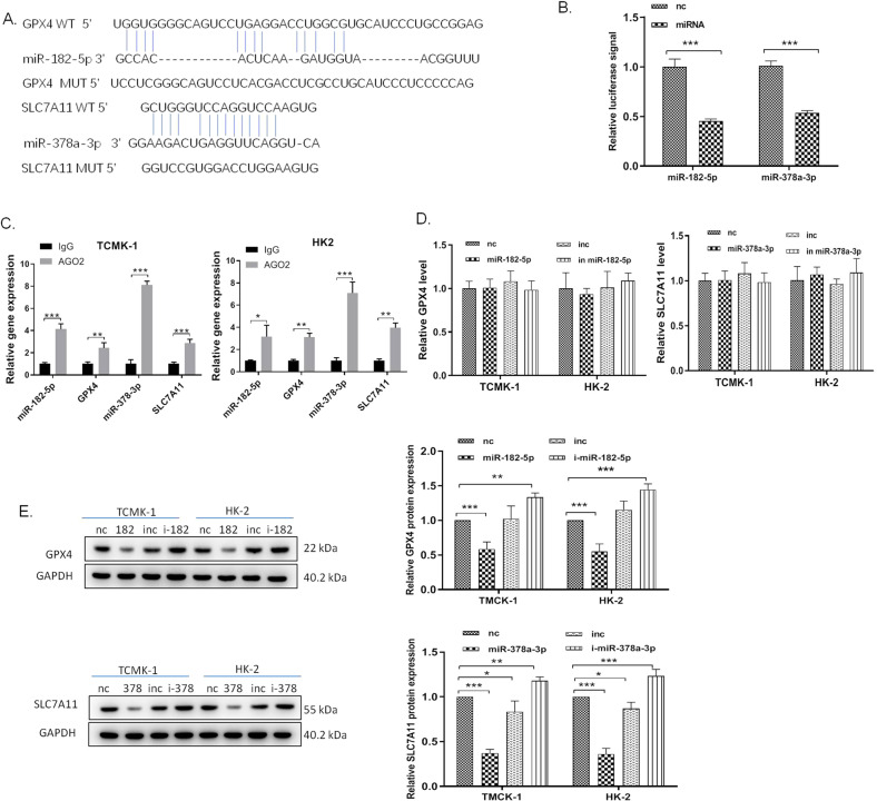
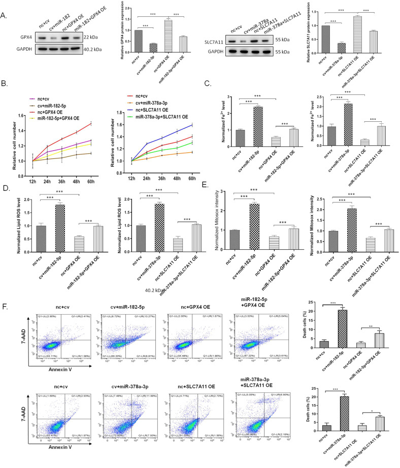
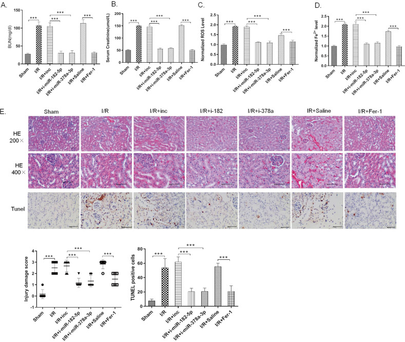
Verification
Claims the paper makes
The claims and statistics of this paper haven’t been checked yet.
Run the check — free, no account needed. It reads the open-access text and checks the sources it cites.
Claims other papers make about this one
Each is checked against this paper’s own text: does it say what the citing paper says it does?
In the citing paper
“For example, miR-182-5p and miR-378a-3p modulate ferroptosis in an ischemia/reperfusion (I/R)-induced renal injury by affecting the mRNA expression of GPX4 and SLC7A11 (Ding et al., 2020).”
From MiR-378a-3p/ SLC7A11 regulate ferroptosis in nerve injury induced by lead exposure (2022) · cites this paper as Ding et al., 2020
In the citing paper
“MiRNAs are also involved in regulation of ferroptosis.”
From MiR-378a-3p/ SLC7A11 regulate ferroptosis in nerve injury induced by lead exposure (2022) · cites this paper as Ding et al., 2020
In the citing paper
“Coincidentally, a study by Ding also revealed that miR-378a-3p decreases the expression of SLC7A11 by binding to the 3 ′ -UTR of SLC7A11 mRNA in the mouse kidney epithelial cell line TCMK-1 (Ding et al., 2020).”
From MiR-378a-3p/ SLC7A11 regulate ferroptosis in nerve injury induced by lead exposure (2022) · cites this paper as Ding et al., 2020
In the citing paper
“Ding et al. also showed that miR-378a-3p directly binds to the 3 ′ -UTR of SLC7A11 (Ding et al., 2020).”
From MiR-378a-3p/ SLC7A11 regulate ferroptosis in nerve injury induced by lead exposure (2022) · cites this paper as Ding et al., 2020
In the citing paper
“The results showed that the co-transfection of miR- 378a-3p with the wild-type SLC7A11 luciferase reporter reduced luciferase activity compared to that of the mutant SLC7A11 luciferase reporter, suggesting that miR-378a-3p directly binds to the 3 ′ -UTR of SLC7A11(Fig.5 C and Fig. 5D).”
From MiR-378a-3p/ SLC7A11 regulate ferroptosis in nerve injury induced by lead exposure (2022) · cites this paper as Ding et al., 2020
Self-check
Percentages, sums and reported differences re-computed from the paper’s own numbers.
The self-check needs the paper’s full text.
Citations
Cites
Regulation of Ferroptotic Cancer Cell Death by GPX4
Yang, W. S., SriRamaratnam, R., Welsch, M. E., Shimada, K., Skouta, R., Viswanathan, V. S., Cheah, J. H., Clemons, P. A., Shamji, A. F., Clish, C. B., et al. (2014) · Cell · doi:10.1016/j.cell.2013.12.010 · Cited by 7,713
ACSL4 dictates ferroptosis sensitivity by shaping cellular lipid composition
Doll, S., Proneth, B., Tyurina, Y. Y., Panzilius, E., Kobayashi, S., Ingold, I., Irmler, M., Beckers, J., Aichler, M., Walch, A., et al. (2017) · Nature Chemical Biology · doi:10.1038/nchembio.2239 · Cited by 4,293
Silencing of microRNAs in vivo with ‘antagomirs’
Krützfeldt, J., Rajewsky, N., Braich, R., Rajeev, K. G., Tuschl, T., Manoharan, M., & Stoffel, M. (2005) · Nature · doi:10.1038/nature04303 · Cited by 3,949
Inactivation of the ferroptosis regulator Gpx4 triggers acute renal failure in mice
Friedmann Angeli, J. P., Schneider, M., Proneth, B., Tyurina, Y. Y., Tyurin, V. A., Hammond, V. J., Herbach, N., Aichler, M., Walch, A., Eggenhofer, E., et al. (2014) · Nature Cell Biology · doi:10.1038/ncb3064 · Cited by 3,732
Ferroptosis: process and function
Xie, Y., Hou, W., Song, X., Yu, Y., Huang, J., Sun, X., Kang, R., & Tang, D. (2016) · Cell Death & Differentiation · doi:10.1038/cdd.2015.158 · Cited by 3,727
Lu, T. X., & Rothenberg, M. E. (2018) · Journal of Allergy and Clinical Immunology · doi:10.1016/j.jaci.2017.08.034 · Cited by 2,425
Ferrostatins Inhibit Oxidative Lipid Damage and Cell Death in Diverse Disease Models
Skouta, R., Dixon, S. J., Wang, J., Dunn, D. E., Orman, M., Shimada, K., Rosenberg, P. A., Lo, D. C., Weinberg, J. M., Linkermann, A., et al. (2014) · Journal of the American Chemical Society · doi:10.1021/ja411006a · Cited by 1,146
Research works printed but not linked: 51
- Schrier, R. W., Wang, W., Poole, B. & Mitra, A. Acute renal failure: definitions, diagnosis, pathogenesis, and therapy. J. Clin. Invest 114, 5–14 (2004). · DOI
- Chertow, G. M., Burdick, E., Honour, M., Bonventre, J. V. & Bates, D. W. Acute kidney injury, mortality, length of stay, and costs in hospitalized patients. J. Am. Soc. Nephrol. 16, 3365–3370 (2005). · DOI
- Ishani, A. et al. Acute kidney injury increases risk of ESRD among elderly. J. Am. Soc. Nephrol. 20, 223–228 (2009). · DOI
- Mehta, R. L. Outcomes research in acute renal failure. Semin. Nephrol. 23, 283–294 (2003). · DOI
- Guan, X. et al. Autophagy protects renal tubular cells against ischemia/reperfusion injury in a time-dependent manner. Cell Physiol. Biochem. 36, 285–298 (2015). · DOI
- Migita, H., Yoshitake, S., Tange, Y., Choijookhuu, N. & Hishikawa, Y. Hyperbaric oxygen therapy suppresses apoptosis and promotes renal tubular regeneration after renal ischemia/reperfusion injury in rats. Nephrourol. Mon. 8, e34421 (2016). · DOI
- Winterberg, P. D. & Lu, C. Y. Acute kidney injury: the beginning of the end of the dark ages. Am. J. Med. Sci. 344, 318–325 (2012). · DOI
- Wu, M. Y. et al. Current mechanistic concepts in ischemia and reperfusion injury. Cell Physiol. Biochem. 46, 1650–1667 (2018). · DOI
- Bonventre, J. V. & Yang, L. Cellular pathophysiology of ischemic acute kidney injury. J. Clin. Invest. 121, 4210–4221 (2011). · DOI
- Dixon, S. J. et al. Ferroptosis: an iron-dependent form of nonapoptotic cell death. Cell 149, 1060–1072 (2012). · DOI · Paper page
- Lachaier, E. et al. Sorafenib induces ferroptosis in human cancer cell lines originating from different solid tumors. Anticancer Res. 34, 6417–6422 (2014).
- Dzikiewicz-Krawczyk, A. et al. ZDHHC11 and ZDHHC11B are critical novel components of the oncogenic MYC-miR-150-MYB network in Burkitt lymphoma. Leukemia 31, 1470–1473 (2017). · DOI
- Masaldan, S., Bush, A. I., Devos, D., Rolland, A. S. & Moreau, C. Striking while the iron is hot: iron metabolism and ferroptosis in neurodegeneration. Free Radic. Biol. Med. 133, 221–233 (2019). · DOI
- Tuo, Q. Z. et al. Tau-mediated iron export prevents ferroptotic damage after ischemic stroke. Mol. Psychiatry 22, 1520–1530 (2017). · DOI
- Sheng, X. et al. Theoretical insights into the mechanism of ferroptosis suppression via inactivation of a lipid peroxide radical by liproxstatin-1. Phys. Chem. Chem. Phys. 19, 13153–13159 (2017). · DOI
- Fang, X. et al. Loss of cardiac ferritin H facilitates cardiomyopathy via Slc7a11-mediated ferroptosis. Circ. Res. 127, 486–501 (2020). · DOI
- Guerrero-Hue, M. et al. Curcumin reduces renal damage associated with rhabdomyolysis by decreasing ferroptosis-mediated cell death. FASEB J. 33, 8961–8975 (2019). · DOI
- Ma, D. et al. Inhibition of ferroptosis attenuates acute kidney injury in rats with severe acute pancreatitis. Dig. Dis. Sci. 2020, https://doi.org/10.1007/s10620-020-06225-2 (2020). · DOI
- Linkermann, A. et al. Synchronized renal tubular cell death involves ferroptosis. Proc. Natl Acad. Sci. USA 111, 16836–16841 (2014). · DOI · Paper page
- Lu, T. X. et al. MicroRNA-21 limits in vivo immune response-mediated activation of the IL-12/IFN-gamma pathway, Th1 polarization, and the severity of delayed-type hypersensitivity. J. Immunol. 187, 3362–3373 (2011). · DOI
- Zhu, H. et al. Regulation of autophagy by a beclin 1-targeted microRNA, miR-30a, in cancer cells. Autophagy 5, 816–823 (2009). · DOI
- Chen, J. F. et al. The role of microRNA-1 and microRNA-133 in skeletal muscle proliferation and differentiation. Nat. Genet 38, 228–233 (2006). · DOI
- Johnson, C. D. et al. The let-7 microRNA represses cell proliferation pathways in human cells. Cancer Res. 67, 7713–7722 (2007). · DOI · Paper page
- Lynam-Lennon, N., Maher, S. G. & Reynolds, J. V. The roles of microRNA in cancer and apoptosis. Biol. Rev. Camb. Philos. Soc. 84, 55–71 (2009). · DOI
- Zhang, X. et al. Circular RNA circNRIP1 acts as a microRNA-149-5p sponge to promote gastric cancer progression via the AKT1/mTOR pathway. Mol. Cancer 18, 20 (2019). · DOI
- Drobna, M. et al. hsa-miR-20b-5p and hsa-miR-363-3p affect expression of PTEN and BIM tumor suppressor genes and modulate survival of T-ALL cells in vitro. Cells 9, 1137 (2020). · DOI
- Xia, L. et al. miR-15b and miR-16 modulate multidrug resistance by targeting BCL2 in human gastric cancer cells. Int. J. Cancer 123, 372–379 (2008). · DOI
- Hao, J. et al. Induction of microRNA-17-5p by p53 protects against renal ischemia-reperfusion injury by targeting death receptor 6. Kidney Int. 91, 106–118 (2017). · DOI
- Wu, H. et al. MiR-155 is involved in renal ischemia-reperfusion injury via direct targeting of FoxO3a and regulating renal tubular cell pyroptosis. Cell Physiol. Biochem. 40, 1692–1705 (2016). · DOI
- Fontecha-Barriuso, M. et al. Molecular pathways driving omeprazole nephrotoxicity. Redox Biol. 32, 101464 (2020). · DOI
- Wang, D. et al. The long noncoding RNA Arrl1 inhibits neurite outgrowth by functioning as a competing endogenous RNA during neuronal regeneration in rats. J. Biol. Chem. 295, 8374–8386 (2020). · DOI
- Qin, Y. et al. Tanshinone A inhibits VSMC inflammation and proliferation in vivo and in vitro by downregulating miR-712-5p expression. Eur. J. Pharmacol. 880, 173140 (2020). · DOI
- Eckstein, M. et al. Co-staining of microRNAs and their target proteins by miRNA in situ hybridization and immunohistofluorescence on prostate cancer tissue microarrays. Lab Invest. 99, 1527–1534 (2019). · DOI
- Silahtaroglu, A. N. et al. Detection of microRNAs in frozen tissue sections by fluorescence in situ hybridization using locked nucleic acid probes and tyramide signal amplification. Nat. Protoc. 2, 2520–2528 (2007). · DOI
- Tan, X. et al. Postconditioning ameliorates mitochondrial DNA damage and deletion after renal ischemic injury. Nephrol. Dial. Transpl. 28, 2754–2765 (2013). · DOI
- Tan, X. H. et al. Fibroblast growth factor 2 protects against renal ischaemia/reperfusion injury by attenuating mitochondrial damage and proinflammatory signalling. J. Cell Mol. Med. 21, 2909–2925 (2017). · DOI
- Ren, X. P. et al. MicroRNA-320 is involved in the regulation of cardiac ischemia/reperfusion injury by targeting heat-shock protein 20. Circulation 119, 2357–2366 (2009). · DOI
- Lu, Y. et al. MicroRNA-328 contributes to adverse electrical remodeling in atrial fibrillation. Circulation 122, 2378–2387 (2010). · DOI
- Zhou, W. et al. Predominant role for C5b-9 in renal ischemia/reperfusion injury. J. Clin. Invest. 105, 1363–1371 (2000). · DOI
- Yu, Y. et al. The ferroptosis inducer erastin enhances sensitivity of acute myeloid leukemia cells to chemotherapeutic agents. Mol. Cell Oncol. 2, e1054549 (2015). · DOI
- Chen, Y., Jiang, S., Zou, J., Zhong, Y. & Ding, X. Silencing HIF-1alpha aggravates growth inhibition and necrosis of proximal renal tubular epithelial cell under hypoxia. Ren. Fail 38, 1726–1734 (2016). · DOI
- Chen, L. et al. Erastin sensitizes glioblastoma cells to temozolomide by restraining xCT and cystathionine-gamma-lyase function. Oncol. Rep. 33, 1465–1474 (2015). · DOI
- Zhang, Y. H. et al. alpha-Lipoic acid improves abnormal behavior by mitigation of oxidative stress, inflammation, ferroptosis, and tauopathy in P301S Tau transgenic mice. Redox Biol. 14, 535–548 (2018). · DOI
- Gao, M., Monian, P., Quadri, N., Ramasamy, R. & Jiang, X. Glutaminolysis and transferrin regulate ferroptosis. Mol. Cell 59, 298–308 (2015). · DOI · Paper page
- Xue, J. et al. miR-182-5p induced by STAT3 activation promotes glioma tumorigenesis. Cancer Res. 76, 4293–4304 (2016). · DOI
- Wilflingseder, J. et al. miR-182-5p inhibition ameliorates ischemic acute kidney injury. Am. J. Pathol. 187, 70–79 (2017). · DOI
- Wilflingseder, J. et al. Molecular pathogenesis of post-transplant acute kidney injury: assessment of whole-genome mRNA and miRNA profiles. PLoS ONE 9, e104164 (2014). · DOI
- Ikeda, K. et al. miR-378a-3p modulates tamoxifen sensitivity in breast cancer MCF-7 cells through targeting GOLT1A. Sci. Rep. 5, 13170 (2015). · DOI
- Zou, Y. F. et al. Urinary MicroRNA-30c-5p and MicroRNA-192-5p as potential biomarkers of ischemia-reperfusion-induced kidney injury. Exp. Biol. Med. 242, 657–667 (2017). · DOI
- Banjac, A. et al. The cystine/cysteine cycle: a redox cycle regulating susceptibility versus resistance to cell death. Oncogene 27, 1618–1628 (2008). · DOI
- Shibasaki, T. et al. Aggravation of ischemia-reperfusion-triggered acute renal failure in xCT-deficient mice. Arch. Biochem. Biophys. 490, 63–69 (2009). · DOI
Cited by
Sorted by citations
Iron Homeostasis and Ferroptosis in Human Diseases: Mechanisms and Therapeutic Prospects
Ru, Q., Li, Y., Chen, L., Wu, Y., Min, J., & Wang, F. (2024) · Signal Transduction and Targeted Therapy · doi:10.1038/s41392-024-01969-z · Cited by 724
MiR-378a-3p/ SLC7A11 regulate ferroptosis in nerve injury induced by lead exposure
Wang, W., Shi, F., Cui, J., Pang, S., Zheng, G., & Zhang, Y. (2022) · Ecotoxicology and Environmental Safety · doi:10.1016/j.ecoenv.2022.113639 · Cited by 47
Metadata
- Authors
- Ding, C., Ding, X., Zheng, J., Wang, B., Li, Y., Xiang, H., Dou, M., Qiao, Y., Tian, P., & Xue, W.
- ORCID
- Qiao, Yuxi
- Venue
- Cell Death & Disease
- Published
- 2020-10-28
- Volume
- 11
- Issue
- 10
- ISSN
- 2041-4889
- PMC
- PMC7595188
- Cited by
- 197
- References
- 7
- Language
- en
About these checks
Registry, retraction, Cites and statistics results are computed, not judged, with one exception: a printed reference that no index lists is read once by a model before it counts as a citation error. Claims are extracted by a model and kept only when their quote appears verbatim in the text; each cited source is then checked, with a model’s help, for whether it says what the claim relies on. Those verdicts can be wrong — read the claim’s quote and the cited source before relying on one.
Statistics and arithmetic are recomputed, not judged.
Confidence bands are shown only for fields calibrated on our annotated test set. None is calibrated yet, so no band is shown.
What counts as a claim ›How we measure confidence ›How we check this ›
Are you an author, or do you see a problem with this page? Request a review
