

The DOI matches its Crossref record; 186 of the 186 references it cites were found in a registry or an index.
Liver macrophages: development, dynamics, and functions
Open accessAbstract
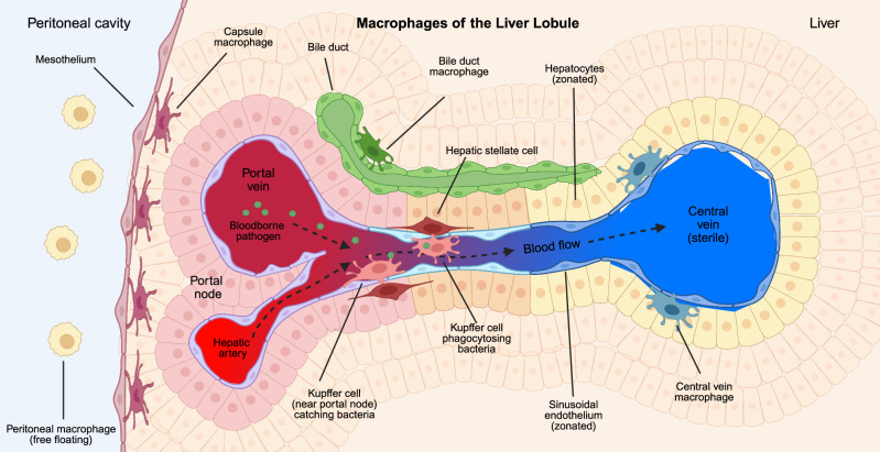
The liver is a sizeable visceral organ whose primary functions involve nutrient metabolism, clearance of toxins, and energy storage. Besides these critical functions, the liver is also a major immunological site. It is populated by several specialized resident immune cells, including B cells, T Cells, dendritic cells, and several populations of macrophages. It is also the site for the production and release of acute-phase proteins during inflammation. One reason for garrisoning these immune sentinels and effectors in the liver is its relative location in the circulatory system. The liver is the first significant organ downstream of the intestine, where blood originating from the intestine enters the liver through the portal vein. This organization facilitates the liver’s uptake and processing of nutrient-rich blood directly from the intestinal source. However, the intestine is also home to trillions of microbes, many of which are commensals but also represent potential pathogens. As such, the portal blood supply represents an avenue for systemic infection. To sterilize the portal blood, the liver immune system filters pathogens, which is primarily accomplished by liver macrophages. Here, we will discuss the major populations of macrophages resident in the liver, their location, functions, development, and role in maintaining the liver in the face of injury and infection.
Overview
The claims and statistics of this paper haven’t been checked yet.
Free: it reads the open-access text and checks the first 5 claims that cite a source we can check. Create a free account to check every claim.
Integrity signals
Registry
Cites
Statistics
Figures
Verification
Claims the paper makes
The claims and statistics of this paper haven’t been checked yet.
Free: it reads the open-access text and checks the first 5 claims that cite a source we can check. Create a free account to check every claim.
Claims other papers make about this one
Each is checked against this paper’s own text: does it say what the citing paper says it does?
The source backs part of the claim, not all of it — or backs it for a different population, condition or comparison.
In the citing paper
“In addition to competing for survival signals like CSF-1 (9, 33, 34), infiltrating monocytes actively replace embryonic KCs (EmKCs) during injury (35–37).”
From Macrophage duality in liver diseases: heterogeneity, plasticity, and the quest for precision therapeutics (2026) · cites this paper as [9]
Experimental — accuracy not yet measuredThe cited source says what the claim attributes to it.
In the citing paper
“Following hepatic insult, however, MoMFs infiltrate the liver in large numbers and can adopt functionally divergent phenotypes, driving either pro-inflammatory tissue injury or anti-inflammatory repair (9).”
From Macrophage duality in liver diseases: heterogeneity, plasticity, and the quest for precision therapeutics (2026) · cites this paper as [9]
Self-check
Percentages, sums and reported differences re-computed from the paper’s own numbers.
The self-check needs the paper’s full text.
Citations
Cites
Mizgerd, J. P., Kubo, H., Kutkoski, G. J., Bhagwan, S. D., Scharffetter-Kochanek, K., Beaudet, A. L., & Doerschuk, C. M. (1997) · The Journal of Experimental Medicine · doi:10.1084/jem.186.8.1357 · Cited by 7,306
Macrophage biology in development, homeostasis and disease
Wynn, T. A., Chawla, A., & Pollard, J. W. (2013) · Nature · doi:10.1038/nature12034 · Cited by 4,684 · cited 1×
“Classically, macrophages across organs express markers such as F4/80, CD64, MerTK, and Iba1 [57 – 61].”
MAFLD: a consensus-driven proposed nomenclature for metabolic associated fatty liver disease
Eslam, M., Sanyal, A. J., George, J., Sanyal, A., Neuschwander-Tetri, B., Tiribelli, C., Kleiner, D. E., Brunt, E., Bugianesi, E., Yki-Järvinen, H., et al. (2020) · Gastroenterology · doi:10.1053/j.gastro.2019.11.312 · Cited by 3,286
Byrne, C. D., & Targher, G. (2015) · Journal of Hepatology · doi:10.1016/j.jhep.2014.12.012 · Cited by 3,042
Mechanisms of hepatic stellate cell activation
Tsuchida, T., & Friedman, S. L. (2017) · Nature Reviews Gastroenterology & Hepatology · doi:10.1038/nrgastro.2017.38 · Cited by 2,909
Tissue-resident macrophages originate from yolk-sac-derived erythro-myeloid progenitors
Gomez Perdiguero, E., Klapproth, K., Schulz, C., Busch, K., Azzoni, E., Crozet, L., Garner, H., Trouillet, C., de Bruijn, M. F., Geissmann, F., et al. (2015) · Nature · doi:10.1038/nature13989 · Cited by 2,339
Resolving the fibrotic niche of human liver cirrhosis at single-cell level
Ramachandran, P., Dobie, R., Wilson-Kanamori, J. R., Dora, E. F., Henderson, B. E. P., Luu, N. T., Portman, J. R., Matchett, K. P., Brice, M., Marwick, J. A., et al. (2019) · Nature · doi:10.1038/s41586-019-1631-3 · Cited by 1,784
Hepatotoxicity and Mechanism of Action of Haloalkanes: Carbon Tetrachloride as a Toxicological Model
Weber, L. W. D., Boll, M., & Stampfl, A. (2003) · Critical Reviews in Toxicology · doi:10.1080/713611034 · Cited by 1,777
Willingham, S. B., Volkmer, J.-P., Gentles, A. J., Sahoo, D., Dalerba, P., Mitra, S. S., Wang, J., Contreras-Trujillo, H., Martin, R., Cohen, J. D., et al. (2012) · Proceedings of the National Academy of Sciences · doi:10.1073/pnas.1121623109 · Cited by 1,666
THE ORIGIN AND KINETICS OF MONONUCLEAR PHAGOCYTES
van Furth, R., & Cohn, Z. A. (1968) · The Journal of Experimental Medicine · doi:10.1084/jem.128.3.415 · Cited by 1,563
Cited by
Metabolic dysfunction and alcohol-associated liver disease (MetALD)
Gao, B., Arab, J. P., Liangpunsakul, S., Ding, W., Szabó, G., Mehal, W. Z., Wang, H., He, Y., Stärkel, P., Llorente, C., et al. (2025) · eGastroenterology · doi:10.1136/egastro-2025-100319 · Cited by 53
The Gut‒Liver Axis in Liver Disease: Molecular Mechanisms and Therapeutic Targets
Chen, Z., Liao, S., Wu, S., Chen, S., Tang, Q., Zhou, L., Li, X., Hu, H., Xu, J., Zhang, A., et al. (2025) · MedComm · doi:10.1002/mco2.70458 · Cited by 34
Tsomidis, I., Tsakou, A., Voumvouraki, A., & Kouroumalis, E. (2026) · Biomedicines · doi:10.3390/biomedicines14010151 · Cited by 24
Macrophages in chronic infections: regulation and remodeling
Cui, H., Wang, M., Jiao, S., Tian, S., Liu, H., & Luo, B. (2025) · Frontiers in Immunology · doi:10.3389/fimmu.2025.1594988 · Cited by 14
Wang, W., Yi, Z., Yang, Z., Huang, Y., Chen, H., Li, Y., Jing, L., Yin, S., & Zhang, F. (2025) · Journal of Translational Medicine · doi:10.1186/s12967-025-07376-4 · Cited by 10
Metadata
- Authors
- Nusse, Y., & Kubes, P.
- Affiliations
- University of Calgary (CA)
- Corresponding author
- Kubes, Paul
- Venue
- Cellular & Molecular Immunology
- Published
- 2025-06-03
- Volume
- 22
- Issue
- 10
- Pages
- 1178-1189
- ISSN
- 1672-7681 (ISSN-L)
- 2042-0226
- PMC
- PMC12480653
- OpenAlex
- W4410974132
- Cited by
- 50
- References
- 184
- Citations by year
- 2026: 43 · 2025: 7
- FWCI
- 7.31
- Type
- journal article
- Language
- en
- Open access
- hybrid
- doi.org
- Licence
- cc-by
- creativecommons.org/licenses/by/4.0 (tdm)
- creativecommons.org/licenses/by/4.0 (vor)
- Locations
- Cellular and Molecular Immunology (publishedVersion, cc-by)
- Europe PMC (PubMed Central) (publishedVersion, cc-by)
- PubMed (publishedVersion)
- PubMed Central (publishedVersion, cc-by)
- Topics
- Health Sciences › Medicine › Hepatology › Liver physiology and pathology
- Life Sciences › Immunology and Microbiology › Immunology › Immune cells in cancer
- Life Sciences › Immunology and Microbiology › Immunology › Phagocytosis and Immune Regulation
- Keywords
- hepatic macrophages, Kupffer cells, liver immunity, macrophage development, portal blood, systemic infection, intestinal commensals, acute phase proteins, liver injury
- MeSH terms
- Animals, Humans, Liver, Macrophages
About these checks
Registry, retraction, Cites and statistics results are computed, not judged, with one exception: a printed reference that no index lists is read once by a model before it counts as a citation error. Claims are extracted by a model and kept only when their quote appears verbatim in the text; each cited source is then checked, with a model’s help, for whether it says what the claim relies on. Those verdicts can be wrong — read the claim’s quote and the cited source before relying on one.
Statistics and arithmetic are recomputed, not judged.
Confidence bands are shown only for fields calibrated on our annotated test set. None is calibrated yet, so no band is shown.
What counts as a claim ›How we measure confidence ›How we check this ›
Are you an author, or do you see a problem with this page? Request a review
How to cite
Cite CoChat for the checks on this page, and the paper for its own findings.
- This page
- CoChat. (2026). Citation check: Liver macrophages: development, dynamics, and functions. https://app.cochat.ai/papers/10.1038/s41423-025-01298-3
- The paper
- Nusse, Y., & Kubes, P. (2025). Liver macrophages: development, dynamics, and functions. Cellular & Molecular Immunology. https://doi.org/10.1038/s41423-025-01298-3
Data sources
Bibliographic data: Semantic Scholar, OpenAlex, Crossref.
Contains information from the Semantic Scholar Academic Graph Datasets, made available under the ODC Attribution License (ODC-BY 1.0).
Semantic Scholar’s data is described in: Kinney, R., et al. (2023). The Semantic Scholar Open Data Platform. arXiv:2301.10140.
