

The DOI matches its Crossref record; 42 of the 42 references it cites were found in a registry or an index.
Genome editing abrogates angiogenesis in vivo
Open accessAbstract
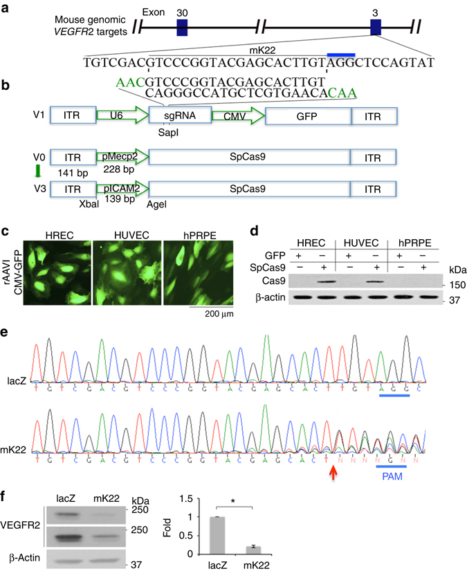
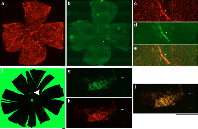
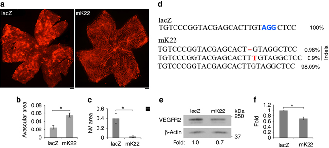
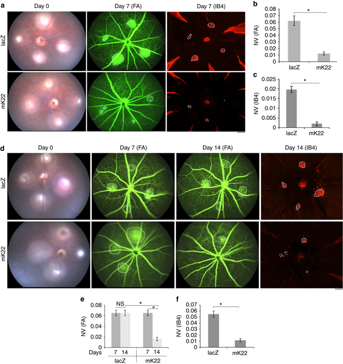
Angiogenesis, in which vascular endothelial growth factor receptor (VEGFR) 2 plays an essential role, is associated with a variety of human diseases including proliferative diabetic retinopathy and wet age-related macular degeneration. Here we report that a system of adeno-associated virus (AAV)-mediated clustered regularly interspaced short palindromic repeats (CRISPR)-associated endonuclease (Cas)9 from Streptococcus pyogenes (SpCas9) is used to deplete VEGFR2 in vascular endothelial cells (ECs), whereby the expression of SpCas9 is driven by an endothelial-specific promoter of intercellular adhesion molecule 2. We further show that recombinant AAV serotype 1 (rAAV1) transduces ECs of pathologic vessels, and that editing of genomic VEGFR2 locus using rAAV1-mediated CRISPR/Cas9 abrogates angiogenesis in the mouse models of oxygen-induced retinopathy and laser-induced choroid neovascularization. This work establishes a strong foundation for genome editing as a strategy to treat angiogenesis-associated diseases.
Overview
The claims and statistics of this paper haven’t been checked yet.
Run the check — free, no account needed. It reads the open-access text and checks the sources it cites.
Integrity signals
Registry
Cites
Statistics
Figures
Verification
Claims the paper makes
The claims and statistics of this paper haven’t been checked yet.
Run the check — free, no account needed. It reads the open-access text and checks the sources it cites.
Claims other papers make about this one
Each is checked against this paper’s own text: does it say what the citing paper says it does?
The source backs part of the claim, not all of it — or backs it for a different population, condition or comparison.
In the citing paper
“Recent efforts to address this problem have employed gene editing strategies like targeting VEGFA using CRISPR-Cas9 (43, 45).”
From Characterization of RNA editing and gene therapy with a compact CRISPR-Cas13 in the retina (2024) · cites this paper as [45]
Experimental — accuracy not yet measuredIn the citing paper
“A study aimed to use genome editing to treat angiogenesis-associated diseases by packaging Cas9 and gRNA targeting vascular endothelial growth factor receptor 2 (VEGFR2) in AAV1 and delivering it by intravitreal injections to the eye of WT C57BL/6J mice.They analyzed toxicity and inflammation by imaging the fundus as well as immunolabeling the retina for inflammation markers but reported no damage”
From Immune responses to gene editing by viral and non-viral delivery vectors used in retinal gene therapy (2022) · cites this paper as [36]
Self-check
Percentages, sums and reported differences re-computed from the paper’s own numbers.
The self-check needs the paper’s full text.
Citations
Cites
A Programmable Dual-RNA–Guided DNA Endonuclease in Adaptive Bacterial Immunity
Jinek, M., Chylinski, K., Fonfara, I., Hauer, M., Doudna, J. A., & Charpentier, E. (2012) · Science · doi:10.1126/science.1225829 · Cited by 17,116
Angiogenesis in cancer, vascular, rheumatoid and other disease
Folkman, J. (1995) · Nature Medicine · doi:10.1038/nm0195-27 · Cited by 7,627
Cillà, S. D., Farruggio, S., Vujosevic, S., Raina, G., Filippini, D. A., Gatti, V., Clemente, N., Mary, D., Vezzola, D., Casini, G., et al. (2017) · Cellular Physiology and Biochemistry · doi:10.1159/000479441 · Cited by 5,154
Tumor Cells Secrete a Vascular Permeability Factor That Promotes Accumulation of Ascites Fluid
Senger, D. R., Galli, S. J., Dvořàk, A. M., Perruzzi, C. A., Harvey, V. S., & Dvorak, H. F. (1983) · Science · doi:10.1126/science.6823562 · Cited by 3,969
Matrix metalloproteinase-9 triggers the angiogenic switch during carcinogenesis
Bergers, G., Brekken, R., McMahon, G., Vu, T. H., Itoh, T., Tamaki, K., Tanzawa, K., Thorpe, P., Itohara, S., Werb, Z., et al. (2000) · Nature Cell Biology · doi:10.1038/35036374 · Cited by 2,797
Safety and Efficacy of Gene Transfer for Leber's Congenital Amaurosis
Maguire, A. M., Simonelli, F., Pierce, E. A., Pugh, E. N., Mingozzi, F., Bennicelli, J., Banfi, S., Marshall, K. A., Testa, F., Surace, E. M., et al. (2008) · New England Journal of Medicine · doi:10.1056/nejmoa0802315 · Cited by 2,145
Effect of gene therapy on visual function in Leber’s congenital amaurosis
Bainbridge, J. W. B., Smith, A. J., Barker, S. S., Robbie, S., Henderson, R., Balaggan, K., Viswanathan, A., Holder, G. E., Stockman, A., Tyler, N., et al. (2008) · New England Journal of Medicine · doi:10.1056/nejmoa0802268 · Cited by 1,924
Efficacy of Intravitreal Bevacizumab for Stage 3+ Retinopathy of Prematurity
Mintz-Hittner, H. A., Kennedy, K. A., & Chuang, A. Z. (2011) · New England Journal of Medicine · doi:10.1056/nejmoa1007374 · Cited by 1,451
Therapeutic genome editing: prospects and challenges
Cox, D. B. T., Platt, R. J., & Zhang, F. (2015) · Nature Medicine · doi:10.1038/nm.3793 · Cited by 1,336
Self-citationTherapeutic Potential of Ex Vivo Expanded Endothelial Progenitor Cells for Myocardial Ischemia
Kawamoto, A., Gwon, H.-C., Iwaguro, H., Yamaguchi, J.-I., Uchida, S., Masuda, H., Silver, M., Ma, H., Kearney, M., Isner, J. M., et al. (2001) · Circulation · doi:10.1161/01.cir.103.5.634 · Cited by 1,219
Holmes, K., Roberts, O. L., Thomas, A. M., & Cross, M. J. (2007) · Cellular Signalling · doi:10.1016/j.cellsig.2007.05.013 · Cited by 1,053
In vivo gene editing in dystrophic mouse muscle and muscle stem cells
Tabebordbar, M., Zhu, K., Cheng, J. K. W., Chew, W. L., Widrick, J. J., Yan, W. X., Maesner, C., Wu, E. Y., Xiao, R., Ran, F. A., et al. (2016) · Science · doi:10.1126/science.aad5177 · Cited by 1,046
Self-citationHauswirth, W. W., Aleman, T. S., Kaushal, S., Cideciyan, A. V., Schwartz, S. B., Wang, L., Conlon, T. J., Boye, S. L., Flotte, T. R., Byrne, B. J., et al. (2008) · Human Gene Therapy · doi:10.1089/hum.2008.107 · Cited by 954
In vivo interrogation of gene function in the mammalian brain using CRISPR-Cas9
Swiech, L., Heidenreich, M., Banerjee, A., Habib, N., Li, Y., Trombetta, J., Sur, M., & Zhang, F. (2015) · Nature Biotechnology · doi:10.1038/nbt.3055 · Cited by 772
Self-citationJessup, M., Greenberg, B., Mancini, D., Cappola, T., Pauly, D. F., Jaski, B., Yaroshinsky, A., Zsebo, K. M., Dittrich, H., & Hajjar, R. J. (2011) · Circulation · doi:10.1161/circulationaha.111.022889 · Cited by 760
Chakravarthy, U., Harding, S. P., Rogers, C. A., Downes, S. M., Lotery, A. J., Culliford, L. A., & Reeves, B. C. (2013) · The Lancet · doi:10.1016/s0140-6736(13)61501-9 · Cited by 748
Connor, K. M., Krah, N. M., Dennison, R. J., Aderman, C. M., Chen, J., Guerin, K. I., Sapieha, P. S., Stahl, A., Willett, K. L., & Smith, L. E. H. (2009) · Nature Protocols · doi:10.1038/nprot.2009.187 · Cited by 742
Giraudo, E., Inoue, M., & Hanahan, D. (2004) · Journal of Clinical Investigation · doi:10.1172/jci200422087 · Cited by 700
The Mouse Retina as an Angiogenesis Model
Stahl, A., Connor, K. M., Sapieha, P. S., Chen, J., Dennison, R. J., Krah, N. M., Seaward, M. R., Willett, K. L., Aderman, C. M., Guerin, K. I., et al. (2010) · Investigative Ophthalmology & Visual Science · doi:10.1167/iovs.10-5176 · Cited by 638
Production and characterization of adeno-associated viral vectors
Grieger, J. C., Choi, V. W., & Samulski, R. J. (2006) · Nature Protocols · doi:10.1038/nprot.2006.207 · Cited by 580
An essential receptor for adeno-associated virus infection
Pillay, S., Meyer, N. L., Puschnik, A. S., Davulcu, O., Diep, J., Ishikawa, Y., Jae, L. T., Wosen, J. E., Nagamine, C. M., Chapman, M. S., et al. (2016) · Nature · doi:10.1038/nature16465 · Cited by 532
Epidemiology of diabetic retinopathy and macular oedema: A systematic review
Williams, R., Airey, M., Baxter, H., Forrester, J., Kennedy-Martin, T., & Girach, A. (2004) · Eye · doi:10.1038/sj.eye.6701476 · Cited by 472
Muller, Y. A., Li, B., Christinger, H. W., Wells, J. A., Cunningham, B. C., & de Vos, A. M. (1997) · Proceedings of the National Academy of Sciences · doi:10.1073/pnas.94.14.7192 · Cited by 467
Laser-induced choroidal neovascularization model to study age-related macular degeneration in mice
Lambert, V., Lecomte, J., Hansen, S., Blacher, S., Gonzalez, M.-L. A., Struman, I., Sounni, N. E., Rozet, E., Tullio, P. D., Foidart, J. M., et al. (2013) · Nature Protocols · doi:10.1038/nprot.2013.135 · Cited by 420
Gaudet, D., Méthot, J., Déry, S., Brisson, D., Essiembre, C., Tremblay, G., Tremblay, K., de Wal, J., Twisk, J., van den Bulk, N., et al. (2013) · Gene Therapy · doi:10.1038/gt.2012.43 · Cited by 404
Shima, D. T., Adamis, A. P., Ferrara, N., Yeo, K.-T., Yeo, T.-K., Allende, R., Folkman, J., & D'Amore, P. A. (1995) · Molecular Medicine · doi:10.1007/bf03401566 · Cited by 353
Self-citationEllis, B. L., Hirsch, M. L., Barker, J. C., Connelly, J. P., Steininger, R. J., & Porteus, M. H. (2013) · Virology Journal · doi:10.1186/1743-422x-10-74 · Cited by 252
Axl is essential for VEGF‐A‐dependent activation of PI3K/Akt
Ruan, G.-X., & Kazlauskas, A. (2012) · The EMBO Journal · doi:10.1038/emboj.2012.21 · Cited by 229
Adeno-associated virus vector integration.
Deyle, D. R., & Russell, D. W. (2009) · PubMed · Cited by 206
Gene therapy for lipoprotein lipase deficiency
Gaudet, D., Méthot, J., & Kastelein, J. (2012) · Current Opinion in Lipidology · doi:10.1097/mol.0b013e3283555a7e · Cited by 178
Suzuki, M., Nagai, N., Izumi‐Nagai, K., Shinoda, H., Koto, T., Uchida, A., Mochimaru, H., Yuki, K., Sasaki, M., Tsubota, K., et al. (2014) · British Journal of Ophthalmology · doi:10.1136/bjophthalmol-2013-304670 · Cited by 100
Giani, A., Thanos, A., Roh, M., Connolly, E., Trichonas, G., Kim, I. K., Gragoudas, E. S., Vavvas, D. G., & Miller, J. W. (2011) · Investigative Ophthalmology & Visual Science · doi:10.1167/iovs.10-6266 · Cited by 99
PGC-1α Regulates Normal and Pathological Angiogenesis in the Retina
Saint‐Geniez, M., Jiang, A., Abend, S., Liu, L., Sweigard, H., Connor, K. M., & Arany, Z. P. (2012) · American Journal Of Pathology · doi:10.1016/j.ajpath.2012.09.003 · Cited by 70
Lei, H., Romeo, G. R., & Kazlauskas, A. (2004) · Circulation Research · doi:10.1161/01.res.0000124979.46214.e3 · Cited by 65
Self-citationEditing VEGFR2 Blocks VEGF-Induced Activation of Akt and Tube Formation
Huang, X., Zhou, G., Wu, W., Ma, G., D'Amore, P. A., Mukai, S., & Lei, H. (2017) · Investigative Ophthalmology & Visual Science · doi:10.1167/iovs.16-20537 · Cited by 61
Self-citationChitranshi, N., Gupta, V., Kumar, S., & Graham, S. L. (2015) · International Journal of Molecular Sciences · doi:10.3390/ijms160921087 · Cited by 49
Update on treatments for diabetic macular edema
Fraser‐Bell, S., Kaines, A., & Hykin, P. (2008) · Current Opinion in Ophthalmology · doi:10.1097/icu.0b013e3282fb7c45 · Cited by 49
CRISPR/Cas9: Prospects and Challenges
Zhang, F. (2015) · Human Gene Therapy · doi:10.1089/hum.2015.29002.fzh · Cited by 42
Self-citationDuan, Y., Ma, G., Huang, X., D'Amore, P. A., Zhang, F., & Lei, H. (2016) · Journal of Biological Chemistry · doi:10.1074/jbc.m116.729467 · Cited by 31
Self-citationLei, H., Qian, C. X., Lei, J., Haddock, L. J., Mukai, S., & Kazlauskas, A. (2015) · Molecular and Cellular Biology · doi:10.1128/mcb.01248-14 · Cited by 27
Self-citationIdentification of Synthetic Endothelial Cell-Specific Promoters by Use of a High-Throughput Screen
Dai, C., McAninch, R., & Sutton, R. (2004) · Journal of Virology · doi:10.1128/jvi.78.12.6209-6221.2004 · Cited by 17
Jiang, A., Abend, S., D'Amore, P. A., Saint‐Geniez, M., & Arany, Z. P. (2011) · Investigative Ophthalmology & Visual Science · Cited by 3
Self-citation
Cited by
Sorted by citations
Gene therapy for neurological disorders: progress and prospects
Deverman, B. E., Ravina, B. M., Bankiewicz, K. S., Paul, S. M., & Sah, D. W. Y. (2018) · Nature Reviews Drug Discovery · doi:10.1038/nrd.2018.110 · Cited by 306
Delivery of CRISPR-Cas tools for in vivo genome editing therapy: Trends and challenges
Taha, E. A., Lee, J., & Hotta, A. (2022) · Journal of Controlled Release · doi:10.1016/j.jconrel.2022.01.013 · Cited by 259
Erratum: Gene therapy for neurological disorders: progress and prospects
Deverman, B. E., Ravina, B. M., Bankiewicz, K. S., Paul, S. M., & Sah, D. W. Y. (2018) · Nature Reviews Drug Discovery · doi:10.1038/nrd.2018.158 · Cited by 248
Delivery of CRISPR/Cas systems for cancer gene therapy and immunotherapy
Song, X., Liu, C., Wang, N., Huang, H., He, S., Gong, C., & Wei, Y. (2020) · Advanced Drug Delivery Reviews · doi:10.1016/j.addr.2020.04.010 · Cited by 217
Diabetic retinopathy: a complex pathophysiology requiring novel therapeutic strategies
Whitehead, M. A., Wickremasinghe, S., Osborne, A., van Wijngaarden, P., & Martin, K. R. G. (2018) · Expert Opinion on Biological Therapy · doi:10.1080/14712598.2018.1545836 · Cited by 209
Metadata
- Authors
- Huang, X., Zhou, G., Wu, W., Duan, Y., Ma, G., Song, J., Xiao, R., Vandenberghe, L., Zhang, F., D’Amore, P. A., & Lei, H.
- Affiliations
- Massachusetts Eye and Ear Infirmary (US)
- Broad Institute (US)
- Corresponding author
- Lei, Hetian
- ORCID
- Venue
- Nature Communications
- Published
- 2017-07-24
- Volume
- 8
- Issue
- 1
- Pages
- 112-112
- ISSN
- 2041-1723 (ISSN-L)
- PMC
- PMC5524639
- OpenAlex
- W2735017428
- Cited by
- 142
- References
- 42
- Citations by year
- 2026: 6 · 2025: 14 · 2024: 14 · 2023: 17 · 2022: 20 · 2021: 19 · 2020: 19 · 2019: 8 · 2018: 21 · 2017: 4
- FWCI
- 5.81
- Type
- journal article
- Language
- en
- Open access
- gold
- doi.org
- Locations
- Nature Communications (publishedVersion, cc-by)
- PubMed Central (publishedVersion, cc-by)
- PubMed (publishedVersion)
- RePEc: Research Papers in Economics (submittedVersion)
- DOAJ (DOAJ: Directory of Open Access Journals) (submittedVersion, cc-by-sa)
- DSpace@MIT (Massachusetts Institute of Technology) (submittedVersion, cc-by-nc)
- Nature
- Funders
- National Institutes of Health
- National Eye Institute (R01EY012509, P30EY003790)
- Topics
- Life Sciences › Biochemistry, Genetics and Molecular Biology › Molecular Biology › CRISPR and Genetic Engineering
- Life Sciences › Biochemistry, Genetics and Molecular Biology › Molecular Biology › Angiogenesis and VEGF in Cancer
- Health Sciences › Medicine › Ophthalmology › Retinal Development and Disorders
- Keywords
- angiogenesis, VEGFR2, CRISPR/Cas9 genome editing, vascular endothelial cells, oxygen-induced retinopathy, diabetic retinopathy, age-related macular degeneration, gene silencing
- MeSH terms
- Gene Editing, Dependovirus, Animals, Cells, Cultured, Female, Humans, Male, Mice, Inbred C57BL, Microscopy, Fluorescence, Neovascularization, Pathologic, Blotting, Western, Retinal Neovascularization, Choroidal Neovascularization, Vascular Endothelial Growth Factor Receptor-2, Endothelial Cells, CRISPR-Cas Systems
About these checks
Registry, retraction, Cites and statistics results are computed, not judged, with one exception: a printed reference that no index lists is read once by a model before it counts as a citation error. Claims are extracted by a model and kept only when their quote appears verbatim in the text; each cited source is then checked, with a model’s help, for whether it says what the claim relies on. Those verdicts can be wrong — read the claim’s quote and the cited source before relying on one.
Statistics and arithmetic are recomputed, not judged.
Confidence bands are shown only for fields calibrated on our annotated test set. None is calibrated yet, so no band is shown.
What counts as a claim ›How we measure confidence ›How we check this ›
Are you an author, or do you see a problem with this page? Request a review
