

The DOI matches its Crossref record; 66 of the 66 references it cites were found in a registry or an index.
Identification of rare sequence variation underlying heritable pulmonary arterial hypertension
Open accessPublished version of a preprint
Abstract
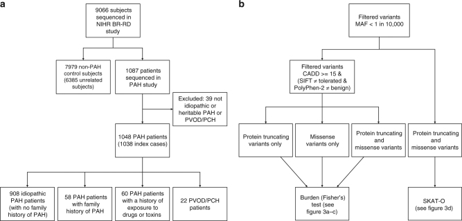
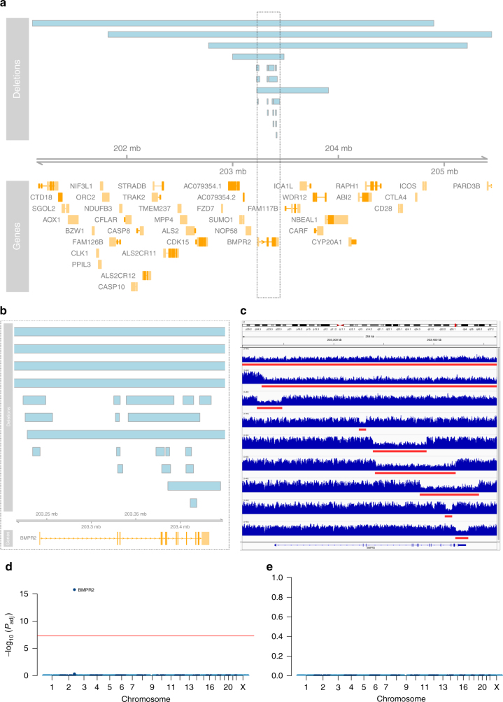
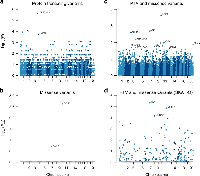
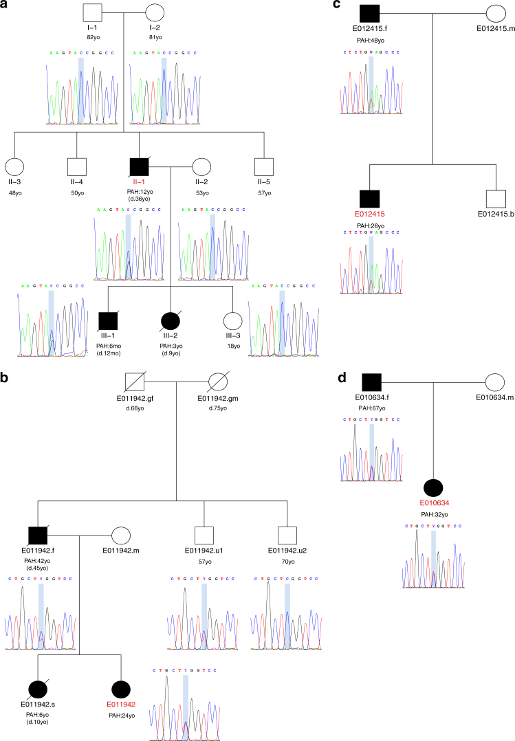
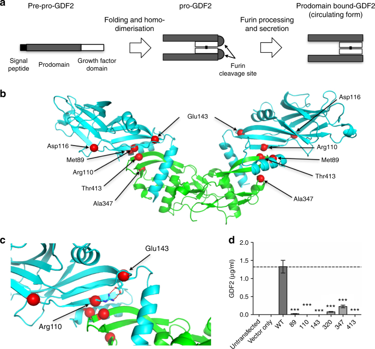
Pulmonary arterial hypertension (PAH) is a rare disorder with a poor prognosis. Deleterious variation within components of the transforming growth factor-β pathway, particularly the bone morphogenetic protein type 2 receptor ( BMPR2 ), underlies most heritable forms of PAH. To identify the missing heritability we perform whole-genome sequencing in 1038 PAH index cases and 6385 PAH-negative control subjects. Case-control analyses reveal significant overrepresentation of rare variants in ATP13A3, AQP1 and SOX17 , and provide independent validation of a critical role for GDF2 in PAH. We demonstrate familial segregation of mutations in SOX17 and AQP1 with PAH. Mutations in GDF2 , encoding a BMPR2 ligand, lead to reduced secretion from transfected cells. In addition, we identify pathogenic mutations in the majority of previously reported PAH genes, and provide evidence for further putative genes. Taken together these findings contribute new insights into the molecular basis of PAH and indicate unexplored pathways for therapeutic intervention.
Overview
The claims and statistics of this paper haven’t been checked yet.
Run the check — free, no account needed. It reads the open-access text and checks the sources it cites.
Integrity signals
Registry
Cites
Statistics
Figures
Verification
Claims the paper makes
The claims and statistics of this paper haven’t been checked yet.
Run the check — free, no account needed. It reads the open-access text and checks the sources it cites.
Claims other papers make about this one
Each is checked against this paper’s own text: does it say what the citing paper says it does?
The cited source says what the claim attributes to it.
In the citing paper
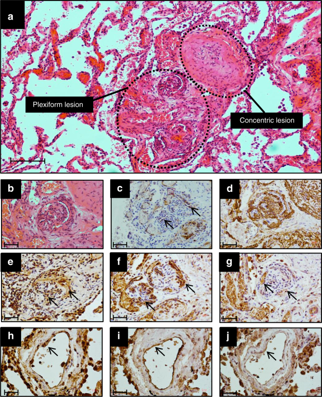
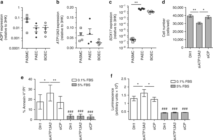
“Protein-truncating variants in this gene have previously been associated with pulmonary artery hypertension. Functional studies have linked the loss of ATP13A3 to inhibition of proliferation and stimulation of apoptosis in endothelial cells25.”
From Integrative genomic analyses identify candidate causal genes for calcific aortic valve stenosis involving tissue-specific regulation (2024) · cites this paper as [25]
Self-check
Percentages, sums and reported differences re-computed from the paper’s own numbers.
The self-check needs the paper’s full text.
Citations
Cites
The Sequence Alignment/Map format and SAMtools
Li, H., Handsaker, B., Wysoker, A., Fennell, T., Ruan, J., Homer, N., Marth, G., Abecasis, G., Durbin, R., & 1000 Genome Project Data Processing Subgroup (2009) · Bioinformatics · doi:10.1093/bioinformatics/btp352 · Cited by 62,041
PLINK: a tool set for whole-genome association and population-based linkage analyses.
Purcell, S., Neale, B., Todd-Brown, K., Thomas, L., Ferreira, M. A. R., Bender, D., Maller, J., Sklar, P., de Bakker, P. I. W., Daly, M. J., et al. (2007) · The American Journal of Human Genetics · doi:10.1086/519795 · Cited by 34,089
A global reference for human genetic variation
The 1000 Genomes Project Consortium, authors, C., Auton, A., Abecasis, G. R., Steering committee, Altshuler, D. M., Durbin, R. M., Abecasis, G. R., Bentley, D. R., Chakravarti, A., et al. (2015) · Nature · doi:10.1038/nature15393 · Cited by 18,247
Self-citationA method and server for predicting damaging missense mutations.
Adzhubei, I. A., Schmidt, S., Peshkin, L., Ramensky, V. E., Gerasimova, A., Bork, P., Kondrashov, A. S., & Sunyaev, S. R. (2010) · Nature Methods · doi:10.1038/nmeth0410-248 · Cited by 13,194
A framework for variation discovery and genotyping using next-generation DNA sequencing data
DePristo, M. A., Banks, E., Poplin, R. E., Garimella, K. V., Maguire, J., Hartl, C., Philippakis, A., del Angel, G., Rivas, M. A., Hanna, M., et al. (2011) · Nature Genetics · doi:10.1038/ng.806 · Cited by 11,457
Analysis of protein-coding genetic variation in 60,706 humans
Exome Aggregation Consortium, Lek, M., Karczewski, K. J., Minikel, E. V., Samocha, K. E., Banks, E., Fennell, T., O’Donnell-Luria, A. H., Ware, J. S., Hill, A. J., et al. (2016) · Nature · doi:10.1038/nature19057 · Cited by 9,587
Integrative Genomics Viewer (IGV): high-performance genomics data visualization and exploration
Thorvaldsdottir, H., Robinson, J. T., & Mesirov, J. P. (2013) · Briefings in Bioinformatics · doi:10.1093/bib/bbs017 · Cited by 9,049
A general framework for estimating the relative pathogenicity of human genetic variants
Kircher, M., Witten, D. M., Jain, P., O'Roak, B. J., Cooper, G. M., & Shendure, J. (2014) · Nature Genetics · doi:10.1038/ng.2892 · Cited by 6,183
Predicting Deleterious Amino Acid Substitutions
Ng, P. C., & Henikoff, S. (2001) · Genome Research · doi:10.1101/gr.176601 · Cited by 2,368
Cited by
Sorted by citations
2022 ESC/ERS Guidelines for the diagnosis and treatment of pulmonary hypertension
Humbert, M. J. C., Kovács, G., Hoeper, M. M., Badagliacca, R., Berger, R. M. F., Brida, M., Carlsen, J., Coats, A. J. S., Subías, P. E., Ferrari, P., et al. (2022) · European Heart Journal · doi:10.1093/eurheartj/ehac237 · Cited by 4,286
Self-citation2022 ESC/ERS Guidelines for the diagnosis and treatment of pulmonary hypertension
Humbert, M. J. C., Kovács, G., Hoeper, M. M., Badagliacca, R., Berger, R. M. F., Brida, M., Carlsen, J., Coats, A. J. S., Subías, P. E., Ferrari, P., et al. (2022) · European Respiratory Journal · doi:10.1183/13993003.00879-2022 · Cited by 2,388
Self-citationTGF-β signaling in health and disease
Massagué, J., & Sheppard, D. (2023) · Cell · doi:10.1016/j.cell.2023.07.036 · Cited by 890
Pulmonary Arterial Hypertension
Hassoun, P. M. (2021) · New England Journal of Medicine · doi:10.1056/nejmra2000348 · Cited by 714
Endothelial to Mesenchymal Transition in Cardiovascular Disease
Kovacic, J. C., Dimmeler, S., Harvey, R. P., Finkel, T., Aïkawa, E., Krenning, G., & Baker, A. H. (2019) · Journal of the American College of Cardiology · doi:10.1016/j.jacc.2018.09.089 · Cited by 639
Whole-genome sequencing of patients with rare diseases in a national health system
Turro, E., Astle, W. J., Megy, K., Gräf, S., Greene, D., Shamardina, O., Allen, H. L., Sanchis-Juan, A., Frontini, M., Thys, C., et al. (2020) · Nature · doi:10.1038/s41586-020-2434-2 · Cited by 554
Self-citationGenetics and genomics of pulmonary arterial hypertension
Morrell, N. W., Aldred, M. A., Chung, W. K., Elliott, C. G., Nichols, W. C., Soubrier, F., Trembath, R. C., & Loyd, J. E. (2018) · European Respiratory Journal · doi:10.1183/13993003.01899-2018 · Cited by 496
Self-citationMolecular genetic framework underlying pulmonary arterial hypertension
Southgate, L., Machado, R. D., Gräf, S., & Morrell, N. W. (2019) · Nature Reviews Cardiology · doi:10.1038/s41569-019-0242-x · Cited by 302
Self-citationACTRIIA-Fc rebalances activin/GDF versus BMP signaling in pulmonary hypertension
Yung, L., Yang, P. B., Joshi, S. R., Augur, Z. M., Kim, S. S., Bocobo, G. A., Dinter, T., Troncone, L., Chen, P.-S., McNeil, M. E., et al. (2020) · Science Translational Medicine · doi:10.1126/scitranslmed.aaz5660 · Cited by 259
Self-citationBone morphogenetic protein receptor signal transduction in human disease
Gomez‐Puerto, M. C., Iyengar, P. V., de Vinuesa, A. G., ten Dijke, P., & Sánchez‐Duffhues, G. (2018) · The Journal of Pathology · doi:10.1002/path.5170 · Cited by 239
The quiescent endothelium: signalling pathways regulating organ-specific endothelial normalcy
Ricard, N., Bailly, S., Guignabert, C., & Simons, M. (2021) · Nature Reviews Cardiology · doi:10.1038/s41569-021-00517-4 · Cited by 242
Rhodes, C. J., Batai, K., Bleda, M., Haimel, M., Southgate, L., Germain, M., Pauciulo, M. W., Hadinnapola, C., Aman, J., Girerd, B., et al. (2018) · The Lancet Respiratory Medicine · doi:10.1016/s2213-2600(18)30409-0 · Cited by 207
Self-citationPulmonary Hypertension: A Contemporary Review
Johnson, S. W., Sommer, N., Cox, K., Weißmann, N., Ventetuolo, C. E., & Maron, B. A. (2023) · American Journal of Respiratory and Critical Care Medicine · doi:10.1164/rccm.202302-0327so · Cited by 210
Clinical trial design and new therapies for pulmonary arterial hypertension
Sitbon, O., Gomberg‐Maitland, M., Granton, J. T., Lewis, M. I., Mathai, S. C., Rainisio, M., Stockbridge, N., Wilkins, M. R., Zamanian, R. T., & Rubin, L. J. (2018) · European Respiratory Journal · doi:10.1183/13993003.01908-2018 · Cited by 196
Self-citationTargeting transforming growth factor-β receptors in pulmonary hypertension
Guignabert, C., & Humbert, M. J. C. (2020) · European Respiratory Journal · doi:10.1183/13993003.02341-2020 · Cited by 203
Self-citationPathology and pathobiology of pulmonary hypertension: current insights and future directions
Guignabert, C., Aman, J., Bonnet, S., Dorfmüller, P., Olschewski, A., Pullamsetti, S. S., Rabinovitch, M., SCHERMULY, R. T., Humbert, M. J. C., & Stenmark, K. R. (2024) · European Respiratory Journal · doi:10.1183/13993003.01095-2024 · Cited by 178
Self-citationCurrent and future treatments of pulmonary arterial hypertension
Sommer, N., Ghofrani, H. A., Pak, O., Bonnet, S., Provencher, S., Sitbon, O., Rosenkranz, S., Hoeper, M. M., & Kiely, D. G. (2020) · British Journal of Pharmacology · doi:10.1111/bph.15016 · Cited by 165
Self-citationSex, Gender, and Sex Hormones in Pulmonary Hypertension and Right Ventricular Failure
Hester, J., Ventetuolo, C. E., & Lahm, T. (2019) · Comprehensive physiology · doi:10.1002/cphy.c190011 · Cited by 183
Zhu, N., Welch, C. L., Wang, J., Allen, P. M., Gonzaga-Jauregui, C. G., Ma, L., King, A. K., Krishnan, U., Rosenzweig, E. B., Ivy, D. D., et al. (2018) · Genome Medicine · doi:10.1186/s13073-018-0566-x · Cited by 162
Pathophysiology and new advances in pulmonary hypertension
Bousseau, S., Fais, R. S., Gu, S., Frump, A. L., & Lahm, T. (2023) · BMJ Medicine · doi:10.1136/bmjmed-2022-000137 · Cited by 171
Endothelial Dysfunction in Pulmonary Hypertension: Cause or Consequence?
Kurakula, K., Smolders, V. F. E. D., Tura-Ceide, O., Jukema, J. W., Quax, P. H. A., & Goumans, M. (2021) · Biomedicines · doi:10.3390/biomedicines9010057 · Cited by 148
Systems Analysis of the Human Pulmonary Arterial Hypertension Lung Transcriptome
Stearman, R. S., Bui, Q. M., Speyer, G., Handen, A. L., Cornelius, A. R., Graham, B. B., Kim, S., Mickler, E. A., Tuder, R. M., Chan, S. Y., et al. (2018) · American Journal of Respiratory Cell and Molecular Biology · doi:10.1165/rcmb.2018-0368oc · Cited by 140
Galambos, C., Mullen, M. P., Shieh, J. T. C., Schwerk, N., Kielt, M. J., Ullmann, N., Boldrini, R., Gantar, I. Š., Haass, C., Bansal, M., et al. (2019) · European Respiratory Journal · doi:10.1183/13993003.01965-2018 · Cited by 139
The 6th World Symposium on Pulmonary Hypertension: what’s old is new
Condon, D. F., Nickel, N. P., Anderson, R. J., Mirza, S., & Pérez, V. D. J. (2019) · F1000Research · doi:10.12688/f1000research.18811.1 · Cited by 138
GermlineBMP9mutation causes idiopathic pulmonary arterial hypertension
Wang, X., Lian, T., Jiang, X., Liu, S., Li, S.-Q., Jiang, R., Wu, W., Ye, J., Cheng, C., Du, Y., et al. (2018) · European Respiratory Journal · doi:10.1183/13993003.01609-2018 · Cited by 136
Zhu, N., Pauciulo, M. W., Welch, C. L., Lutz, K. A., Coleman, A. W., Gonzaga-Jauregui, C. G., Wang, J., Grimes, J. M., Martin, L. J., He, H., et al. (2019) · Genome Medicine · doi:10.1186/s13073-019-0685-z · Cited by 136
Selective BMP-9 Inhibition Partially Protects Against Experimental Pulmonary Hypertension
Tu, L., Desroches‐Castan, A., Mallet, C., Guyon, L., Cumont, A., Phan, C., Robert, F., Thuillet, R., Bordenave, J., Sekine, A., et al. (2019) · Circulation Research · doi:10.1161/circresaha.118.313356 · Cited by 130
Self-citationBMPR2 Mutation and Metabolic Reprogramming in Pulmonary Arterial Hypertension
Cuthbertson, I., Morrell, N. W., & Caruso, P. L. (2023) · Circulation Research · doi:10.1161/circresaha.122.321554 · Cited by 129
Self-citationCharacterization of GDF2 Mutations and Levels of BMP9 and BMP10 in Pulmonary Arterial Hypertension
Hodgson, J., Swietlik, E. M., Salmon, R. M., Hadinnapola, C., Nikolić, I., Wharton, J., Guo, J., Liley, J., Haimel, M., Bleda, M., et al. (2019) · American Journal of Respiratory and Critical Care Medicine · doi:10.1164/rccm.201906-1141oc · Cited by 123
Self-citationMechanisms and treatment of pulmonary arterial hypertension
Ghofrani, H. A., Gomberg‐Maitland, M., Zhao, L., & Grimminger, F. (2024) · Nature Reviews Cardiology · doi:10.1038/s41569-024-01064-4 · Cited by 134
Self-citationPotus, F., Pauciulo, M. W., Cook, E. K., Zhu, N., Hsieh, A., Welch, C. L., Shen, Y., Tian, L., Lima, P., Mewburn, J. D., et al. (2020) · Circulation · doi:10.1161/circulationaha.119.044320 · Cited by 124
The role of bone morphogenetic protein signaling in vascular calcification
Yang, P. B., Troncone, L., Augur, Z. M., Kim, S. S., McNeil, M. E., & Yu, P. B. (2020) · Bone · doi:10.1016/j.bone.2020.115542 · Cited by 127
Sánchez‐Duffhues, G., Williams, E., Goumans, M., Heldin, C., & ten Dijke, P. (2020) · Bone · doi:10.1016/j.bone.2020.115472 · Cited by 126
Widening the landscape of heritable pulmonary hypertension mutations in paediatric and adult cases
Eyries, M., Montani, D., Nadaud, S., Girerd, B., Lévy, M., BOURDIN, A. A., Trésorier, R., Chaouat, A., Cottin, V., Sanfiorenzo, C., et al. (2018) · European Respiratory Journal · doi:10.1183/13993003.01371-2018 · Cited by 115
Self-citationChromatin accessibility landscape and regulatory network of high-altitude hypoxia adaptation
Xin, J., Zhang, H., He, Y., Duren, Z., Bai, C., Chen, L., Luo, X., Yan, D.-S., Zhang, C., Zhu, X., et al. (2020) · Nature Communications · doi:10.1038/s41467-020-18638-8 · Cited by 103
Wagner, K., Unger, L., Salman, M. M., Kitchen, P., Bill, R. M., & Yool, A. J. (2022) · International Journal of Molecular Sciences · doi:10.3390/ijms23031388 · Cited by 97
ATP13A3 is a major component of the enigmatic mammalian polyamine transport system
Hamouda, N. N., Van den Haute, C., Vanhoutte, R., Sannerud, R., Azfar, M., Mayer, R., Cortés Calabuig, Á., Swinnen, J. V., Agostinis, P., Baekelandt, V., et al. (2021) · Journal of Biological Chemistry · doi:10.1074/jbc.ra120.013908 · Cited by 85
Ion Channels in Pulmonary Hypertension: A Therapeutic Interest?
Lambert, M., Capuano, V., Olschewski, A., Sabourin, J., Nagaraj, C., Girerd, B., Weatherald, J. C., Humbert, M. J. C., & Antigny, F. (2018) · International Journal of Molecular Sciences · doi:10.3390/ijms19103162 · Cited by 93
Self-citationZhu, N., Swietlik, E. M., Welch, C. L., Pauciulo, M. W., Hagen, J., Zhou, X., Guo, Y., Karten, J., Pandya, D., Tilly, T., et al. (2021) · Genome Medicine · doi:10.1186/s13073-021-00891-1 · Cited by 87
Self-citationRichardson, T. G., Hemani, G., Gaunt, T. R., Relton, C. L., & Smith, G. D. (2020) · Nature Communications · doi:10.1038/s41467-019-13921-9 · Cited by 84
Defining the clinical validity of genes reported to cause pulmonary arterial hypertension
Welch, C. L., Aldred, M. A., Balachandar, S., Dooijes, D., Eichstaedt, C. A., Gräf, S., Houweling, A. C., Machado, R. D., Pandya, D., Prapa, M., et al. (2023) · Genetics in Medicine · doi:10.1016/j.gim.2023.100925 · Cited by 92
Self-citationReyes‐Palomares, A., Gu, M., Grubert, F., Berest, I., Sa, S., Kasowski, M. M., Arnold, C., Mao, S., Srivas, R., Miao, S., et al. (2020) · Nature Communications · doi:10.1038/s41467-020-15463-x · Cited by 81
Zheng, W., He, Y., Guo, Y., Yue, T., Zhang, H., Li, J., Zhou, B., Zeng, X., Li, L., Wang, B., et al. (2023) · Genome biology · doi:10.1186/s13059-023-02912-1 · Cited by 81
Screening strategies for pulmonary arterial hypertension
Kiely, D. G., Lawrie, A., & Humbert, M. J. C. (2019) · European Heart Journal Supplements · doi:10.1093/eurheartj/suz204 · Cited by 80
Self-citationBMP10-mediated ALK1 signaling is continuously required for vascular development and maintenance
Capasso, T. L., Li, B., Volek, H. J., Khalid, W., Rochon, E. R., Anbalagan, A., Herdman, C., Yost, H. J., Villanueva, F. S., Kim, K., et al. (2019) · Angiogenesis · doi:10.1007/s10456-019-09701-0 · Cited by 77
Balistrieri, A., Makino, A., & Yuan, J. X. ‐. (2022) · Physiological Reviews · doi:10.1152/physrev.00030.2021 · Cited by 77
Circulating BMP9 Protects the Pulmonary Endothelium during Inflammation-induced Lung Injury in Mice
Li, W., Long, L., Yang, X., Tong, Z., Southwood, M. R., King, R., Caruso, P. L., Upton, P. D., Yang, P. B., Bocobo, G. A., et al. (2020) · American Journal of Respiratory and Critical Care Medicine · doi:10.1164/rccm.202005-1761oc · Cited by 77
Self-citationBouvard, C., Tu, L., Rossi, M., Desroches‐Castan, A., Berrebeh, N., Helfer, E., Roelants, C., Liu, H., Ouarné, M., Chaumontel, N., et al. (2021) · Cardiovascular Research · doi:10.1093/cvr/cvab187 · Cited by 76
Self-citationBMP9 and BMP10 : Two close vascular quiescence partners that stand out
Desroches‐Castan, A., Tillet, E., Bouvard, C., & Bailly, S. (2021) · Developmental Dynamics · doi:10.1002/dvdy.395 · Cited by 85
Aldred, M. A., Morrell, N. W., & Guignabert, C. (2022) · Circulation Research · doi:10.1161/circresaha.122.320084 · Cited by 75
Self-citationPhenotype and outcome of pulmonary arterial hypertension patients carrying aTBX4mutation
Thoré, P., Girerd, B., Jaïs, X., Savale, L., Ghigna, M., Eyries, M., Lévy, M., Ovaert, C., Servettaz, A., Guillaumot, A., et al. (2020) · European Respiratory Journal · doi:10.1183/13993003.02340-2019 · Cited by 74
Self-citationSOXopathies: growing family of developmental disorders due to SOX mutations
Angelozzi, M., & Lefebvre, V. (2019) · Trends in Genetics · doi:10.1016/j.tig.2019.06.003 · Cited by 74
Whole-Blood RNA Profiles Associated with Pulmonary Arterial Hypertension and Clinical Outcome
Rhodes, C. J., Otero-Núñez, P., Wharton, J., Swietlik, E. M., Kariotis, S., Harbaum, L., Dunning, M., Elinoff, J. M., Errington, N., Thompson, A. A. R., et al. (2020) · American Journal of Respiratory and Critical Care Medicine · doi:10.1164/rccm.202003-0510oc · Cited by 69
Self-citationErrington, N., Iremonger, J., Pickworth, J. A., Kariotis, S., Rhodes, C. J., Rothman, A. M. K., Condliffe, R., Elliot, C. A., Kiely, D. G., Howard, L., et al. (2021) · EBioMedicine · doi:10.1016/j.ebiom.2021.103444 · Cited by 71
Self-citationFamilial pulmonary arterial hypertension byKDRheterozygous loss of function
Eyries, M., Montani, D., Girerd, B., Favrolt, N., Riou, M., Faivre, L. O., Manaud, G., Perros, F., Gräf, S., Morrell, N. W., et al. (2020) · European Respiratory Journal · doi:10.1183/13993003.02165-2019 · Cited by 65
Self-citationThe hallmarks of severe pulmonary arterial hypertension: the cancer hypothesis—ten years later
Cool, C. D., Kuebler, W. M., Bogaard, H. J., Spiekerkoetter, E. F., Nicolls, M. R., & Voelkel, N. F. (2020) · American Journal of Physiology-Lung Cellular and Molecular Physiology · doi:10.1152/ajplung.00476.2019 · Cited by 63
Self-citationEvolving Concepts in Endothelial Pathobiology of Pulmonary Arterial Hypertension
Cober, N. D., VandenBroek, M. M., Ormiston, M. L., & Stewart, D. J. (2022) · Hypertension · doi:10.1161/hypertensionaha.122.18261 · Cited by 61
Balachandar, S., Graves, T. J., Shimonty, A., Kerr, K., Kilner, J., Xiao, S., Slade, R., Sroya, M., Alikian, M., Curetean, E., et al. (2021) · American Journal of Medical Genetics Part A · doi:10.1002/ajmg.a.62584 · Cited by 61
Self-citationHarnessing Big Data to Advance Treatment and Understanding of Pulmonary Hypertension
Rhodes, C. J., Sweatt, A. J., & Maron, B. A. (2022) · Circulation Research · doi:10.1161/circresaha.121.319969 · Cited by 61
Self-citationEichstaedt, C. A., Belge, C., Chung, W. K., Gräf, S., Grünig, E., Montani, D., Quarck, R. A., Tenorio, J. A., Soubrier, F., Trembath, R. C., et al. (2022) · European Respiratory Journal · doi:10.1183/13993003.01471-2022 · Cited by 60
Self-citationUnited States Pulmonary Hypertension Scientific Registry
Badlam, J. B., Badesch, D. B., Austin, E. D., Benza, R. L., Chung, W. K., Farber, H. W., Feldkircher, K., Frost, A., Poms, A. D., Lutz, K. A., et al. (2020) · CHEST Journal · doi:10.1016/j.chest.2020.07.088 · Cited by 60
Pulmonary arterial hypertension
Boucly, A., Gerges, C., Savale, L., Jaïs, X., Jevnikar, M., Montani, D., Sitbon, O., & Humbert, M. J. C. (2023) · La Presse Médicale · doi:10.1016/j.lpm.2023.104168 · Cited by 61
Self-citationOwen, N. E., Alexander, G., Sen, S., Bunclark, K., Polwarth, G. J., Pepke‐Żaba, J., Davenport, A. P., Morrell, N. W., & Upton, P. D. (2020) · EBioMedicine · doi:10.1016/j.ebiom.2020.102794 · Cited by 58
Self-citationXiao, Y., Chen, P., Zhou, R.-L., Zhang, Y., Tian, Z., & Zhang, S. (2020) · Aging and Disease · doi:10.14336/ad.2020.0111 · Cited by 59
Kariotis, S., Jammeh, E., Swietlik, E. M., Pickworth, J. A., Rhodes, C. J., Otero, P., Wharton, J., Iremonger, J., Dunning, M., Pandya, D., et al. (2021) · Nature Communications · doi:10.1038/s41467-021-27326-0 · Cited by 56
Self-citationVascular–Parenchymal Cross-Talk Promotes Lung Fibrosis through BMPR2 Signaling
Yanagihara, T., Tsubouchi, K., Zhou, Q., Chong, M., Otsubo, K., Isshiki, T., Schupp, J. C., Sato, S., Scallan, C. J., Upagupta, C., et al. (2023) · American Journal of Respiratory and Critical Care Medicine · doi:10.1164/rccm.202109-2174oc · Cited by 56
The Genetic Epidemiology of Pediatric Pulmonary Arterial Hypertension
Haarman, M. G., Kerstjens‐Frederikse, W. S., Vissia-Kazemier, T. R., Breeman, K. T. N., Timens, W., Vos, Y. J., Roofthooft, M. T. R., Hillege, H. L., & Berger, R. M. F. (2020) · The Journal of Pediatrics · doi:10.1016/j.jpeds.2020.05.051 · Cited by 57
Genetics and precision genomics approaches to pulmonary hypertension
Austin, E. D., Aldred, M. A., Alotaibi, M., Gräf, S., Nichols, W. C., Trembath, R. C., & Chung, W. K. (2024) · European Respiratory Journal · doi:10.1183/13993003.01370-2024 · Cited by 66
Self-citationSox17 Deficiency Promotes Pulmonary Arterial Hypertension via HGF/c-Met Signaling
Park, C. S., Kim, S. H., Yang, H. Y., Kim, J., SCHERMULY, R. T., Cho, Y. S., Kang, H., Park, J., Lee, E., Park, H., et al. (2022) · Circulation Research · doi:10.1161/circresaha.122.320845 · Cited by 55
Thomas, C. A., Anderson, R. J., Condon, D. F., & Pérez, V. D. J. (2019) · Pulmonary Therapy · doi:10.1007/s41030-019-00105-5 · Cited by 53
Harbaum, L., Rhodes, C. J., Wharton, J., Lawrie, A., Karnes, J. H., Desai, A. A., Nichols, W. C., Humbert, M. J. C., Montani, D., Girerd, B., et al. (2022) · American Journal of Respiratory and Critical Care Medicine · doi:10.1164/rccm.202109-2106oc · Cited by 50
Self-citationAndré, P., Joshi, S. R., Briscoe, S. D., Alexander, M. J., Li, G., & Kumar, R. (2022) · Frontiers in Medicine · doi:10.3389/fmed.2021.814222 · Cited by 53
Novel Mechanisms Targeted by Drug Trials in Pulmonary Arterial Hypertension
Condon, D. F., Agarwal, S., Chakraborty, A., Auer, N., Vazquez, R., Patel, H. J., Zamanian, R. T., & Pérez, V. D. J. (2021) · CHEST Journal · doi:10.1016/j.chest.2021.10.010 · Cited by 50
ATP13A3 facilitates polyamine transport in human pancreatic cancer cells
Sekhar, V., Andl, T., & Phanstiel, O. (2022) · Scientific Reports · doi:10.1038/s41598-022-07712-4 · Cited by 50
Swietlik, E. M., Greene, D. J., Zhu, N., Mégy, K., Cogliano, M., Rajaram, S., Pandya, D., Tilly, T., Lutz, K. A., Welch, C. L., et al. (2020) · Circulation Genomic and Precision Medicine · doi:10.1161/circgen.120.003155 · Cited by 49
Self-citationThériault, S., Li, Z., Abner, E., Luan, J., Manikpurage, H. D., Houessou, U., Zamani, P., Briend, M., Estonian Biobank Research Team, Boudreau, D. K., et al. (2024) · Nature Communications · doi:10.1038/s41467-024-46639-4 · Cited by 49
Pulmonary Arterial Hypertension and Hereditary Haemorrhagic Telangiectasia
Vorselaars, V. M. M., Hosman, A. E., Westermann, C. J. J., Snijder, R. J., Mager, J. J., Goumans, M., & Post, M. C. (2018) · International Journal of Molecular Sciences · doi:10.3390/ijms19103203 · Cited by 47
An emerging phenotype of pulmonary arterial hypertension patients carryingSOX17variants
Montani, D., Lechartier, B., Girerd, B., Eyries, M., Ghigna, M., Savale, L., Jaïs, X., Seferian, A., Jevnikar, M., Boucly, A., et al. (2022) · European Respiratory Journal · doi:10.1183/13993003.00656-2022 · Cited by 46
Self-citationTargeting RUNX1 as a novel treatment modality for pulmonary arterial hypertension
Jeong, E.-M., Pereira, M. G., So, E. Y., Wu, K. Q., Tatto, M. D., Wen, S., Dooner, M. S., Dubielecka, P. M., Reginato, A. M., Ventetuolo, C. E., et al. (2022) · Cardiovascular Research · doi:10.1093/cvr/cvac001 · Cited by 45
Wang, X., Xu, X.-Q., Sun, K., Liu, K., Li, S., Jiang, X., Zhao, Q., Wang, L., Peng, F.-H., Ye, J., et al. (2020) · JAMA Cardiology · doi:10.1001/jamacardio.2020.0479 · Cited by 45
Genetics and Genomics of Pediatric Pulmonary Arterial Hypertension
Welch, C. L., & Chung, W. K. (2020) · Genes · doi:10.3390/genes11101213 · Cited by 44
Mendelian randomisation analysis of red cell distribution width in pulmonary arterial hypertension
Ulrich, A., Wharton, J., Thayer, T. E., Swietlik, E. M., Assad, T. R., Desai, A. A., Gräf, S., Harbaum, L., Humbert, M. J. C., Morrell, N. W., et al. (2019) · European Respiratory Journal · doi:10.1183/13993003.01486-2019 · Cited by 42
Self-citationDunmore, B. J., Jones, R. J., Toshner, M. R., Upton, P. D., & Morrell, N. W. (2020) · Cardiovascular Research · doi:10.1093/cvr/cvaa350 · Cited by 43
Self-citationHodgson, J. M., Ruiz‐Llorente, L., McDonald, J., Quarrell, O., Ugonna, K., Bentham, J. R., Mason, R., Martin, J. M., Moore, D., Bergstrom, K. L., et al. (2021) · Molecular Genetics & Genomic Medicine · doi:10.1002/mgg3.1685 · Cited by 52
Self-citationGenetic Delivery and Gene Therapy in Pulmonary Hypertension
Rai, N., Shihan, M., Seeger, W., SCHERMULY, R. T., & Novoyatleva, T. (2021) · International Journal of Molecular Sciences · doi:10.3390/ijms22031179 · Cited by 44
Self-citationDhoble, S., Patravale, V. B., Weaver, E., Lamprou, D. A., & Patravale, T. (2022) · International Journal of Pharmaceutics · doi:10.1016/j.ijpharm.2022.121792 · Cited by 43
Gene panel diagnostics reveals new pathogenic variants in pulmonary arterial hypertension
Eichstaedt, C. A., Saßmannshausen, Z., Shaukat, M., Cao, D., Xanthouli, P., Gall, H. J., Sommer, N., Ghofrani, H.-A., Seyfarth, H., Lerche, M., et al. (2022) · Respiratory Research · doi:10.1186/s12931-022-01987-x · Cited by 39
Self-citationPrapa, M., Lago‐Docampo, M., Swietlik, E. M., Montani, D., Eyries, M., Humbert, M. J. C., Welch, C. L., Chung, W. K., Berger, R. M. F., Bogaard, H. J., et al. (2022) · American Journal of Respiratory and Critical Care Medicine · doi:10.1164/rccm.202203-0485oc · Cited by 39
Self-citationGenetic Evaluation in a Cohort of 126 Dutch Pulmonary Arterial Hypertension Patients
van den Heuvel, L. M., Jansen, S. M. A., Alsters, S., Post, M. C., van der Smagt, J. J., de Man, F. S., van Tintelen, J. P., Gille, H., Christiaans, I., Noordegraaf, A. V., et al. (2020) · Genes · doi:10.3390/genes11101191 · Cited by 38
Self-citationMolecular Function and Contribution of TBX4 in Development and Disease
Karolak, J. A., Welch, C. L., Mosimann, C., Bzdęga, K., West, J. D., Montani, D., Eyries, M., Mullen, M. P., Abman, S. H., Prapa, M., et al. (2022) · American Journal of Respiratory and Critical Care Medicine · doi:10.1164/rccm.202206-1039tr · Cited by 38
Self-citationSOX17 Deficiency Mediates Pulmonary Hypertension: At the Crossroads of Sex, Metabolism, and Genetics
Sangam, S., Sun, X., Schwantes-An, T.-H. L., Yegambaram, M., Lü, Q., Shi, Y., Cook, T. G., Fisher, A. J., Frump, A. L., Coleman, A. W., et al. (2023) · American Journal of Respiratory and Critical Care Medicine · doi:10.1164/rccm.202203-0450oc · Cited by 38
The pathophysiological role of novel pulmonary arterial hypertension geneSOX17
Wu, Y., Wharton, J., Walters, R., Vasilaki, E., Aman, J., Zhao, L., Wilkins, M. R., & Rhodes, C. J. (2021) · European Respiratory Journal · doi:10.1183/13993003.04172-2020 · Cited by 37
Self-citationRole of biomarkers in evaluation, treatment and clinical studies of pulmonary arterial hypertension
Hemnes, A. R., Rothman, A. M. K., Swift, A. J., & Zisman, L. S. (2020) · Pulmonary Circulation · doi:10.1177/2045894020957234 · Cited by 36
DNA Damage and Repair in Pulmonary Arterial Hypertension
Sharma, S., & Aldred, M. A. (2020) · Genes · doi:10.3390/genes11101224 · Cited by 38
Self-citationPulmonary Hypertension and ATP-Sensitive Potassium Channels
McClenaghan, C., Woo, K. V., & Nichols, C. G. (2019) · Hypertension · doi:10.1161/hypertensionaha.119.12992 · Cited by 38
P5B-ATPases in the mammalian polyamine transport system and their role in disease
Azfar, M., van Veen, S., Houdou, M., Hamouda, N. N., Eggermont, J., & Vangheluwe, P. (2022) · Biochimica et Biophysica Acta (BBA) - Molecular Cell Research · doi:10.1016/j.bbamcr.2022.119354 · Cited by 35
Genetic analyses in a cohort of 191 pulmonary arterial hypertension patients
Yang, H., Zeng, Q., Ma, Y., Liu, B., Chen, Q., Li, W., Xiong, C., & Zhou, Z. (2018) · Respiratory Research · doi:10.1186/s12931-018-0789-9 · Cited by 34
Self-citationSpiekerkoetter, E. F., Goncharova, E. A., Guignabert, C., Stenmark, K. R., Kwapiszewska, G., Rabinovitch, M., Voelkel, N. F., Bogaard, H. J., Graham, B. B., Pullamsetti, S. S., et al. (2019) · Pulmonary Circulation · doi:10.1177/2045894019889775 · Cited by 34
Self-citationGenes that drive the pathobiology of pediatric pulmonary arterial hypertension
Welch, C. L., Austin, E. D., & Chung, W. K. (2020) · Pediatric Pulmonology · doi:10.1002/ppul.24637 · Cited by 34
Genetics and Other Omics in Pediatric Pulmonary Arterial Hypertension
Welch, C. L., & Chung, W. K. (2020) · CHEST Journal · doi:10.1016/j.chest.2020.01.013 · Cited by 35
Metadata
- Authors
- Gräf, S., Haimel, M., Bleda, M., Hadinnapola, C., Southgate, L., Li, W., Hodgson, J., Liu, B., Salmon, R. M., Southwood, M., Machado, R. D., Martin, J. M., Treacy, C. M., Yates, K., Daugherty, L. C., Shamardina, O., Whitehorn, D., Holden, S., Aldred, M., Bogaard, H. J., Church, C., Coghlan, G., Condliffe, R., Corris, P. A., Danesino, C., Eyries, M., Gall, H., Ghio, S., Ghofrani, H.-A., Gibbs, J. S. R., Girerd, B., Houweling, A. C., Howard, L., Humbert, M., Kiely, D. G., Kovacs, G., MacKenzie Ross, R. V., Moledina, S., Montani, D., Newnham, M., Olschewski, A., Olschewski, H., Peacock, A. J., Pepke-Zaba, J., Prokopenko, I., Rhodes, C. J., Scelsi, L., Seeger, W., Soubrier, F., Stein, D. F., Suntharalingam, J., Swietlik, E. M., Toshner, M. R., van Heel, D. A., Vonk Noordegraaf, A., Waisfisz, Q., Wharton, J., Wort, S. J., Ouwehand, W. H., Soranzo, N., Lawrie, A., Upton, P. D., Wilkins, M. R., Trembath, R. C., & Morrell, N. W.
- Affiliations
- University of Cambridge (GB)
- St George's, University of London (GB)
- Royal Papworth Hospital (GB)
- Addenbrooke's Hospital (GB)
- Cleveland Clinic (US)
- Amsterdam UMC Location Vrije Universiteit Amsterdam (NL)
- Golden Jubilee National Hospital (GB)
- The Royal Free Hospital (GB)
- Royal Hallamshire Hospital (GB)
- Newcastle University (GB)
- University of Pavia (IT)
- Inserm (FR)
- Justus-Liebig-Universität Gießen (DE)
- Policlinico San Matteo Fondazione (IT)
- Imperial College London (GB)
- Université Paris-Sud (FR)
- Great Ormond Street Hospital (GB)
- Ludwig Boltzmann Institute for Lung Vascular Research (AT)
- Medical University of Graz (AT)
- Royal United Hospital (GB)
- Royal Brompton Hospital (GB)
- University of Sheffield (GB)
- King's College London (GB)
- Corresponding author
- Gräf, Stefan
- Morrell, Nicholas W.
- ORCID
- Gräf, Stefan
- Haimel, Matthias
- Bleda, Marta
- Hadinnapola, Charaka
- Southgate, Laura
- Li, Wei
- Hodgson, Joshua
- Liu, Bin
- Salmon, Richard M.
- Southwood, Mark
- Machado, Rajiv D.
- Martin, Jennifer M.
- Daugherty, Louise C.
- Shamardina, Olga
- Holden, Simon
- Aldred, Micheala
- Bogaard, Harm J.
- Church, Colin
- Coghlan, Gerry
- Condliffe, Robin
- Danesino, Cesare
- Eyries, Mélanie
- Gall, Henning
- Ghio, Stefano
- Ghofrani, Hossein-Ardeschir
- Gibbs, J. Simon R.
- Houweling, Arjan C.
- Howard, Luke
- Humbert, Marc
- Kiely, David G.
- Moledina, Shahin
- Montani, David
- Newnham, Michael
- Olschewski, Andrea
- Olschewski, Horst
- Peacock, Andrew J.
- Prokopenko, Inga
- Rhodes, Christopher J.
- Scelsi, Laura
- Seeger, Werner
- Soubrier, Florent
- Stein, Dan F.
- Suntharalingam, Jay
- Swietlik, Emilia M.
- Toshner, Mark R.
- van Heel, David A.
- Waisfisz, Quinten
- Wharton, John
- Wort, Stephen J.
- Ouwehand, Willem H.
- Soranzo, Nicole
- Lawrie, Allan
- Upton, Paul D.
- Wilkins, Martin R.
- Trembath, Richard C.
- Morrell, Nicholas W.
- Venue
- Nature Communications
- Published
- 2018-04-12
- Volume
- 9
- Issue
- 1
- Pages
- 1416-1416
- ISSN
- 2041-1723 (ISSN-L)
- PMC
- PMC5897357
- OpenAlex
- W2797926700
- Cited by
- 380
- References
- 66
- Citations by year
- 2026: 22 · 2025: 36 · 2024: 36 · 2023: 50 · 2022: 55 · 2021: 56 · 2020: 60 · 2019: 36 · 2018: 28
- FWCI
- 33.91
- Type
- journal article
- Language
- en
- Open access
- gold
- doi.org
- Locations
- Nature Communications (publishedVersion, cc-by)
- Europe PMC (PubMed Central) (publishedVersion, other-oa)
- PubMed Central (publishedVersion, cc-by)
- PubMed (publishedVersion)
- Pure Amsterdam UMC (publishedVersion, cc-by)
- Amsterdam UMC (VU Amsterdam) - Institutional Repository (publishedVersion, other-oa)
- University of Birmingham Research Portal (University of Birmingham) (publishedVersion, cc-by)
- White Rose Research Online (University of Leeds, The University of Sheffield, University of York) (acceptedVersion)
- St George's Online Research Archive (St George's University of London) (acceptedVersion)
- Apollo (University of Cambridge) (submittedVersion, cc-by)
- HAL (submittedVersion)
- RePEc: Research Papers in Economics (submittedVersion)
- DOAJ (DOAJ: Directory of Open Access Journals) (submittedVersion)
- DOAJ (DOAJ: Directory of Open Access Journals) (submittedVersion, cc-by-sa)
- UCL Discovery (University College London) (submittedVersion)
- eScholarship (California Digital Library) (submittedVersion)
- eScholarship (California Digital Library) (submittedVersion, cc-by)
- Research Portal (King's College London) (submittedVersion, cc-by)
- Queen Mary Research Online (Queen Mary University of London) (submittedVersion)
- Spiral (Imperial College London) (submittedVersion, cc-by)
- Data Archiving and Networked Services (DANS) (submittedVersion, other-oa)
- Apollo (cc-by)
- Funders
- Pulmonary Hypertension Association
- Wellcome Trust (204809/Z/16/Z, 204809, 204809/Z/16)
- National Institute for Health and Care Research (NF-SI-0514-10086, 204809/Z/16/Z)
- British Heart Foundation (PG/17/58/33134, RG/13/4/30107, PG/12/54/29734, FS/13/48/30453, RG/08/006/25302, PG/15/39/31519, SP/12/12/29836, PG/17/1/32532, FS/18/52/33808, FS/15/59/31839)
- University of Cambridge
- ZonMw
- Cambridge University Hospitals (BRC 2012-2017, 146281)
- Nederlandse Federatie van Universitair Medische Centra
- Dinosaur Trust
- NIHR BioResource
- Medical Research Council (MR/K020919/1, 204809/Z/16/Z)
- St. George's, University of London
- NIHR Cambridge Biomedical Research Centre
- National Heart, Lung, and Blood Institute (R01HL098199)
- Topics
- Health Sciences › Medicine › Pulmonary and Respiratory Medicine › Pulmonary Hypertension Research and Treatments
- Health Sciences › Medicine › Genetics (clinical) › Vascular Anomalies and Treatments
- Life Sciences › Biochemistry, Genetics and Molecular Biology › Genetics › Genomics and Rare Diseases
- Keywords
- heritable pulmonary arterial hypertension, missing heritability, TGF-beta signaling, BMPR2, BMP9, SOX17, aquaporin-1, ATP13A3, family segregation, pathogenic variants, whole genome sequencing
- MeSH terms
- Whole Genome Sequencing, Adenosine Triphosphatases, Adult, Base Sequence, Female, Gene Expression Regulation, Humans, Male, Models, Molecular, Mutation, Prognosis, Signal Transduction, Case-Control Studies, Transforming Growth Factor beta, Genetic Predisposition to Disease, Membrane Transport Proteins, Aquaporin 1, Bone Morphogenetic Protein Receptors, Type II, Growth Differentiation Factors, Growth Differentiation Factor 2, SOXF Transcription Factors, HEK293 Cells, Familial Primary Pulmonary Hypertension
About these checks
Registry, retraction, Cites and statistics results are computed, not judged, with one exception: a printed reference that no index lists is read once by a model before it counts as a citation error. Claims are extracted by a model and kept only when their quote appears verbatim in the text; each cited source is then checked, with a model’s help, for whether it says what the claim relies on. Those verdicts can be wrong — read the claim’s quote and the cited source before relying on one.
Statistics and arithmetic are recomputed, not judged.
Confidence bands are shown only for fields calibrated on our annotated test set. None is calibrated yet, so no band is shown.
What counts as a claim ›How we measure confidence ›How we check this ›
Are you an author, or do you see a problem with this page? Request a review
