Cited by 179

The DOI matches its Crossref record; 7 of the 7 references it cites were found in a registry or an index.

m5C modification of mRNA serves a DNA damage code to promote homologous recombination

Chen, Hao, Yang, Haibo, Zhu, Xiaolan, Yadav, Tribhuwan, Ouyang, Jian, Truesdell, Samuel S., Tan, Jun, Wang, Yumin, Duan, Meihan, Wei, Leizhen, Zou, Lee, Levine, Arthur S., Vasudevan, Shobha, Lan, Li·2020·Nature Communications

Abstract

Recruitment of DNA repair proteins to DNA damage sites is a critical step for DNA repair. Post-translational modifications of proteins at DNA damage sites serve as DNA damage codes to recruit specific DNA repair factors. Here, we show that mRNA is locally modified by m 5 C at sites of DNA damage. The RNA methyltransferase TRDMT1 is recruited to DNA damage sites to promote m 5 C induction. Loss of TRDMT1 compromises homologous recombination (HR) and increases cellular sensitivity to DNA double-strand breaks (DSBs). In the absence of TRDMT1, RAD51 and RAD52 fail to localize to sites of reactive oxygen species (ROS)-induced DNA damage. In vitro, RAD52 displays an increased affinity for DNA:RNA hybrids containing m 5 C-modified RNA. Loss of TRDMT1 in cancer cells confers sensitivity to PARP inhibitors in vitro and in vivo. These results reveal an unexpected TRDMT1-m 5 C axis that promotes HR, suggesting that post-transcriptional modifications of RNA can also serve as DNA damage codes to regulate DNA repair.

Overview

The claims and statistics of this paper haven’t been checked yet.

Run the check — free, no account needed. It reads the open-access text and checks the sources it cites.

A few free checks a day · papers already verified open instantly · results are public

Integrity signals

Registry

Crossref · 10.1038/s41467-020-16722-7

Cites

7 of 7 references verified0 citation errors · 0 not checkable

Statistics

Figures

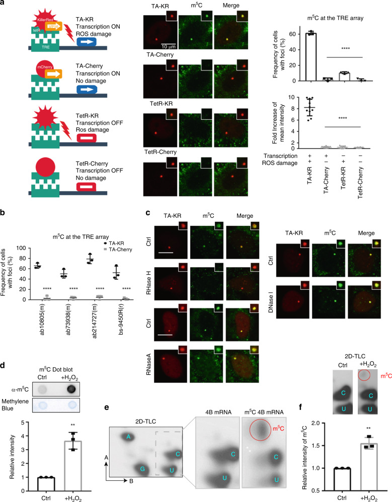
Fig. 1 m5C mRNA methylation is enriched at transcriptionally active sites with DNA damage. a U2OS-TRE cells transfected with TA-KR/TA-Cherry/tetR-KR/tetR-Cherry plasmids were exposed to light for 30 min for KR activation and allowed to recover for 1 h before harvest (scale bar: 10 μm). Quantification of frequency of cells in 500 cells with m5C foci from three independent experiments, mean ± SD (upper right). Fold increase of m5C mean intensity = mean intensity of m5C at TA-KR/mean intensity of background (n = 20, mean ± SD) (lower right). b U2OS-TRE cells were transfected with TA-KR/TA-Cherry to induce local oxidative damage or for the control condition. Cells were then stained for m5C with four different anti-m5C antibodies. Frequency of m5C-positive cells in 500 cells was quantified (n = 3, mean ± SD). c U2OS-TRE cells transfected with TA-KR were digested with RNaseH1, RNaseA, or DNase I and stained for m5C quantification (scale bar: 10 μm). d The mRNA from Flp-in 293 cells treated with or without 2 mM H2O2 for 40 min was used for m5C measurement via dot blot. Quantification of m5C levels (mean ± SD) from three independent experiments normalized with Ctrl and methylene blue is shown. e 32P-labeled mRNA monophosphate nucleosides were run on 2D gels for 2D-TLC analysis. In vitro-transcribed 4B mRNA with or without m5C was run in parallel. Representative images from three sets of independent experiments are shown with arrows showing the directions of each solvent run. Position of each nucleotide and m5C are labeled (Left). f 32P-labeled mRNA monophosphate nucleosides from U2OS cells with or without 2 mM H2O2 for 40 min were run on 2D gels for 2D-TLC analysis. Representative images from three sets of independent experiments. Associated quantification of relative increase in m5C in peroxide-treated cells compared to control, normalized to nucleotide C (right). Statistical analysis was performed with the unpaired two tailed Student’s t-test. *p < 0.05; **p < 0.01; ***p < 0.001; ****p < 0.0001.
Fig. 1 m5C mRNA methylation is enriched at transcriptionally active sites with DNA damage. a U2OS-TRE cells transfected with TA-KR/TA-Cherry/tetR-KR/tetR-Cherry plasmids were exposed to light for 30 min for KR activation and allowed to recover for 1 h before harvest (scale bar: 10 μm). Quantification of frequency of cells in 500 cells with m5C foci from three independent experiments, mean ± SD (upper right). Fold increase of m5C mean intensity = mean intensity of m5C at TA-KR/mean intensity of background (n = 20, mean ± SD) (lower right). b U2OS-TRE cells were transfected with TA-KR/TA-Cherry to induce local oxidative damage or for the control condition. Cells were then stained for m5C with four different anti-m5C antibodies. Frequency of m5C-positive cells in 500 cells was quantified (n = 3, mean ± SD). c U2OS-TRE cells transfected with TA-KR were digested with RNaseH1, RNaseA, or DNase I and stained for m5C quantification (scale bar: 10 μm). d The mRNA from Flp-in 293 cells treated with or without 2 mM H2O2 for 40 min was used for m5C measurement via dot blot. Quantification of m5C levels (mean ± SD) from three independent experiments normalized with Ctrl and methylene blue is shown. e 32P-labeled mRNA monophosphate nucleosides were run on 2D gels for 2D-TLC analysis. In vitro-transcribed 4B mRNA with or without m5C was run in parallel. Representative images from three sets of independent experiments are shown with arrows showing the directions of each solvent run. Position of each nucleotide and m5C are labeled (Left). f 32P-labeled mRNA monophosphate nucleosides from U2OS cells with or without 2 mM H2O2 for 40 min were run on 2D gels for 2D-TLC analysis. Representative images from three sets of independent experiments. Associated quantification of relative increase in m5C in peroxide-treated cells compared to control, normalized to nucleotide C (right). Statistical analysis was performed with the unpaired two tailed Student’s t-test. *p < 0.05; **p < 0.01; ***p < 0.001; ****p < 0.0001.CC BY · PubMed Central
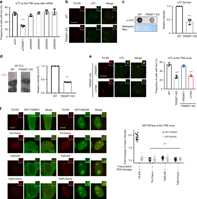
Fig. 2 TRDMT1 mediates m5C mRNA methylation at DNA damage sites. a U2OS-TRE cells pre-treated with the indicated siRNA were transfected with TA-KR to induce local oxidative damage and stained for m5C. The frequency of cells in 500 cells with m5C foci (n = 3, mean ± SD) is shown. b The U2OS-TRE WT and TRDMT1 KO cells were transfected with TA-KR, stained for m5C, and quantified (scale bar: 10 μm) (n = 3, mean ± SD). c WT U2OS and TRDMT1 KO cells were treated with 2 mM H2O2 for 40 min. The mRNA was then extracted from the cell lysates for m5C measurement via dot blot. Quantification of m5C levels (mean ± SD) from three independent experiments normalized with Ctrl and methylene blue is shown. d 32P-labeled monophosphate nucleosides from mRNA from WT U2OS and TRDMT1 KO cells with or without 2 mM H2O2 for 40 min were run on 2D gels for 2D-TLC analysis. Representative images from three sets of independent experiments. Associated quantification of relative increases in m5C from H2O2-treated cells were compared to the control and normalized to nucleotide C (n = 3, mean ± SD). e The TRDMT1 KO and stably rescued cell lines expressing WT TRDMT1 or the C79A mutant were transfected with TA-KR, stained for m5C, and quantified (scale bar: 10 μm) (n = 3, mean ± SD). f U2OS-TRE cells transfected with TA-KR/TA-Cherry/tetR-KR/tetR-Cherry and GFP-TRDMT1/GFP-NSUN2 plasmids were light-irradiated and allowed to recover for 1 h before fixation and were quantified (scale bar: 10 μm) (n = 10, mean ± SD). Statistical analysis was performed with the unpaired two tailed Student’s t-test. *p < 0.05; **p < 0.01; ***p < 0.001; ****p < 0.0001.
Fig. 2 TRDMT1 mediates m5C mRNA methylation at DNA damage sites. a U2OS-TRE cells pre-treated with the indicated siRNA were transfected with TA-KR to induce local oxidative damage and stained for m5C. The frequency of cells in 500 cells with m5C foci (n = 3, mean ± SD) is shown. b The U2OS-TRE WT and TRDMT1 KO cells were transfected with TA-KR, stained for m5C, and quantified (scale bar: 10 μm) (n = 3, mean ± SD). c WT U2OS and TRDMT1 KO cells were treated with 2 mM H2O2 for 40 min. The mRNA was then extracted from the cell lysates for m5C measurement via dot blot. Quantification of m5C levels (mean ± SD) from three independent experiments normalized with Ctrl and methylene blue is shown. d 32P-labeled monophosphate nucleosides from mRNA from WT U2OS and TRDMT1 KO cells with or without 2 mM H2O2 for 40 min were run on 2D gels for 2D-TLC analysis. Representative images from three sets of independent experiments. Associated quantification of relative increases in m5C from H2O2-treated cells were compared to the control and normalized to nucleotide C (n = 3, mean ± SD). e The TRDMT1 KO and stably rescued cell lines expressing WT TRDMT1 or the C79A mutant were transfected with TA-KR, stained for m5C, and quantified (scale bar: 10 μm) (n = 3, mean ± SD). f U2OS-TRE cells transfected with TA-KR/TA-Cherry/tetR-KR/tetR-Cherry and GFP-TRDMT1/GFP-NSUN2 plasmids were light-irradiated and allowed to recover for 1 h before fixation and were quantified (scale bar: 10 μm) (n = 10, mean ± SD). Statistical analysis was performed with the unpaired two tailed Student’s t-test. *p < 0.05; **p < 0.01; ***p < 0.001; ****p < 0.0001.CC BY · PubMed Central
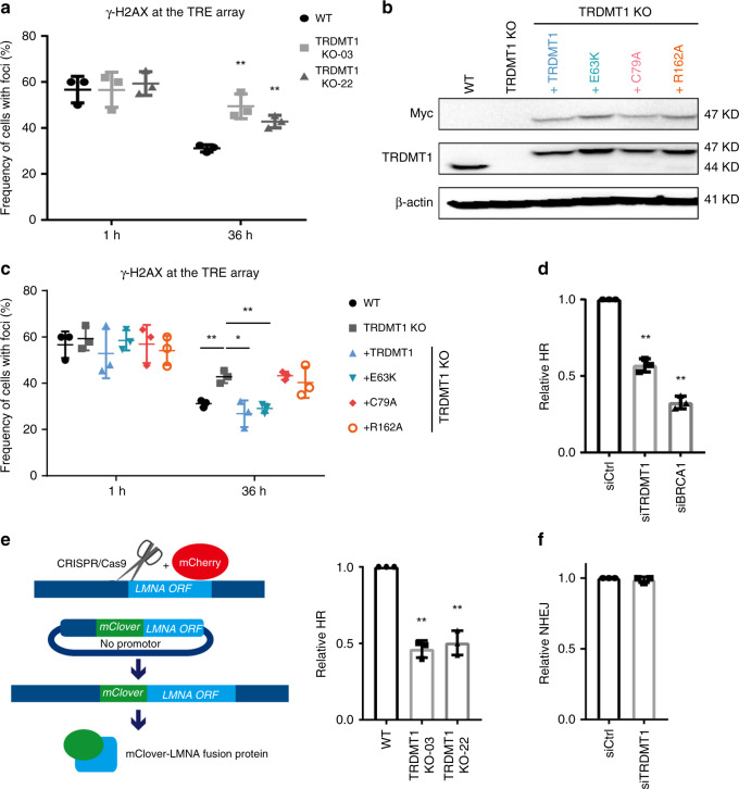
Fig. 3 TRDMT1 is required for damage removal and HR. a WT or TRDMT1 KO U2OS-TRE cells were transfected with TA-KR and stained for γH2AX at the indicated timepoint after damage (n = 3, mean ± SD). b Stable expression of Myc-tagged TRDMT1 in TRDMT1 KO U2OS-TRE cell lines shown in Western blots. Expression of β-actin is shown as a positive control. c U2OS-TRE WT, TRDMT1 KO, and TRDMT1 stably expressing cells were stained for γH2AX after damage caused by TA-KR at the indicated timepoint (n = 3, mean ± SD). d DR-GFP cells were pre-treated with siTRDMT1, siBRCA1, or control siRNA and then transfected with the NLS-I-SceI plasmid to induce DSBs. The GFP-positive population was analyzed by flow cytometry (n = 3, mean ± SD). e WT or TRDMT1 KO U2OS cells were co-transfected with CRISPR/Cas9-sgLMNA, LMNA-mClover, and mCherry plasmids. The fraction of mClover-positive cells in the mCherry-positive population was analyzed by flow cytometry (n = 3, mean ± SD). f EJ5-GFP cells were pre-treated with siTRDMT1 or control siRNA and then transfected with the NLS-I-SceI plasmid to induce DSBs. The GFP-positive population was analyzed by flow cytometry (n = 3, mean ± SD). Statistical analysis was performed with the unpaired two tailed Student’s t-test. *p < 0.05; **p < 0.01; ***p < 0.001; ****p < 0.0001.
Fig. 3 TRDMT1 is required for damage removal and HR. a WT or TRDMT1 KO U2OS-TRE cells were transfected with TA-KR and stained for γH2AX at the indicated timepoint after damage (n = 3, mean ± SD). b Stable expression of Myc-tagged TRDMT1 in TRDMT1 KO U2OS-TRE cell lines shown in Western blots. Expression of β-actin is shown as a positive control. c U2OS-TRE WT, TRDMT1 KO, and TRDMT1 stably expressing cells were stained for γH2AX after damage caused by TA-KR at the indicated timepoint (n = 3, mean ± SD). d DR-GFP cells were pre-treated with siTRDMT1, siBRCA1, or control siRNA and then transfected with the NLS-I-SceI plasmid to induce DSBs. The GFP-positive population was analyzed by flow cytometry (n = 3, mean ± SD). e WT or TRDMT1 KO U2OS cells were co-transfected with CRISPR/Cas9-sgLMNA, LMNA-mClover, and mCherry plasmids. The fraction of mClover-positive cells in the mCherry-positive population was analyzed by flow cytometry (n = 3, mean ± SD). f EJ5-GFP cells were pre-treated with siTRDMT1 or control siRNA and then transfected with the NLS-I-SceI plasmid to induce DSBs. The GFP-positive population was analyzed by flow cytometry (n = 3, mean ± SD). Statistical analysis was performed with the unpaired two tailed Student’s t-test. *p < 0.05; **p < 0.01; ***p < 0.001; ****p < 0.0001.CC BY · PubMed Central
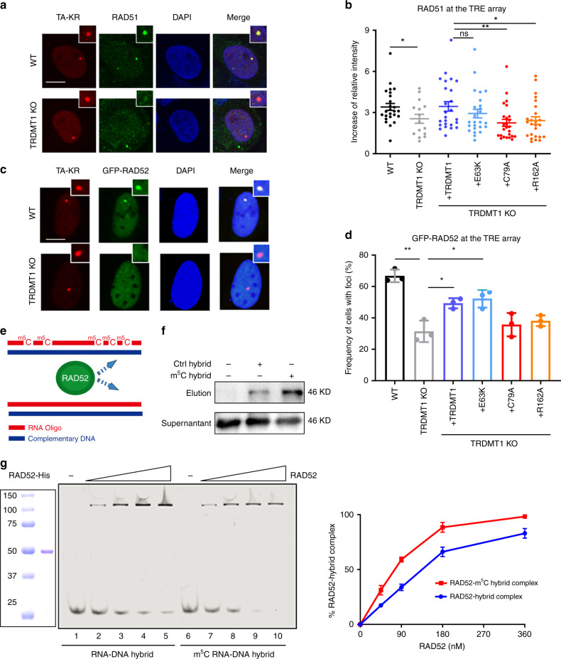
Fig. 4 RAD52 is a m5C reader. a U2OS-TRE WT and TRDMT1 KO cells were transfected with TA-KR and stained for RAD51 1 h after light irradiation. Representative figures were shown (scale bar: 10 μm). b U2OS-TRE WT, TRDMT1 KO, and TRDMT1 stably rescued cell lines were transfected with TA-KR. and stained for RAD51 1 h after light irradiation. Fold increase of RAD51 foci intensity was calculated (n = 25, mean ± SD). c U2OS-TRE WT and TRDMT1 KO cells were transfected with TA-KR and GFP-RAD52 and fixed 1 h after light irradiation. Representative figures were shown (scale bar: 10 μm). d U2OS-TRE WT, TRDMT1 KO, and TRDMT1 stably rescued cell lines were co-transfected with GFP-RAD52 and TA-KR and quantified for RAD52 foci frequency (n = 3, mean ± SD). e RNA oligos with or without m5C modification and their complementary DNA oligos were chemically synthesized for RAD52 binding experiments. f RAD52 protein in the cell lysate was pulled down by biotin-labeled DNA:RNA hybrids, with or without m5C RNA modification, coated on streptavidin magnetic beads. g The binding of RAD52 protein to DNA:RNA hybrids with or without m5C RNA modification was measured in electrophoretic mobility shift assays and quantified (n = 3, mean ± SD). Statistical analysis was performed with the unpaired two tailed Student’s t-test. *p < 0.05; **p < 0.01; ***p < 0.001; ****p < 0.0001.
Fig. 4 RAD52 is a m5C reader. a U2OS-TRE WT and TRDMT1 KO cells were transfected with TA-KR and stained for RAD51 1 h after light irradiation. Representative figures were shown (scale bar: 10 μm). b U2OS-TRE WT, TRDMT1 KO, and TRDMT1 stably rescued cell lines were transfected with TA-KR. and stained for RAD51 1 h after light irradiation. Fold increase of RAD51 foci intensity was calculated (n = 25, mean ± SD). c U2OS-TRE WT and TRDMT1 KO cells were transfected with TA-KR and GFP-RAD52 and fixed 1 h after light irradiation. Representative figures were shown (scale bar: 10 μm). d U2OS-TRE WT, TRDMT1 KO, and TRDMT1 stably rescued cell lines were co-transfected with GFP-RAD52 and TA-KR and quantified for RAD52 foci frequency (n = 3, mean ± SD). e RNA oligos with or without m5C modification and their complementary DNA oligos were chemically synthesized for RAD52 binding experiments. f RAD52 protein in the cell lysate was pulled down by biotin-labeled DNA:RNA hybrids, with or without m5C RNA modification, coated on streptavidin magnetic beads. g The binding of RAD52 protein to DNA:RNA hybrids with or without m5C RNA modification was measured in electrophoretic mobility shift assays and quantified (n = 3, mean ± SD). Statistical analysis was performed with the unpaired two tailed Student’s t-test. *p < 0.05; **p < 0.01; ***p < 0.001; ****p < 0.0001.CC BY · PubMed Central
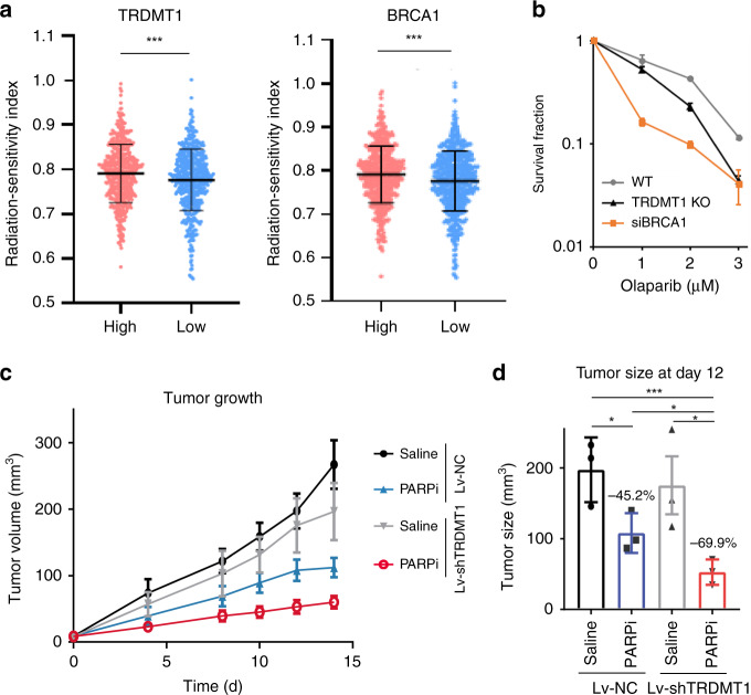
Fig. 5 TRDMT1 inhibition sensitizes cells to radiation and PARPi. a Clinical and RNA-seq gene expression data were downloaded from TCGA. RSI was calculated by a rank-based linear regression algorithm. The median value of BRCA1 and TRDMT1 TPM was defined as the cutoff to classify patients into high expression and low expression groups (n = 1091, mean ±SD). b WT, TRDMT1 KO U2OS, or siBRCA1 cells were treated with Olaparib at the indicated dose. The survival rate was measured via the colony formation assay (n = 3, mean ± SEM). c Tumor volume in mice injected with LV-shTRDMT1 or LV-NC pre-treated MDA-MB-231 cells at the indicated time (n = 3, mean ± SEM). One week after injection, 50 mg/kg Olaparib or saline was intraperitoneally injected into the xenograft tumors once every other day. d Tumor volume in mice injected with LV-shTRDMT1 or LV-NC pre-treated MDA-MB-231 cells at day 12 after PARPi treatment (n = 3, mean ± SEM). Statistical analysis was performed with the unpaired two tailed Student’s t-test. *p < 0.05; **p < 0.01; ***p < 0.001; ****p < 0.0001.
Fig. 5 TRDMT1 inhibition sensitizes cells to radiation and PARPi. a Clinical and RNA-seq gene expression data were downloaded from TCGA. RSI was calculated by a rank-based linear regression algorithm. The median value of BRCA1 and TRDMT1 TPM was defined as the cutoff to classify patients into high expression and low expression groups (n = 1091, mean ±SD). b WT, TRDMT1 KO U2OS, or siBRCA1 cells were treated with Olaparib at the indicated dose. The survival rate was measured via the colony formation assay (n = 3, mean ± SEM). c Tumor volume in mice injected with LV-shTRDMT1 or LV-NC pre-treated MDA-MB-231 cells at the indicated time (n = 3, mean ± SEM). One week after injection, 50 mg/kg Olaparib or saline was intraperitoneally injected into the xenograft tumors once every other day. d Tumor volume in mice injected with LV-shTRDMT1 or LV-NC pre-treated MDA-MB-231 cells at day 12 after PARPi treatment (n = 3, mean ± SEM). Statistical analysis was performed with the unpaired two tailed Student’s t-test. *p < 0.05; **p < 0.01; ***p < 0.001; ****p < 0.0001.CC BY · PubMed Central

Verification

Claims the paper makes

The claims and statistics of this paper haven’t been checked yet.

Run the check — free, no account needed. It reads the open-access text and checks the sources it cites.

A few free checks a day · papers already verified open instantly · results are public

Claims other papers make about this one

Each is checked against this paper’s own text: does it say what the citing paper says it does?

Self-check

Percentages, sums and reported differences re-computed from the paper’s own numbers.

The self-check needs the paper’s full text.

Citations

Cites

Show all 7 references

Research works printed but not linked: 39

  1. Pankotai, T., Bonhomme, C., Chen, D. & Soutoglou, E. DNAPKcs-dependent arrest of RNA polymerase II transcription in the presence of DNA breaks. Nat. Struct. Mol. Biol. 19, 276–282 (2012). · DOI · Paper page
  2. Keskin, H. et al. Transcript-RNA-templated DNA recombination and repair. Nature 515, 436–439 (2014). · DOI
  3. Wang, X. et al. Induced ncRNAs allosterically modify RNA-binding proteins in cis to inhibit transcription. Nature 454, 126–130 (2008). · DOI
  4. Lu, W.-T. et al. Drosha drives the formation of DNA:RNA hybrids around DNA break sites to facilitate DNA repair. Nat. Commun. 9, 532 (2018). · DOI
  5. Durdevic, Z., Mobin, M. B., Hanna, K., Lyko, F. & Schaefer, M. The RNA methyltransferase Dnmt2 is required for efficient Dicer-2-dependent siRNA pathway activity in Drosophila. Cell Rep. 4, 931–937 (2013). · DOI
  6. Wei, L. et al. DNA damage during the G0/G1 phase triggers RNA-templated, Cockayne syndrome B-dependent homologous recombination. Proc. Natl Acad. Sci. USA 112, E3495–E3504 (2015). · DOI
  7. Teng, Y. et al. ROS-induced R loops trigger a transcription-coupled but BRCA1/2-independent homologous recombination pathway through CSB. Nat. Commun. 9, 4115 (2018). · DOI
  8. Yasuhara, T. et al. Human Rad52 promotes XPG-mediated R-loop processing to initiate transcription-associated homologous recombination repair. Cell 175, 558–570 (2018). · DOI
  9. Crossley, M. P., Bocek, M. & Cimprich, K. A. R-loops as cellular regulators and genomic threats. Mol. Cell 73, 398–411 (2019). · DOI
  10. Ayoub, N., Jeyasekharan, A. D., Bernal, J. A. & Venkitaraman, A. R. HP1-beta mobilization promotes chromatin changes that initiate the DNA damage response. Nature 453, 682–686 (2008). · DOI
  11. Fernandez-Capetillo, O., Allis, C. D. & Nussenzweig, A. Phosphorylation of histone H2B at DNA double-strand breaks. J. Exp. Med. 199, 1671–1677 (2004). · DOI
  12. Khoury-Haddad, H., Nadar-Ponniah, P. T., Awwad, S. & Ayoub, N. The emerging role of lysine demethylases in DNA damage response: dissecting the recruitment mode of KDM4D/JMJD2D to DNA damage sites. Cell Cycle 14, 950–958 (2015). · DOI
  13. Jurkowski, T. P. et al. Human DNMT2 methylates tRNA(Asp) molecules using a DNA methyltransferase-like catalytic mechanism. RNA 14, 1663–1670 (2008). · DOI
  14. Elhardt, W., Shanmugam, R., Jurkowski, T. P. & Jeltsch, A. Somatic cancer mutations in the DNMT2 tRNA methyltransferase alter its catalytic properties. Biochimie 112, 66–72 (2015). · DOI
  15. Rai, K. et al. Dnmt2 functions in the cytoplasm to promote liver, brain, and retina development in zebrafish. Genes Dev. 21, 261–266 (2007). · DOI
  16. Grosjean, H., Keith, G. & Droogmans, L. Detection and quantification of modified nucleotides in RNA using thin-layer chromatography. Methods Mol. Biol. 265, 357–391 (2004).
  17. Xiang, Y. et al. Corrigendum: RNA m(6)A methylation regulates the ultraviolet-induced DNA damage response. Nature 552, 430 (2017). · DOI
  18. Lan, L. et al. The ACF1 complex is required for DNA double-strand break repair in human cells. Mol. Cell 40, 976–987 (2010). · DOI
  19. Schapira, M. Structural chemistry of human RNA methyltransferases. ACS Chem. Biol. 11, 575–582 (2016). · DOI
  20. Khoddami, V. & Cairns, B. R. Identification of direct targets and modified bases of RNA cytosine methyltransferases. Nat. Biotechnol. 31, 458–464 (2013). · DOI
  21. Mazina, O. M., Keskin, H., Hanamshet, K., Storici, F. & Mazin, A. V. Rad52 inverse strand exchange drives RNA-templated DNA double-strand break repair. Mol. Cell 67, 19–29 (2017). · DOI
  22. Welty, S. et al. RAD52 is required for RNA-templated recombination repair in post-mitotic neurons. J. Biol. Chem. 293, 1353–1362 (2017). · DOI
  23. Yang, X. et al. 5-methylcytosine promotes mRNA export—NSUN2 as the methyltransferase and ALYREF as an m(5)C reader. Cell Res 27, 606–625 (2017). · DOI · Paper page
  24. Robb, R. et al. Inhibiting BRAF oncogene-mediated radioresistance effectively radiosensitizes BRAF(V600E)-mutant thyroid cancer cells by constraining DNA double-strand break repair. Clin. Cancer Res. 25, 4749–4760 (2019). · DOI
  25. De Cecco, L., Bossi, P., Locati, L., Canevari, S. & Licitra, L. Comprehensive gene expression meta-analysis of head and neck squamous cell carcinoma microarray data defines a robust survival predictor. Ann. Oncol. 25, 1628–1635 (2014). · DOI
  26. Seton-Rogers, S. Targeted therapies: expanding the use of PARP inhibitors. Nat. Rev. Cancer 17, 397 (2017).
  27. Ceccaldi, R. et al. Homologous-recombination-deficient tumours are dependent on Poltheta-mediated repair. Nature 518, 258–262 (2015). · DOI
  28. Bekker-Jensen, S. et al. Spatial organization of the mammalian genome surveillance machinery in response to DNA strand breaks. J. Cell Biol. 173, 195–206 (2006). · DOI
  29. Huen, M. S. et al. RNF8 transduces the DNA-damage signal via histone ubiquitylation and checkpoint protein assembly. Cell 131, 901–914 (2007). · DOI
  30. Xie, A. et al. Distinct roles of chromatin-associated proteins MDC1 and 53BP1 in mammalian double-strand break repair. Mol. Cell 28, 1045–1057 (2007). · DOI
  31. Aguilera, A. & Gomez-Gonzalez, B. DNA-RNA hybrids: the risks of DNA breakage during transcription. Nat. Struct. Mol. Biol. 24, 439–443 (2017). · DOI
  32. Cohen, S. et al. Senataxin resolves RNA:DNA hybrids forming at DNA double-strand breaks to prevent translocations. Nat. Commun. 9, 533 (2018). · DOI
  33. Xiang, Y. et al. RNA m6A methylation regulates the ultraviolet-induced DNA damage response. Nature 543, 573–576 (2017). · DOI
  34. Liu, L. et al. Decomposition of RNA methylome reveals co-methylation patterns induced by latent enzymatic regulators of the epitranscriptome. Mol. Biosyst. 11, 262–274 (2015). · DOI
  35. Bukhari, S. I. A. et al. A specialized mechanism of translation mediated by FXR1a-associated microRNP in cellular quiescence. Mol. Cell 61, 760–773 (2016). · DOI
  36. Zhong, S. et al. MTA is an Arabidopsis messenger RNA adenosine methylase and interacts with a homolog of a sex-specific splicing factor. Plant Cell 20, 1278–1288 (2008). · DOI
  37. Lan, L. et al. Novel method for site-specific induction of oxidative DNA damage reveals differences in recruitment of repair proteins to heterochromatin and euchromatin. Nucleic Acids Res. 42, 2330–2345 (2014). · DOI
  38. Nguyen, H. D. et al. Functions of replication protein A as a sensor of R loops and a regulator of RNaseH1. Mol. Cell 65, 832–847.e834 (2017). · DOI
  39. Pinder, J., Salsman, J. & Dellaire, G. Nuclear domain ‘knock-in’ screen for the evaluation and identification of small molecule enhancers of CRISPR-based genome editing. Nucleic Acids Res. 43, 9379–9392 (2015). · DOI

Cited by

See all 179

Metadata

Authors
Chen, H., Yang, H., Zhu, X., Yadav, T., Ouyang, J., Truesdell, S. S., Tan, J., Wang, Y., Duan, M., Wei, L., Zou, L., Levine, A. S., Vasudevan, S., & Lan, L.
Venue
Nature Communications
Published
2020-06-05
Volume
11
Issue
1
ISSN
2041-1723
Cited by
179
References
7
Language
en
Funders
U.S. Department of Health & Human Services | National Institutes of Health (GM118833)

About these checks

Registry, retraction, Cites and statistics results are computed, not judged, with one exception: a printed reference that no index lists is read once by a model before it counts as a citation error. Claims are extracted by a model and kept only when their quote appears verbatim in the text; each cited source is then checked, with a model’s help, for whether it says what the claim relies on. Those verdicts can be wrong — read the claim’s quote and the cited source before relying on one.

Statistics and arithmetic are recomputed, not judged.

Confidence bands are shown only for fields calibrated on our annotated test set. None is calibrated yet, so no band is shown.

What counts as a claim ›How we measure confidence ›How we check this ›

Are you an author, or do you see a problem with this page? Request a review

How to cite

Cite CoChat for the checks on this page, and the paper for its own findings.

This page
CoChat. (2026). Citation check: m5C modification of mRNA serves a DNA damage code to promote homologous recombination. https://app.cochat.ai/papers/10.1038/s41467-020-16722-7
The paper
Chen, H., Yang, H., Zhu, X., Yadav, T., Ouyang, J., Truesdell, S. S., Tan, J., Wang, Y., Duan, M., Wei, L., Zou, L., Levine, A. S., Vasudevan, S., & Lan, L. (2020). m5C modification of mRNA serves a DNA damage code to promote homologous recombination. Nature Communications. https://doi.org/10.1038/s41467-020-16722-7