

The DOI matches its Crossref record; 12 of the 12 references it cites were found in a registry or an index.
A systematic review and meta-analysis of gene therapy with hematopoietic stem and progenitor cells for monogenic disorders
Published version of a preprint
Abstract
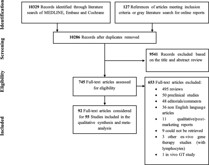
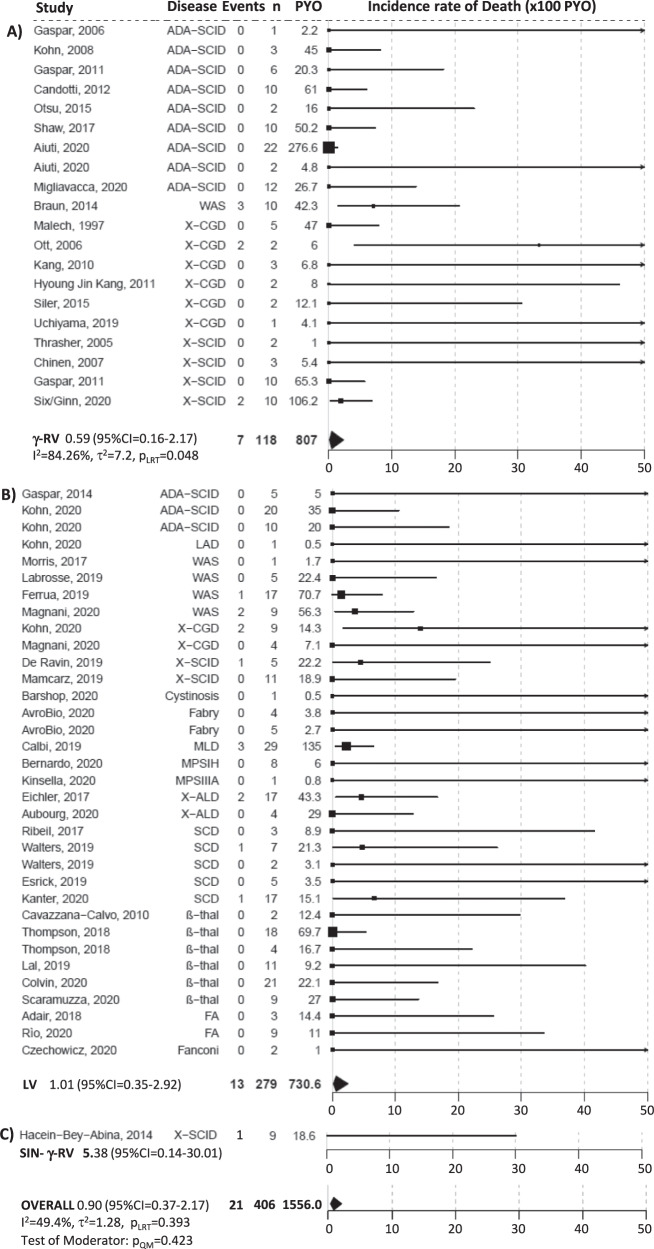
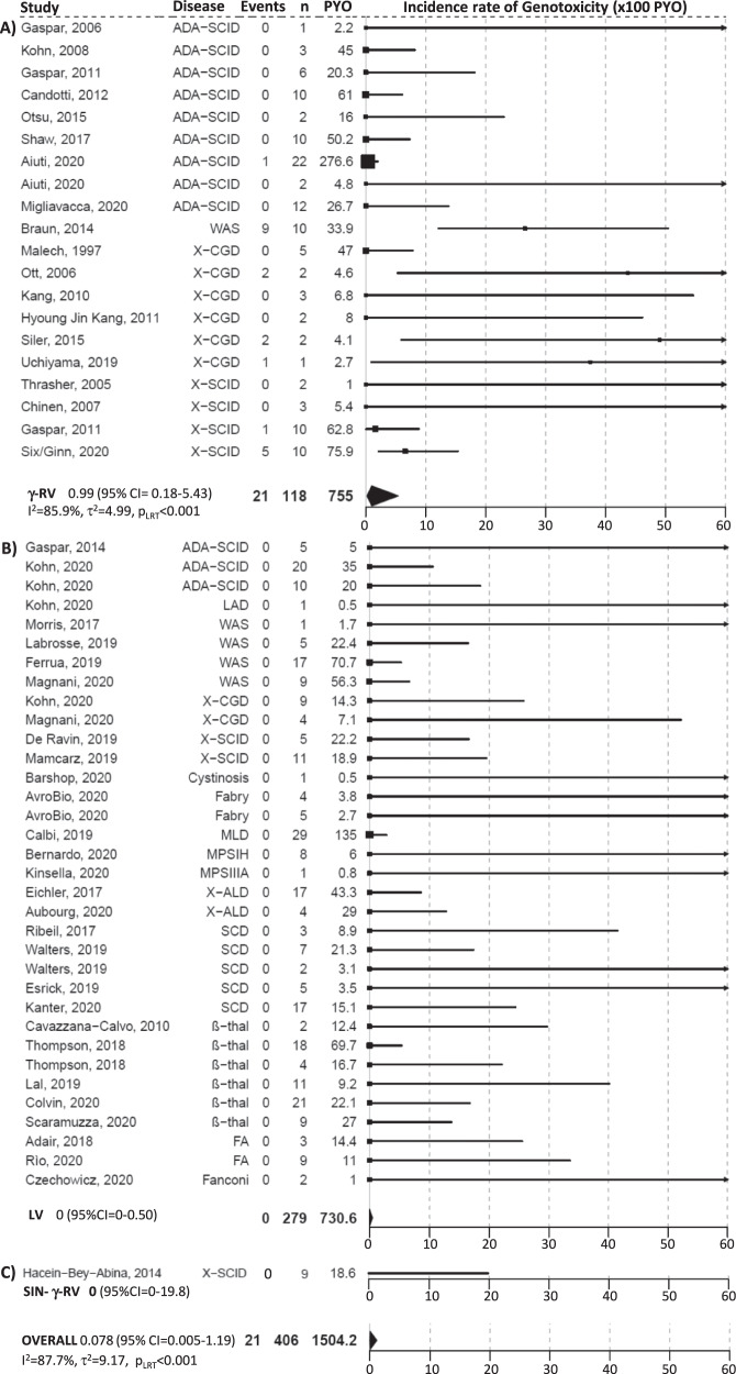
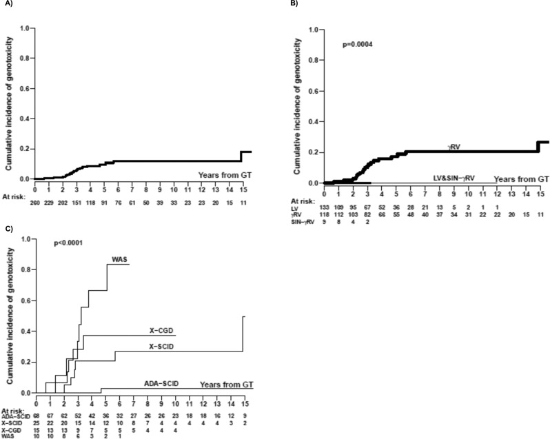
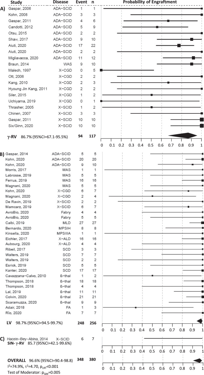
Ex-vivo gene therapy (GT) with hematopoietic stem and progenitor cells (HSPCs) engineered with integrating vectors is a promising treatment for monogenic diseases, but lack of centralized databases is hampering an overall outcomes assessment. Here we aim to provide a comprehensive assessment of the short and long term safety of HSPC-GT from trials using different vector platforms. We review systematically the literature on HSPC-GT to describe survival, genotoxicity and engraftment of gene corrected cells. From 1995 to 2020, 55 trials for 14 diseases met inclusion criteria and 406 patients with primary immunodeficiencies (55.2%), metabolic diseases (17.0%), haemoglobinopathies (24.4%) and bone marrow failures (3.4%) were treated with gammaretroviral vector (γRV) (29.1%), self-inactivating γRV (2.2%) or lentiviral vectors (LV) (68.7%). The pooled overall incidence rate of death is 0.9 per 100 person-years of observation (PYO) (95% CI = 0.37–2.17). There are 21 genotoxic events out of 1504.02 PYO, which occurred in γRV trials (0.99 events per 100 PYO, 95% CI = 0.18–5.43) for primary immunodeficiencies. Pooled rate of engraftment is 86.7% (95% CI = 67.1–95.5%) for γRV and 98.7% (95% CI = 94.5–99.7%) for LV HSPC-GT (p = 0.005). Our analyses show stable reconstitution of haematopoiesis in most recipients with superior engraftment and safer profile in patients receiving LV-transduced HSPCs.
Overview
The claims and statistics of this paper haven’t been checked yet.
Run the check — free, no account needed. It reads the open-access text and checks the sources it cites.
Integrity signals
Registry
Cites
Statistics
Figures
Verification
Claims the paper makes
The claims and statistics of this paper haven’t been checked yet.
Run the check — free, no account needed. It reads the open-access text and checks the sources it cites.
Claims other papers make about this one
Each is checked against this paper’s own text: does it say what the citing paper says it does?
In the citing paper
“A systematic review and meta-analysis showed that no cases of insertional oncogenesis have been reported for lentiviral vectors through March 2022.”
From Hematologic Cancer after Gene Therapy for Cerebral Adrenoleukodystrophy (2024) · cites this paper as [11]
Self-check
Percentages, sums and reported differences re-computed from the paper’s own numbers.
The self-check needs the paper’s full text.
Citations
Cites
Insertional oncogenesis in 4 patients after retrovirus-mediated gene therapy of SCID-X1
Hacein-Bey-Abina, S., Garrigue, A., Wang, G. P., Soulier, J., Lim, A., Morillon, E., Clappier, E., Caccavelli, L., Delabesse, E., Beldjord, K., et al. (2008) · Journal of Clinical Investigation · doi:10.1172/jci35700 · Cited by 1,854
Hematopoietic Stem Cell Gene Therapy with a Lentiviral Vector in X-Linked Adrenoleukodystrophy
Cartier, N., Hacein-Bey-Abina, S., Bartholomae, C. C., Veres, G., Schmidt, M., Kutschera, I., Vidaud, M., Abel, U., Dal-Cortivo, L., Caccavelli, L., et al. (2009) · Science · doi:10.1126/science.1171242 · Cited by 1,508
Transfusion independence and HMGA2 activation after gene therapy of human β-thalassaemia
Cavazzana-Calvo, M., Payen, E., Negre, O., Wang, G., Hehir, K., Fusil, F., Down, J., Denaro, M., Brady, T., Westerman, K., et al. (2010) · Nature · doi:10.1038/nature09328 · Cited by 1,276
Stijnen, T., Hamza, T. H., & Özdemir, P. (2010) · Statistics in Medicine · doi:10.1002/sim.4040 · Cited by 757
Gene therapy in a patient with sickle cell disease
Ribeil, J.-A., Hacein-Bey-Abina, S., Payen, E., Magnani, A., Semeraro, M., Magrin, E., Caccavelli, L., Neven, B., Bourget, P., El Nemer, W., et al. (2017) · New England Journal of Medicine · doi:10.1056/nejmoa1609677 · Cited by 696
Gene Therapy in Patients with Transfusion-Dependent β-Thalassemia
Thompson, A. A., Walters, M. C., Kwiatkowski, J., Rasko, J. E. J., Ribeil, J.-A., Hongeng, S., Magrin, E., Schiller, G. J., Payen, E., Semeraro, M., et al. (2018) · New England Journal of Medicine · doi:10.1056/nejmoa1705342 · Cited by 684
Hematopoietic stem-cell gene therapy for cerebral adrenoleukodystrophy
Eichler, F., Duncan, C., Musolino, P. L., Orchard, P. J., De Oliveira, S., Thrasher, A. J., Armant, M., Dansereau, C., Lund, T. C., Miller, W. P., et al. (2017) · New England Journal of Medicine · doi:10.1056/nejmoa1700554 · Cited by 604
Montini, E., Cesana, D., Schmidt, M., Sanvito, F., Bartholomae, C. C., Ranzani, M., Benedicenti, F., Sergi, L. S., Ambrosi, A., Ponzoni, M., et al. (2009) · Journal of Clinical Investigation · doi:10.1172/jci37630 · Cited by 571
Self-citationGene therapy for Wiskott-Aldrich syndrome–long-term efficacy and genotoxicity
Braun, C. J., Boztug, K., Paruzynski, A., Witzel, M., Schwarzer, A., Rothe, M., Modlich, U., Beier, R., Göhring, G., Steinemann, D., et al. (2014) · Science Translational Medicine · doi:10.1126/scitranslmed.3007280 · Cited by 562
Sessa, M., Lorioli, L., Fumagalli, F., Acquati, S., Redaelli, D., Baldoli, C., Canale, S., Lopez, I. D., Morena, F., Calabria, A., et al. (2016) · The Lancet · doi:10.1016/s0140-6736(16)30374-9 · Cited by 502
Self-citationA Modified γ-Retrovirus Vector for X-Linked Severe Combined Immunodeficiency
Hacein-Bey-Abina, S., Pai, S.-Y., Gaspar, H. B., Armant, M., Berry, C. C., Blanche, S., Bleesing, J., Blondeau, J., de Boer, H., Buckland, K. F., et al. (2014) · New England Journal of Medicine · doi:10.1056/nejmoa1404588 · Cited by 412
Lentiviral Gene Therapy Combined with Low-Dose Busulfan in Infants with SCID-X1
Mamcarz, E., Zhou, S., Lockey, T., Abdelsamed, H., Cross, S. J., Kang, G., Ma, Z., Condori, J., Dowdy, J., Triplett, B., et al. (2019) · New England Journal of Medicine · doi:10.1056/nejmoa1815408 · Cited by 263
Research works printed but not linked: 81
- Ferrari, G., Thrasher, A. J. & Aiuti, A. Gene therapy using haematopoietic stem and progenitor cells. Nat. Rev. Genet. https://doi.org/10.1038/s41576-020-00298-5 (2020). · DOI
- Tucci, F., Scaramuzza, S., Aiuti, A. & Mortellaro, A. Update on Clinical Ex vivo hematopoietic stem cell gene therapy for inherited monogenic diseases. Mol. Ther. https://doi.org/10.1016/j.ymthe.2020.11.020 (2020). · DOI
- Cavazzana, M., Bushman, F. D., Miccio, A., Andre-Schmutz, I. & Six, E. Gene therapy targeting haematopoietic stem cells for inherited diseases: progress and challenges. Nat. Rev. Drug Disco. 18, 447–462 (2019). · DOI
- Slatter, M. A. et al. Treosulfan and fludarabine conditioning for hematopoietic stem cell transplantation in children with primary immunodeficiency: UK experience. Biol. Blood Marrow Transpl. 24, 529–536 (2018). · DOI
- Hassan, A. et al. Outcome of hematopoietic stem cell transplantation for adenosine deaminase-deficient severe combined immunodeficiency. Blood 120, 3615–3624 (2012). · DOI
- Fox, T. A. et al. Successful outcome following allogeneic hematopoietic stem cell transplantation in adults with primary immunodeficiency. Blood 131, 917–931 (2018). · DOI
- Albert, M. H. et al. Allogeneic stem cell transplantation in adolescents and young adults with primary immunodeficiencies. J. Allergy Clin. Immunol. Pr. 6, 298–301 e292 (2018). · DOI
- Pai, S. Y. et al. Transplantation outcomes for severe combined immunodeficiency, 2000–2009. N. Engl. J. Med 371, 434–446 (2014). · DOI
- Murillo-Sanjuan, L. et al. Survival and toxicity outcomes of hematopoietic stem cell transplantation for pediatric patients with Fanconi anemia: a unified multicentric national study from the Spanish Working Group for Bone Marrow Transplantation in Children. Bone Marrow Transplant. https://doi.org/10.1038/s41409-020-01172-y (2020). · DOI
- Boucher, A. A. et al. Long-term outcomes after allogeneic hematopoietic stem cell transplantation for metachromatic leukodystrophy: the largest single-institution cohort report. Orphanet J. Rare Dis. 10, 94 (2015). · DOI
- Raymond, G. V. et al. Survival and functional outcomes in boys with cerebral adrenoleukodystrophy with and without hematopoietic stem cell transplantation. Biol. Blood Marrow Transpl. 25, 538–548 (2019). · DOI · Paper page
- Aldenhoven, M. et al. Hematopoietic cell transplantation for mucopolysaccharidosis patients is safe and effective: results after implementation of international guidelines. Biol. Blood Marrow Transpl. 21, 1106–1109 (2015). · DOI
- Strocchio, L. & Locatelli, F. Hematopoietic stem cell transplantation in Thalassemia. Hematol. Oncol. Clin. North Am. 32, 317–328 (2018). · DOI
- Gluckman, E. et al. Sickle cell disease: an international survey of results of HLA-identical sibling hematopoietic stem cell transplantation. Blood 129, 1548–1556 (2017). · DOI
- Mayor, P. C. et al. Cancer in primary immunodeficiency diseases: Cancer incidence in the United States Immune Deficiency Network Registry. J. Allergy Clin. Immunol. 141, 1028–1035 (2018). · DOI
- Hauck, F., Gennery, A. R. & Seidel, M. G. Editorial: The relationship between cancer predisposition and primary immunodeficiency. Front. Immunol. 10, 1781 (2019). · DOI
- Ferrua, F. et al. Lentiviral haemopoietic stem/progenitor cell gene therapy for treatment of Wiskott-Aldrich syndrome: interim results of a non-randomised, open-label, phase 1/2 clinical study. Lancet Haematol. 6, e239–e253 (2019). · DOI
- Marktel, S. et al. Intrabone hematopoietic stem cell gene therapy for adult and pediatric patients affected by transfusion-dependent ss-thalassemia. Nat. Med. 25, 234–241 (2019). · DOI
- Bushman, F. D. Retroviral insertional mutagenesis in humans: evidence for four genetic mechanisms promoting expansion of cell clones. Mol. Ther. 28, 352–356 (2020). · DOI
- Walters M. C. et al. Exploring the drivers of potential clinical benefit in initial patients treated in the Hgb-206 Study of Lentiglobin for Sickle Cell Disease (SCD) Gene Therapy. Blood 2061 (2019). https://doi.org/10.1182/blood-2019-128814 · DOI
- Jones, R. R. & DeBaun, M. R. Leukemia after gene therapy for sickle cell disease: insertional mutagenesis, busulfan, both, or neither. Blood 138, 942–947 (2021). · DOI
- Servick, K. Gene therapy clinical trial halted as cancer risk surfaces. https://doi.org/10.1126/science.abl8782 (2021). · DOI
- Palchaudhuri, R. et al. Non-genotoxic conditioning for hematopoietic stem cell transplantation using a hematopoietic-cell-specific internalizing immunotoxin. Nat. Biotechnol. 34, 738–745 (2016). · DOI
- Kwon, H. S. et al. Anti-human CD117 antibody-mediated bone marrow niche clearance in nonhuman primates and humanized NSG mice. Blood 133, 2104–2108 (2019). · DOI
- Hsieh, M. M. et al. Myelodysplastic syndrome unrelated to lentiviral vector in a patient treated with gene therapy for sickle cell disease. Blood Adv. 4, 2058–2063 (2020). · DOI · Paper page
- Sokolic, R. et al. Myeloid dysplasia and bone marrow hypocellularity in adenosine deaminase-deficient severe combined immune deficiency. Blood 118, 2688–2694 (2011). · DOI
- Regan, S., Yang, X., Finnberg, N. K., El-Deiry, W. S. & Pu, J. J. Occurrence of acute myeloid leukemia in hydroxyurea-treated sickle cell disease patient. Cancer Biol. Ther. 20, 1389–1397 (2019). · DOI
- Aiuti, A. & Naldini, L. Safer conditioning for blood stem cell transplants. Nat. Biotechnol. 34, 721–723 (2016). · DOI
- Agarwal, R. et al. Toxicity-Free Hematopoietic Stem Cell Engraftment Achieved with Anti-CD117 Monoclonal Antibody Conditioning. Biol. Blood Marrow Transpl. 25, S92 (2019). · DOI
- McGrath, E., Chabannon, C., Terwel, S., Bonini, C. & Kuball, J. Opportunities and challenges associated with the evaluation of chimeric antigen receptor T cells in real-life. Curr. Opin. Oncol. 32, 427–433 (2020). · DOI
- Candotti, F. et al. Gene therapy for adenosine deaminase-deficient severe combined immune deficiency: clinical comparison of retroviral vectors and treatment plans. Blood 120, 3635–3646 (2012). · DOI
- Shaw, K. L. et al. Clinical efficacy of gene-modified stem cells in adenosine deaminase-deficient immunodeficiency. J. Clin. Invest. 127, 1689–1699 (2017). · DOI
- Aiuti, A. et al. Correction of ADA-SCID by stem cell gene therapy combined with nonmyeloablative conditioning. Science 296, 2410–2413 (2002). · DOI · Paper page
- Aiuti, A. et al. Gene therapy for immunodeficiency due to adenosine deaminase deficiency. N. Engl. J. Med. 360, 447–458 (2009). · DOI
- Cicalese, M. P. et al. Update on the safety and efficacy of retroviral gene therapy for immunodeficiency due to adenosine deaminase deficiency. Blood 128, 45–54 (2016). · DOI
- Cicalese M. P. et al. Retroviral gene therapy for the treatment of ADA-SCID: long-term follow up and first case of T-cell acute leukaemia due to insertional mutagenesis. HemaSphere 5, https://doi.org/10.1097/hs9.0000000000000566 (2021). · DOI
- Otsu, M. et al. Outcomes in two Japanese adenosine deaminase-deficiency patients treated by stem cell gene therapy with no cytoreductive conditioning. J. Clin. Immunol. 35, 384–398 (2015). · DOI
- Gaspar, H. B. et al. Hematopoietic stem cell gene therapy for adenosine deaminase-deficient severe combined immunodeficiency leads to long-term immunological recovery and metabolic correction. Sci. Transl. Med. 3, 97ra80 (2011). · DOI
- Gaspar, H. B. et al. Successful reconstitution of immunity in ADA-SCID by stem cell gene therapy following cessation of PEG-ADA and use of mild preconditioning. Mol. Ther. 14, 505–513 (2006). · DOI
- Kohn, D. B. et al. T lymphocytes with a normal ADA gene accumulate after transplantation of transduced autologous umbilical cord blood CD34+ cells in ADA-deficient SCID neonates. Nat. Med. 4, 775–780 (1998). · DOI
- Schmidt, M. et al. Clonality analysis after retroviral-mediated gene transfer to CD34+ cells from the cord blood of ADA-deficient SCID neonates. Nat. Med. 9, 463–468 (2003). · DOI
- Boztug, K. et al. Stem-cell gene therapy for the Wiskott-Aldrich syndrome. N. Engl. J. Med. 363, 1918–1927 (2010). · DOI · Paper page
- Malech, H. L. et al. Prolonged production of NADPH oxidase-corrected granulocytes after gene therapy of chronic granulomatous disease. Proc. Natl Acad. Sci. USA 94, 12133–12138 (1997). · DOI
- Siler, U. et al. Successful combination of sequential gene therapy and rescue Allo-HSCT in Two Children with X-CGD - Importance of Timing. Curr. Gene Ther. 15, 416–427 (2015). · DOI
- Ott, M. G. et al. Correction of X-linked chronic granulomatous disease by gene therapy, augmented by insertional activation of MDS1-EVI1, PRDM16 or SETBP1. Nat. Med. 12, 401–409 (2006). · DOI · Paper page
- Kang, E. M. et al. Retrovirus gene therapy for X-linked chronic granulomatous disease can achieve stable long-term correction of oxidase activity in peripheral blood neutrophils. Blood 115, 783–791 (2010). · DOI
- Kang, H. J. et al. Retroviral gene therapy for X-linked chronic granulomatous disease: results from phase I/II trial. Mol. Ther. 19, 2092–2101 (2011). · DOI
- Uchiyama, T. et al. Insertional Oncogenesis in X-CGD Patient after MFGS Retroviral Vector-Mediated Gene Therapy. Mol. Ther. 27, 9 (2019).
- Hacein-Bey-Abina, S. et al. LMO2-associated clonal T cell proliferation in two patients after gene therapy for SCID-X1. Science 302, 415–419 (2003). · DOI · Paper page
- Hacein-Bey-Abina, S. et al. Efficacy of gene therapy for X-linked severe combined immunodeficiency. N. Engl. J. Med. 363, 355–364 (2010). · DOI
- Ginn, S. L. et al. Treatment of an infant with X-linked severe combined immunodeficiency (SCID-X1) by gene therapy in Australia. Med. J. Aust. 182, 458–463 (2005). · DOI
- Six, E. et al. LMO2 Associated Clonal T Cell Proliferation 15 Years After Gamma-Retrovirus Mediated Gene Therapy for SCIDX1. Mol. Ther. 25, 347–348 (2017).
- Gaspar, H. B. et al. Long-term persistence of a polyclonal T cell repertoire after gene therapy for X-linked severe combined immunodeficiency. Sci. Transl. Med. 3, 97ra79 (2011). · DOI
- Chinen, J. et al. Gene therapy improves immune function in preadolescents with X-linked severe combined immunodeficiency. Blood 110, 67–73 (2007). · DOI
- Thrasher, A. J. et al. Failure of SCID-X1 gene therapy in older patients. Blood 105, 4255–4257 (2005). · DOI
- Gaspar, H. B. et al. Immunological and Metabolic Correction After Lentiviral Vector Mediated Haematopoietic Stem Cell Gene Therapy for ADA Deficiency. Mol. Ther. 22, S106 (2014). · DOI
- Kohn, D. B. et al. Lentiviral Gene Therapy with Autologous Hematopoietic Stem and Progenitor Cells (HSPCs) for the Treatment of Severe Combined Immune Deficiency Due to Adenosine Deaminase Deficiency (ADA-SCID): Two Year Follow-Up Results. Mol. Ther. 28, 554–555 (2020).
- Scaramuzza, S. et al. Clinical Outcomes from a Phase I/II Gene Therapy Trial for Patients affected by Severe Transfusion Dependent Beta-Thalassemia: Two Years Follow Up. Mol. Ther. 28, 169 (2020).
- Lal, A. et al. Northstar-3: Interim Results from a Phase 3 Study Evaluating Lentiglobin Gene Therapy in Patients with Transfusion-Dependent β-Thalassemia and Either a β0 or IVS-I-110 Mutation at Both Alleles of the HBB Gene. Blood 134, 815 (2019). · DOI
- Barshop, B. et al. Hematopoietic Stem Cell Gene Therapy for Cystinosis: Initial Results from a Phase I/II Clinical Trial. Mol. Ther. 28, 233 (2020).
- Khan, A. et al. Lentivirus-mediated gene therapy for Fabry disease. Nat. Commun. 12, 1178 (2021). · DOI
- Adair, J. E. et al. Lessons Learned from Two Decades of Clinical Trial Experience in Gene Therapy for Fanconi Anemia. Curr. Gene Ther. 16, 338–348 (2017). · DOI
- Rio, P. et al. Successful engraftment of gene-corrected hematopoietic stem cells in non-conditioned patients with Fanconi anemia. Nat. Med. 25, 1396–1401 (2019). · DOI
- Río, P. et al. Updated Results of a European Gene Therapy Trial in Fanconi Anemia Patients, Subtype A. Mol. Ther. 28, 57 (2020).
- Czechowicz, A. et al. Changing the Natural History of Fanconi Anemia Complementation Group-À with Gene Therapy: Early Results of U.S. Phase I Study of Lentiviral-Mediated Ex-Vivo FANCA Gene Insertion in Human Stem and Progenitor Cells. Biol. Blood Marrow Transplant 26, S39–S40 (2020). · DOI
- Kohn, D. B. et al. Phase 1/2 Study of Lentiviral-Mediated Ex-Vivo Gene Therapy for Pediatric Patients with Severe Leukocyte Adhesion Deficiency-I (LAD-I): Initial Results from the First Treated Patient. Mol. Ther. 28, 56 (2020).
- Biffi, A. et al. Lentiviral hematopoietic stem cell gene therapy benefits metachromatic leukodystrophy. Science 341, 1233158 (2013). · DOI · Paper page
- Fumagalli, F. et al. In 16th Annual WORLD Symposium, Orlando USA (2020).
- Kinsella, J. B. K. et al. Preliminary outcomes of haematopoietic stem cell gene therapy in a patient with Mucopolysaccharidosis IIIA. Mol. Ther. 28, 231–232 (2020).
- Tisdale, J. F. et al. Resolution of Sickle Cell Disease (SCD) Manifestations in Patients Treated with Lentiglobin Gene Therapy: Updated Results from the Phase 1/2 HGB-206 Group C Study. Mol. Ther. 28, 553 (2020).
- Esrick, E. B. et al. Validation of BCL11A As Therapeutic Target in Sickle Cell Disease: Results from the Adult Cohort of a Pilot/Feasibility Gene Therapy Trial Inducing Sustained Expression of Fetal Hemoglobin Using Post-Transcriptional Gene Silencing. Blood 134: https://doi.org/10.1182/blood-2019-132745 (2019). · DOI
- Aiuti, A. et al. Lentiviral hematopoietic stem cell gene therapy in patients with Wiskott-Aldrich syndrome. Science 341, 1233151 (2013). · DOI · Paper page
- Hacein-Bey Abina, S. et al. Outcomes following gene therapy in patients with severe Wiskott-Aldrich syndrome. JAMA 313, 1550–1563 (2015). · DOI · Paper page
- Magnani, A. et al. Long-Term Follow-Up Study after Lentiviral Hematopoietic Stem/Progenitor Cell Gene Therapy for Wiskott-Aldrich Syndrome. Mol. Ther. 28, 58 (2020).
- Morris, E. C. et al. Gene therapy for Wiskott-Aldrich syndrome in a severely affected adult. Blood 130, 1327–1335 (2017). · DOI
- Labrosse, R. et al. Outcome of Hematopoietic Stem Cell Gene Therapy for Wiskott-Aldrich Syndrome. Blood 134, 4629 (2019). · DOI
- Aubourg, P. et al. Continuing Clinical Efficacy and ALDP Gene Expression 5–10 Years after Lentiviral LV-Based CD34+ Cell Gene Therapy in Patients with X-linked Adrenoleukodystrophy (X-ALD). Mol. Ther. 28, 274 (2020).
- Kohn, D. B. et al. Lentiviral gene therapy for X-linked chronic granulomatous disease. Nat. Med 26, 200–206 (2020). · DOI · Paper page
- De Ravin, S. S. et al. Lentiviral hematopoietic stem cell gene therapy for X-linked severe combined immunodeficiency. Sci. Transl. Med 8, 335ra357 (2016). · DOI · Paper page
- De Ravin, S. S. et al. Enhanced Transduction Lentivector Gene Therapy for Treatment of Older Patients with X-Linked Severe Combined Immunodeficiency. Blood 134, 608 (2019). · DOI
- Mamcarz, E. et al. Lentiviral Gene Therapy with Low Dose Busulfan for Infants with X-SCID Results in the Development of a Functional Normal Immune System: Interim Results of an Ongoing Phase I/II Clinical Study. Blood 134, 608 (2019). · DOI
Cited by
Hematologic Cancer after Gene Therapy for Cerebral Adrenoleukodystrophy
Duncan, C. N., Bledsoe, J. R., Grzywacz, B., Beckman, A., Bonner, M., Eichler, F. S., Kühl, J.-S., Harris, M. H., Slauson, S., Colvin, R. A., et al. (2024) · New England Journal of Medicine · doi:10.1056/nejmoa2405541 · Cited by 164
Treatment of cerebral adrenoleukodystrophy: allogeneic transplantation and lentiviral gene therapy
Gupta, A. O., Raymond, G., Pierpont, E. I., Kemp, S., McIvor, R. S., Rayannavar, A., Miller, B., Lund, T. C., & Orchard, P. J. (2022) · Expert Opinion on Biological Therapy · doi:10.1080/14712598.2022.2124857 · Cited by 67
“tional mutagenesis across 55 studies including a total of 406 participants using lentiviral and gammaretroviral vectors [89].”
BackgroundLentiviral vectors for ocular gene therapy
Arsenijevic, Y., Berger, A., Udry, F., & Kostic, C. (2022) · Pharmaceutics · doi:10.3390/pharmaceutics14081605 · Cited by 47
Nanoblades allow high-level genome editing in murine and human organoids
Tiroille, V., Krug, A., Bokobza, E., Kahi, M., Bulcaen, M., Ensinck, M. M., Geurts, M. H., Hendriks, D., Vermeulen, F., Larbret, F., et al. (2023) · Molecular Therapy - Nucleic Acids · doi:10.1016/j.omtn.2023.06.004 · Cited by 25
Metadata
- Authors
- Tucci, F., Galimberti, S., Naldini, L., Valsecchi, M. G., & Aiuti, A.
- Venue
- Nature Communications
- Published
- 2022-03-14
- Volume
- 13
- Issue
- 1
- ISSN
- 2041-1723
- PMC
- PMC8921234
- Cited by
- 145
- References
- 12
- Language
- en
- Funders
- Fondazione Telethon (SR-TIGET core grant)
About these checks
Registry, retraction, Cites and statistics results are computed, not judged, with one exception: a printed reference that no index lists is read once by a model before it counts as a citation error. Claims are extracted by a model and kept only when their quote appears verbatim in the text; each cited source is then checked, with a model’s help, for whether it says what the claim relies on. Those verdicts can be wrong — read the claim’s quote and the cited source before relying on one.
Statistics and arithmetic are recomputed, not judged.
Confidence bands are shown only for fields calibrated on our annotated test set. None is calibrated yet, so no band is shown.
What counts as a claim ›How we measure confidence ›How we check this ›
Are you an author, or do you see a problem with this page? Request a review
