

The DOI matches its Crossref record; 1 of the 1 references it cites were found in a registry or an index.
Emergence of a proton exchange-based isomerization and lactonization mechanism in the plant coumarin synthase COSY
Open accessPublished version of a preprint
Abstract
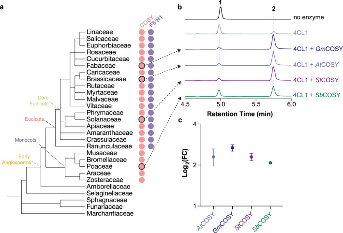
Plants contain rapidly evolving specialized enzymes that support the biosynthesis of functionally diverse natural products. In coumarin biosynthesis, a BAHD acyltransferase-family enzyme COSY was recently discovered to accelerate coumarin formation as the only known BAHD enzyme to catalyze an intramolecular acyl transfer reaction. Here we investigate the structural and mechanistic basis for COSY’s coumarin synthase activity. Our structural analyses reveal an unconventional active-site configuration adapted to COSY’s specialized activity. Through mutagenesis studies and deuterium exchange experiments, we identify a unique proton exchange mechanism at the α-carbon of the o -hydroxylated trans -hydroxycinnamoyl-CoA substrates during the catalytic cycle of COSY. Quantum mechanical cluster modeling and molecular dynamics further support this key mechanism for lowering the activation energy of the rate-limiting trans -to- cis isomerization step in coumarin production. This study unveils an unconventional catalytic mechanism mediated by a BAHD-family enzyme, and sheds light on COSY’s evolutionary origin and its recruitment to coumarin biosynthesis in eudicots.
Overview
The claims and statistics of this paper haven’t been checked yet.
Run the check — free, no account needed. It reads the open-access text and checks the sources it cites.
Integrity signals
Registry
Cites
Statistics
Figures
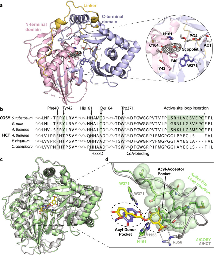
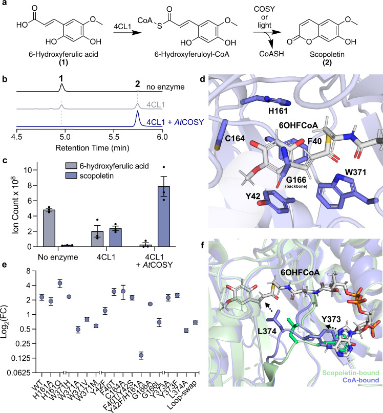
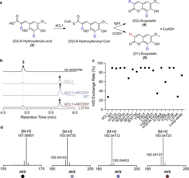
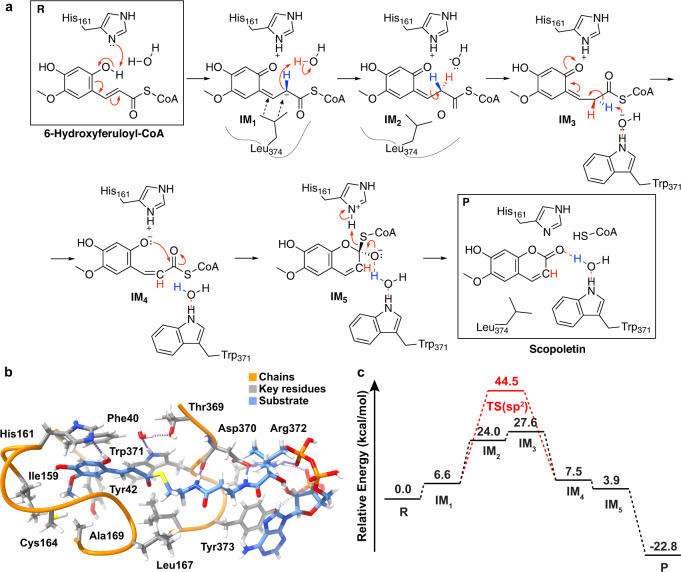
Verification
Claims the paper makes
The claims and statistics of this paper haven’t been checked yet.
Run the check — free, no account needed. It reads the open-access text and checks the sources it cites.
Self-check
Percentages, sums and reported differences re-computed from the paper’s own numbers.
The self-check needs the paper’s full text.
Citations
Cites
Highly accurate protein structure prediction with AlphaFold
Jumper, J., Evans, R., Pritzel, A., Green, T., Figurnov, M., Ronneberger, O., Tunyasuvunakool, K., Bates, R., Žídek, A., Potapenko, A., et al. (2021) · Nature · doi:10.1038/s41586-021-03819-2 · Cited by 43,764
Research works printed but not linked: 68
- Musa, M. A., Cooperwood, J. S. & Khan, M. O. F. A review of coumarin derivatives in pharmacotherapy of breast cancer. Curr. Med. Chem. 15, 2664–2679 (2008). · DOI
- Robe, K., Izquierdo, E., Vignols, F., Rouached, H. & Dubos, C. The coumarins: secondary metabolites playing a primary role in plant nutrition and health. Trends Plant Sci. 26, 248–259 (2021). · DOI
- Stassen, M. J. J., Hsu, S.-H., Pieterse, C. M. J. & Stringlis, I. A. Coumarin communication along the microbiome–root–shoot axis. Trends Plant Sci. 26, 169–183 (2021). · DOI
- Mishra, S., Pandey, A. & Manvati, S. Coumarin: an emerging antiviral agent. Heliyon 6, e03217 (2020). · DOI
- Sahoo, C. R. et al. Coumarin derivatives as promising antibacterial agent(s). Arab. J. Chem. 14, 102922 (2021). · DOI
- Lowenthal, J. & Birnbaum, H. Vitamin K and coumarin anticoagulants: dependence of anticoagulant effect on inhibition of vitamin K transport. Science 164, 181–183 (1969). · DOI
- Kai, K. et al. Scopoletin is biosynthesized via ortho-hydroxylation of feruloyl CoA by a 2-oxoglutarate-dependent dioxygenase in Arabidopsis thaliana. Plant J. 55, 989–999 (2008). · DOI
- Vanholme, R. et al. COSY catalyses trans–cis isomerization and lactonization in the biosynthesis of coumarins. Nat. Plants 5, 1066–1075 (2019). · DOI
- Dudareva, N., D’Auria, J. C., Nam, K. H., Raguso, R. A. & Pichersky, E. Acetyl-CoA:benzylalcohol acetyltransferase–an enzyme involved in floral scent production in Clarkia breweri. Plant J. 14, 297–304 (1998). · DOI
- Fujiwara, H. et al. Anthocyanin 5-aromatic acyltransferase from Gentiana triflora. Purification, characterization and its role in anthocyanin biosynthesis. Eur. J. Biochem. 249, 45–51 (1997). · DOI
- Yang, Q., Reinhard, K., Schiltz, E. & Matern, U. Characterization and heterologous expression of hydroxycinnamoyl/benzoyl-CoA:anthranilate N-hydroxycinnamoyl/benzoyltransferase from elicited cell cultures of carnation, Dianthus caryophyllus L. Plant Mol. Biol. 35, 777–789 (1997). · DOI
- Power, R., Kurz, W. G. & De Luca, V. Purification and characterization of acetylcoenzyme A: deacetylvindoline 4-O-acetyltransferase from Catharanthus roseus. Arch. Biochem. Biophys. 279, 370–376 (1990). · DOI
- D’Auria, J. C. Acyltransferases in plants: a good time to be BAHD. Curr. Opin. Plant Biol. 9, 331–340 (2006). · DOI
- Levsh, O. et al. Dynamic conformational states dictate selectivity toward the native substrate in a substrate-permissive acyltransferase. Biochemistry 55, 6314–6326 (2016). · DOI
- Ma, X., Koepke, J., Panjikar, S., Fritzsch, G. & Stöckigt, J. Crystal structure of vinorine synthase, the first representative of the BAHD superfamily. J. Biol. Chem. 280, 13576–13583 (2005). · DOI
- Yamane, M., Takenoya, M., Yajima, S. & Sue, M. Molecular and structural characterization of agmatine coumaroyltransferase in Triticeae, the key regulator of hydroxycinnamic acid amide accumulation. Phytochemistry 189, 112825 (2021). · DOI
- Wang, L., Chen, K., Zhang, M., Ye, M. & Qiao, X. Catalytic function, mechanism, and application of plant acyltransferases. Crit. Rev. Biotechnol. 42, 125–144 (2022). · DOI
- Wu, D. et al. Structure of human carnitine acetyltransferase. Molecular basis for fatty acyl transfer. J. Biol. Chem. 278, 13159–13165 (2003). · DOI
- Greene, N. P., Crow, A., Hughes, C. & Koronakis, V. Structure of a bacterial toxin-activating acyltransferase. Proc. Natl Acad. Sci. USA 112, E3058–E3066 (2015). · DOI
- Bayoumi, S. A. L., Rowan, M. G., Blagbrough, I. S. & Beeching, J. R. Biosynthesis of scopoletin and scopolin in cassava roots during post-harvest physiological deterioration: the E-Z-isomerisation stage. Phytochemistry 69, 2928–2936 (2008). · DOI
- Bader, R. F. W. In Encyclopedia of Computational Chemistry (ed. von Rague Schleyer, P.) Ch. 1 (Wiley, 2002).
- Mayer, I. Bond order and valence indices: a personal account. J. Comput. Chem. 28, 204–221 (2007). · DOI
- Stringlis, I. A., de Jonge, R. & Pieterse, C. M. J. The age of coumarins in plant-microbe interactions. Plant Cell Physiol. 60, 1405–1419 (2019). · DOI
- Shaw, P. E. Peptidyl-prolyl isomerases: a new twist to transcription. EMBO Rep. 3, 521–526 (2002). · DOI
- Schmidpeter, P. A. M. & Schmid, F. X. Prolyl isomerization and its catalysis in protein folding and protein function. J. Mol. Biol. 427, 1609–1631 (2015). · DOI
- Heipieper, H. J., Meinhardt, F. & Segura, A. The cis-trans isomerase of unsaturated fatty acids in Pseudomonas and Vibrio: biochemistry, molecular biology and physiological function of a unique stress adaptive mechanism. FEMS Microbiol. Lett. 229, 1–7 (2003). · DOI · Paper page
- Hluska, T., Šebela, M., Lenobel, R., Frébort, I. & Galuszka, P. Purification of maize nucleotide pyrophosphatase/phosphodiesterase casts doubt on the existence of zeatin cis–trans isomerase in plants. Front. Plant Sci. 8, 1473 (2017). · DOI
- Bassil, N. V., Mok, D. & Mok, M. C. Partial purification of a cis-trans-isomerase of zeatin from immature seed of Phaseolus vulgaris L. Plant Physiol. 102, 867–872 (1993). · DOI
- Torrens-Spence, M. P. et al. PBS3 and EPS1 complete salicylic acid biosynthesis from isochorismate in Arabidopsis. Mol. Plant 12, 1577–1586 (2019). · DOI
- Torrens-Spence, M. P. et al. Mechanistic basis for the emergence of EPS1 as a catalyst in plant salicylic acid biosynthesis. Preprint at bioRxiv https://doi.org/10.1101/2021.08.21.457228 (2021). · DOI
- Jez, J. M., Bowman, M. E., Dixon, R. A. & Noel, J. P. Structure and mechanism of the evolutionarily unique plant enzyme chalcone isomerase. Nat. Struct. Biol. 7, 786–791 (2000). · DOI
- Waki, T. et al. A conserved strategy of chalcone isomerase-like protein to rectify promiscuous chalcone synthase specificity. Nat. Commun. 11, 870 (2020). · DOI
- Kaltenbach, M. et al. Evolution of chalcone isomerase from a noncatalytic ancestor. Nat. Chem. Biol. 14, 548–555 (2018). · DOI
- Ueda, M., Tashiro, C. & Yamamura, S. cis-p-Coumaroylagmatine, the genuine leaf-opening substance of a nyctinastic plant, Albizzia julibrissin Durazz. Tetrahedron Lett. 38, 3253–3256 (1997). · DOI
- Vanholme, R., De Meester, B., Ralph, J. & Boerjan, W. Lignin biosynthesis and its integration into metabolism. Curr. Opin. Biotechnol. 56, 230–239 (2019). · DOI
- Steenackers, W. et al. cis-Cinnamic acid is a natural plant growth-promoting compound. J. Exp. Bot. 70, 6293–6304 (2019). · DOI
- El Houari, I. et al. Seedling developmental defects upon blocking CINNAMATE-4-HYDROXYLASE are caused by perturbations in auxin transport. N. Phytol. 230, 2275–2291 (2021). · DOI
- Vanholme, B., El Houari, I. & Boerjan, W. Bioactivity: phenylpropanoids’ best kept secret. Curr. Opin Biotechnol. 56, 156–162 (2019). · DOI
- Panjikar, S., Parthasarathy, V., Lamzin, V. S., Weiss, M. S. & Tucker, P. A. Auto-rickshaw: an automated crystal structure determination platform as an efficient tool for the validation of an X-ray diffraction experiment. Acta Crystallogr. D Biol. Crystallogr. 61, 449–457 (2005). · DOI
- Panjikar, S., Parthasarathy, V., Lamzin, V. S., Weiss, M. S. & Tucker, P. A. On the combination of molecular replacement and single-wavelength anomalous diffraction phasing for automated structure determination. Acta Crystallogr. D Biol. Crystallogr. 65, 1089–1097 (2009). · DOI
- Murshudov, G. N. et al. REFMAC5 for the refinement of macromolecular crystal structures. Acta Crystallogr. D Biol. Crystallogr. 67, 355–367 (2011). · DOI
- Emsley, P., Lohkamp, B., Scott, W. G. & Cowtan, K. Features and development of Coot. Acta Crystallogr. D Biol. Crystallogr. 66, 486–501 (2010). · DOI · Paper page
- McCoy, A. J. et al. Phaser crystallographic software. J. Appl. Crystallogr. 40, 658–674 (2007). · DOI · Paper page
- Liebschner, D. et al. Macromolecular structure determination using X-rays, neutrons and electrons: recent developments in Phenix. Acta Crystallogr D Struct. Biol. 75, 861–877 (2019). · DOI · Paper page
- Afonine, P. V. et al. Towards automated crystallographic structure refinement with phenix.refine. Acta Crystallogr. D Biol. Crystallogr. 68, 352–367 (2012). · DOI · Paper page
- Anandakrishnan, R., Aguilar, B. & Onufriev, A. V. H++ 3.0: automating pK prediction and the preparation of biomolecular structures for atomistic molecular modeling and simulations. Nucleic Acids Res. 40, W537–W541 (2012). · DOI
- Gordon, J. C. et al. H: a server for estimating pKas and adding missing hydrogens to macromolecules. Nucleic Acids Res. 33, W368–W371 (2005). · DOI
- Myers, J., Grothaus, G., Narayanan, S. & Onufriev, A. A simple clustering algorithm can be accurate enough for use in calculations of pKs in macromolecules. Proteins 63, 928–938 (2006). · DOI
- Webb, B. & Sali, A. Protein structure modeling with MODELLER. Methods Mol. Biol. 1137, 1–15 (2014). · DOI
- Maier, J. A. et al. ff14SB: improving the accuracy of protein side chain and backbone parameters from ff99SB. J. Chem. Theory Comput. 11, 3696–3713 (2015). · DOI
- Bayly, C. I., Cieplak, P., Cornell, W. & Kollman, P. A. A well-behaved electrostatic potential based method using charge restraints for deriving atomic charges: the RESP model. J. Phys. Chem. 97, 10269–10280 (1993). · DOI
- Hariharan, P. C. & Pople, J. A. The influence of polarization functions on molecular orbital hydrogenation energies. Theor. Chim. Acta 28, 213–222 (1973). · DOI
- Vanquelef, E. et al. R.E.D. Server: a web service for deriving RESP and ESP charges and building force field libraries for new molecules and molecular fragments. Nucleic Acids Res. 39, W511–W517 (2011). · DOI
- Jorgensen, W. L., Chandrasekhar, J., Madura, J. D., Impey, R. W. & Klein, M. L. Comparison of simple potential functions for simulating liquid water. J. Chem. Phys. 79, 926–935 (1983). · DOI
- Götz, A. W. et al. Routine microsecond molecular dynamics simulations with AMBER on GPUs. 1. Generalized born. J. Chem. Theory Comput. 8, 1542–1555 (2012). · DOI
- Salomon-Ferrer, R., Götz, A. W., Poole, D., Le Grand, S. & Walker, R. C. Routine microsecond molecular dynamics simulations with AMBER on GPUs. 2. Explicit solvent particle mesh Ewald. J. Chem. Theory Comput. 9, 3878–3888 (2013). · DOI
- Himo, F. & de Visser, S. P. Status report on the quantum chemical cluster approach for modeling enzyme reactions. Commun. Chem. 5, 29 (2022). · DOI
- Goddard, T. D. et al. UCSF ChimeraX: meeting modern challenges in visualization and analysis. Protein Sci. 27, 14–25 (2018). · DOI · Paper page
- Seritan, S. et al. TeraChem: a graphical processing unit‐accelerated electronic structure package for large‐scale ab initio molecular dynamics. WIREs Comput. Mol. Sci. https://doi.org/10.1002/wcms.1494 (2021). · DOI
- Kulik, H. J., Luehr, N., Ufimtsev, I. S. & Martinez, T. J. Ab initio quantum chemistry for protein structures. J. Phys. Chem. B 116, 12501–12509 (2012). · DOI
- Grimme, S., Antony, J., Ehrlich, S. & Krieg, H. A consistent and accurate ab initio parametrization of density functional dispersion correction (DFT-D) for the 94 elements H-Pu. J. Chem. Phys. 132, 154104 (2010). · DOI · Paper page
- Lange, A. W. & Herbert, J. M. A smooth, nonsingular, and faithful discretization scheme for polarizable continuum models: the switching/Gaussian approach. J. Chem. Phys. 133, 244111 (2010). · DOI
- Bader, R. F. W. ChemInform abstract: a quantum theory of molecular structure and its applications. ChemInform https://doi.org/10.1002/chin.199203324 (2010). · DOI
- Lu, T. & Chen, F. Multiwfn: a multifunctional wavefunction analyzer. J. Comput. Chem. 33, 580–592 (2012). · DOI
- Yang, X.-Y. et al. Measuring the turnover rates of Arabidopsis proteins using deuterium oxide: an auxin signaling case study. Plant J. 63, 680–695 (2010). · DOI
- Edgar, R. C. MUSCLE: multiple sequence alignment with high accuracy and high throughput. Nucleic Acids Res. 32, 1792–1797 (2004). · DOI · Paper page
- Kumar, S., Stecher, G., Li, M., Knyaz, C. & Tamura, K. MEGA X: molecular evolutionary genetics analysis across computing platforms. Mol. Biol. Evol. 35, 1547–1549 (2018). · DOI
- Robert, X. & Gouet, P. Deciphering key features in protein structures with the new ENDscript server. Nucleic Acids Res. 42, W320–W324 (2014). · DOI · Paper page
Metadata
- Authors
- Kim, C. Y., Mitchell, A. J., Kastner, D. W., Albright, C. E., Gutierrez, M. A., Glinkerman, C. M., Kulik, H. J., & Weng, J.-K.
- Affiliations
- Whitehead Institute for Biomedical Research (US)
- Massachusetts Institute of Technology (US)
- Corresponding author
- Weng, Jing-Ke
- ORCID
- Venue
- Nature Communications
- Published
- 2023-02-03
- Volume
- 14
- Issue
- 1
- Pages
- 597-597
- ISSN
- 2041-1723 (ISSN-L)
- PMC
- PMC9898226
- OpenAlex
- W4319078070
- Cited by
- 25
- References
- 1
- Citations by year
- 2026: 7 · 2025: 5 · 2024: 8 · 2023: 4
- FWCI
- 2.13
- Type
- journal article
- Language
- en
- Open access
- gold
- doi.org
- Locations
- Nature Communications (publishedVersion, cc-by)
- bioRxiv (Cold Spring Harbor Laboratory) (acceptedVersion)
- PubMed Central (publishedVersion, cc-by)
- PubMed (publishedVersion)
- DSpace@MIT (Massachusetts Institute of Technology) (submittedVersion, cc-by-nc)
- RePEc: Research Papers in Economics (submittedVersion)
- DOAJ (DOAJ: Directory of Open Access Journals) (submittedVersion, cc-by-sa)
- Funders
- National Science Foundation (1745302)
- Burroughs Wellcome Fund
- W. M. Keck Foundation
- Family Larsson‐Rosenquist Foundation
- Arnold and Mabel Beckman Foundation
- Topics
- Life Sciences › Biochemistry, Genetics and Molecular Biology › Molecular Biology › Plant biochemistry and biosynthesis
- Life Sciences › Biochemistry, Genetics and Molecular Biology › Molecular Biology › Plant Gene Expression Analysis
- Physical Sciences › Chemistry › Organic Chemistry › Physical Organic Chemistry and Reaction Kinetics
- Keywords
- COSY, coumarin biosynthesis, BAHD acyltransferase, trans-cis isomerization, lactonization, active site configuration, hydrogen-deuterium exchange, enzyme mechanism, eudicots
- MeSH terms
- Acyltransferases, Coumarins, Isomerism, Plants, Protons
About these checks
Registry, retraction, Cites and statistics results are computed, not judged, with one exception: a printed reference that no index lists is read once by a model before it counts as a citation error. Claims are extracted by a model and kept only when their quote appears verbatim in the text; each cited source is then checked, with a model’s help, for whether it says what the claim relies on. Those verdicts can be wrong — read the claim’s quote and the cited source before relying on one.
Statistics and arithmetic are recomputed, not judged.
Confidence bands are shown only for fields calibrated on our annotated test set. None is calibrated yet, so no band is shown.
What counts as a claim ›How we measure confidence ›How we check this ›
Are you an author, or do you see a problem with this page? Request a review
