

The DOI matches its Crossref record; 2 of the 2 references it cites were found in a registry or an index.
Structural basis for membrane attack complex inhibition by CD59
Open accessPublished version of a preprint
Abstract
CD59 is an abundant immuno-regulatory receptor that protects human cells from damage during complement activation. Here we show how the receptor binds complement proteins C8 and C9 at the membrane to prevent insertion and polymerization of membrane attack complex (MAC) pores. We present cryo-electron microscopy structures of two inhibited MAC precursors known as C5b8 and C5b9. We discover that in both complexes, CD59 binds the pore-forming β-hairpins of C8 to form an intermolecular β-sheet that prevents membrane perforation. While bound to C8, CD59 deflects the cascading C9 β-hairpins, rerouting their trajectory into the membrane. Preventing insertion of C9 restricts structural transitions of subsequent monomers and indirectly halts MAC polymerization. We combine our structural data with cellular assays and molecular dynamics simulations to explain how the membrane environment impacts the dual roles of CD59 in controlling pore formation of MAC, and as a target of bacterial virulence factors which hijack CD59 to lyse human cells.
Overview
The claims and statistics of this paper haven’t been checked yet.
Run the check — free, no account needed. It reads the open-access text and checks the sources it cites.
Integrity signals
Registry
Cites
Statistics
Figures
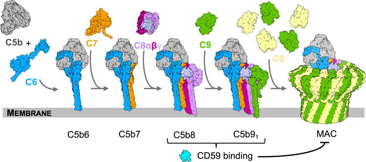
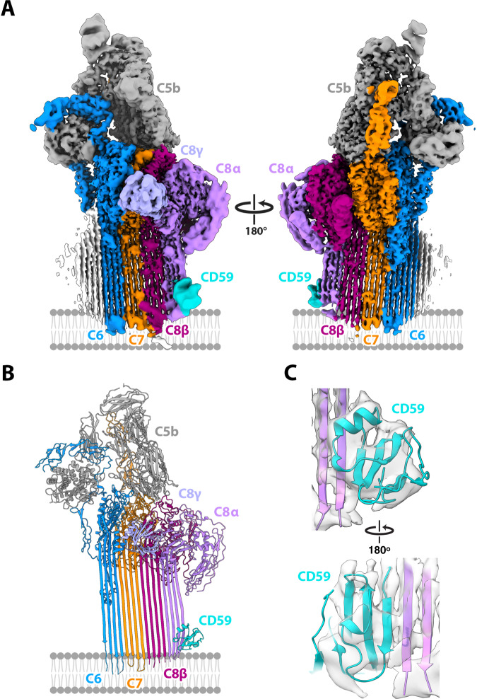
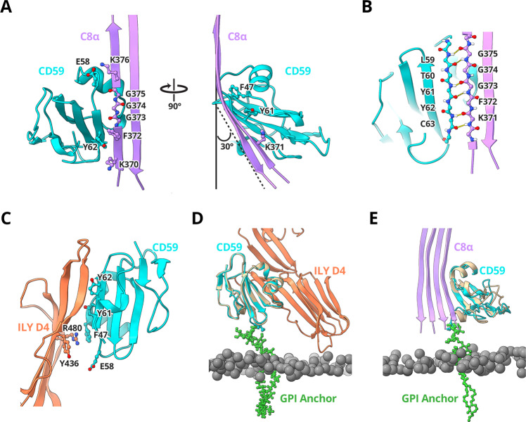
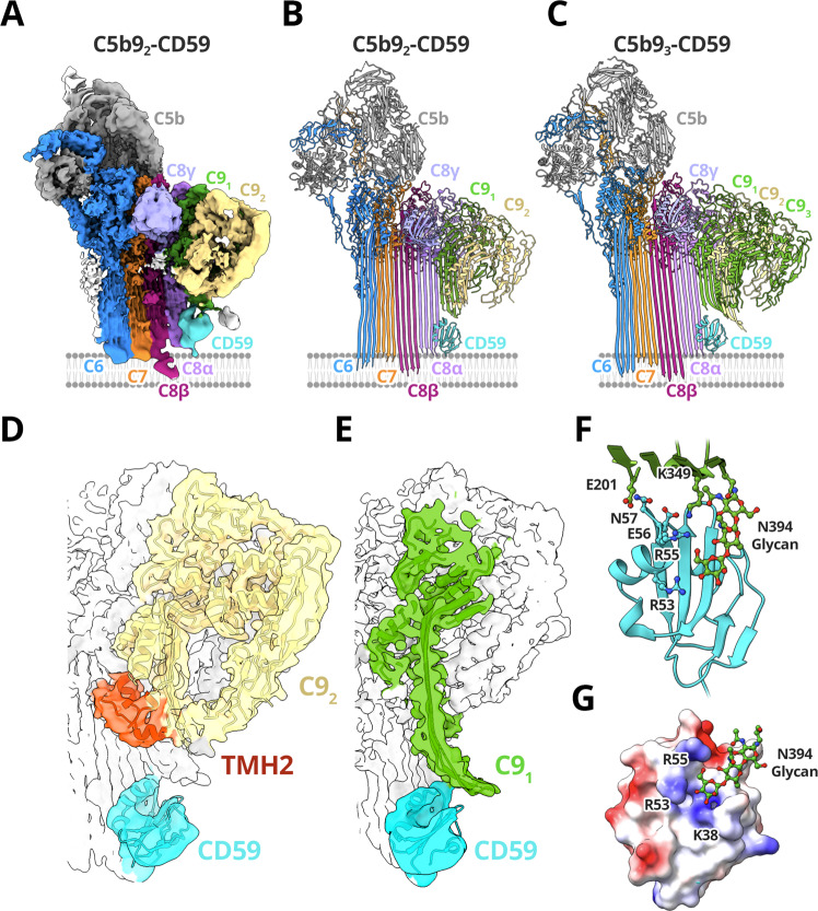
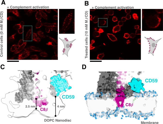
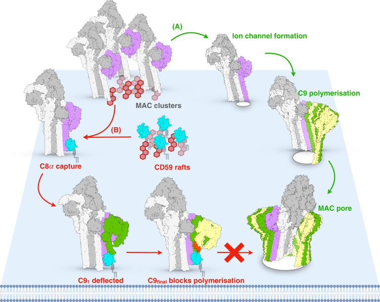
Verification
Claims the paper makes
The claims and statistics of this paper haven’t been checked yet.
Run the check — free, no account needed. It reads the open-access text and checks the sources it cites.
Self-check
Percentages, sums and reported differences re-computed from the paper’s own numbers.
The self-check needs the paper’s full text.
Citations
Cites
Highly accurate protein structure prediction with AlphaFold
Jumper, J., Evans, R., Pritzel, A., Green, T., Figurnov, M., Ronneberger, O., Tunyasuvunakool, K., Bates, R., Žídek, A., Potapenko, A., et al. (2021) · Nature · doi:10.1038/s41586-021-03819-2 · Cited by 43,764
Complement: a key system for immune surveillance and homeostasis
Ricklin, D., Hajishengallis, G., Yang, K., & Lambris, J. D. (2010) · Nature Immunology · doi:10.1038/ni.1923 · Cited by 3,654 · cited 1×
“Particles (1,138,825) of C5b8-CD59 were picked using the general model in crYOLO v1.”
Method
Research works printed but not linked: 84
- Tomlinson, S., Taylor, P. W., Morgan, B. P. & Luzio, J. P. Killing of gram-negative bacteria by complement. Fractionation of cell membranes after complement C5b-9 deposition on to the surface of Salmonella minnesota Re595. Biochem J. 263, 505–511 (1989). · DOI
- Davies, A. et al. CD59, an LY-6-like protein expressed in human lymphoid cells, regulates the action of the complement membrane attack complex on homologous cells. J. Exp. Med. 170, 637–654 (1989). · DOI
- Nevo, Y. et al. CD59 deficiency is associated with chronic hemolysis and childhood relapsing immune-mediated polyneuropathy. Blood 121, 129–135 (2013). · DOI
- Fishelson, Z., Donin, N., Zell, S., Schultz, S. & Kirschfink, M. Obstacles to cancer immunotherapy: expression of membrane complement regulatory proteins (mCRPs) in tumors. Mol. Immunol. 40, 109–123 (2003). · DOI
- Shao, F. et al. Silencing EGFR-upregulated expression of CD55 and CD59 activates the complement system and sensitizes lung cancer to checkpoint blockade. Nat. Cancer 3, 1192–1210 (2022). · DOI
- Podack, E. R. Molecular composition of the tubular structure of the membrane attack complex of complement. J. Biol. Chem. 259, 8641–8647 (1984). · DOI
- Ninomiya, H. & Sims, P. J. The human complement regulatory protein CD59 binds to the alpha-chain of C8 and to the “b“domain of C9. J. Biol. Chem. 267, 13675–13680 (1992). · DOI
- Huang, Y., Qiao, F., Abagyan, R., Hazard, S. & Tomlinson, S. Defining the CD59-C9 binding interaction. J. Biol. Chem. 281, 27398–27404 (2006). · DOI
- Bodian, D. L., Davis, S. J., Morgan, B. P. & Rushmere, N. K. Mutational analysis of the active site and antibody epitopes of the complement-inhibitory glycoprotein, CD59. J. Exp. Med 185, 507–516 (1997). · DOI
- Lockert, D. H. et al. Identity of the segment of human complement C8 recognized by complement regulatory protein CD59. J. Biol. Chem. 270, 19723–19728 (1995). · DOI
- Law, R. H. et al. The structural basis for membrane binding and pore formation by lymphocyte perforin. Nature 468, 447–451 (2010). · DOI
- Rosado, C. J. et al. A common fold mediates vertebrate defense and bacterial attack. Science 317, 1548–1551 (2007). · DOI
- Shatursky, O. et al. The mechanism of membrane insertion for a cholesterol-dependent cytolysin: a novel paradigm for pore-forming toxins. Cell 99, 293–299 (1999). · DOI
- Serna, M., Giles, J. L., Morgan, B. P. & Bubeck, D. Structural basis of complement membrane attack complex formation. Nat. Commun. 7, 10587 (2016). · DOI
- Spicer, B. A. et al. The first transmembrane region of complement component-9 acts as a brake on its self-assembly. Nat. Commun. 9, 3266 (2018). · DOI
- Hadders, M. A., Beringer, D. X. & Gros, P. Structure of C8alpha-MACPF reveals mechanism of membrane attack in complement immune defense. Science 317, 1552–1554 (2007). · DOI
- Lovelace, L. L., Cooper, C. L., Sodetz, J. M. & Lebioda, L. Structure of human C8 protein provides mechanistic insight into membrane pore formation by complement. J. Biol. Chem. 286, 17585–17592 (2011). · DOI
- Giddings, K. S., Zhao, J., Sims, P. J. & Tweten, R. K. Human CD59 is a receptor for the cholesterol-dependent cytolysin intermedilysin. Nat. Struct. Mol. Biol. 11, 1173–1178 (2004). · DOI
- Wickham, S. E. et al. Mapping the intermedilysin-human CD59 receptor interface reveals a deep correspondence with the binding site on CD59 for complement binding proteins C8alpha and C9. J. Biol. Chem. 286, 20952–20962 (2011). · DOI
- Johnson, S., Brooks, N. J., Smith, R. A., Lea, S. M. & Bubeck, D. Structural basis for recognition of the pore-forming toxin intermedilysin by human complement receptor CD59. Cell Rep. 3, 1369–1377 (2013). · DOI
- Farkas, I. et al. CD59 blocks not only the insertion of C9 into MAC but inhibits ion channel formation by homologous C5b-8 as well as C5b-9. J. Physiol. 539, 537–545 (2002). · DOI
- Meri, S. et al. Human protectin (CD59), an 18,000–20,000 MW complement lysis restricting factor, inhibits C5b-8 catalysed insertion of C9 into lipid bilayers. Immunology 71, 1–9 (1990).
- Hill, A. et al. Protection of erythrocytes from human complement-mediated lysis by membrane-targeted recombinant soluble CD59: a new approach to PNH therapy. Blood 107, 2131–2137 (2006). · DOI
- Kastner, B. et al. GraFix: sample preparation for single-particle electron cryomicroscopy. Nat. Methods 5, 53–55 (2008). · DOI
- Ng, S. C., Rao, A. G., Howard, O. M. & Sodetz, J. M. The eighth component of human complement: evidence that it is an oligomeric serum protein assembled from products of three different genes. Biochemistry 26, 5229–5233 (1987). · DOI
- Steckel, E. W., Welbaum, B. E. & Sodetz, J. M. Evidence of direct insertion of terminal complement proteins into cell membrane bilayers during cytolysis. Labeling by a photosensitive membrane probe reveals a major role for the eighth and ninth components. J. Biol. Chem. 258, 4318–4324 (1983). · DOI
- Menny, A. et al. Structural basis of soluble membrane attack complex packaging for clearance. Nat. Commun. 12, 6086 (2021). · DOI
- Leath, K. J. et al. High-resolution structures of bacterially expressed soluble human CD59. Acta Crystallogr Sect. F. Struct. Biol. Cryst. Commun. 63, 648–652 (2007). · DOI
- Menny, A. et al. CryoEM reveals how the complement membrane attack complex ruptures lipid bilayers. Nat. Commun. 9, 5316 (2018). · DOI
- Petranka, J. et al. Structure-function relationships of the complement regulatory protein, CD59. Blood Cells Mol. Dis. 22, 281–296 (1996). · DOI
- Rollins, S. A., Zhao, J., Ninomiya, H. & Sims, P. J. Inhibition of homologous complement by CD59 is mediated by a species-selective recognition conferred through binding to C8 within C5b-8 or C9 within C5b-9. J. Immunol. 146, 2345–2351 (1991). · DOI
- Lawrence, S. L. et al. Structural basis for receptor recognition by the human cd59-responsive cholesterol-dependent cytolysins. Structure 24, 1488–1498 (2016). · DOI
- Nakano, Y. et al. Determination of the active site of CD59 with synthetic peptides. Mol. Immunol. 32, 241–247 (1995). · DOI
- Suzuki, K. G. et al. Transient GPI-anchored protein homodimers are units for raft organization and function. Nat. Chem. Biol. 8, 774–783 (2012). · DOI
- Cragg, M. S. et al. Complement-mediated lysis by anti-CD20 mAb correlates with segregation into lipid rafts. Blood 101, 1045–1052 (2003). · DOI
- Cooper, N. R. & Müller-Eberhard, H. J. The reaction mechanism of human C5 in immune hemolysis. J. Exp. Med 132, 775–793 (1970). · DOI
- Parsons, E. S. et al. Single-molecule kinetics of pore assembly by the membrane attack complex. Nat. Commun. 10, 2066 (2019). · DOI
- Jiang, Y. et al. Membrane-mediated protein interactions drive membrane protein organization. Nat. Commun. 13, 7373 (2022). · DOI
- Koyama-Honda, I. et al. High-speed single-molecule imaging reveals signal transduction by induced transbilayer raft phases. J. Cell Biol. 219, e202006125 (2020). · DOI
- Polekhina, G., Giddings, K. S., Tweten, R. K. & Parker, M. W. Insights into the action of the superfamily of cholesterol-dependent cytolysins from studies of intermedilysin. Proc. Natl Acad. Sci. USA 102, 600–605 (2005). · DOI
- Shah, N. R. et al. Structural basis for tuning activity and membrane specificity of bacterial cytolysins. Nat. Commun. 11, 5818 (2020). · DOI
- Banerjee, P., Wehle, M., Lipowsky, R. & Santer, M. A molecular dynamics model for glycosylphosphatidyl-inositol anchors: “flop down” or “lollipop”? Phys. Chem. Chem. Phys. 20, 29314–29324 (2018). · DOI
- Giddings, K. S., Johnson, A. E. & Tweten, R. K. Redefining cholesterol’s role in the mechanism of the cholesterol-dependent cytolysins. Proc. Natl Acad. Sci. USA 100, 11315–11320 (2003). · DOI
- Cheerla, R. & Ayappa, K. G. Molecular dynamics study of lipid and cholesterol reorganization due to membrane binding and pore formation by listeriolysin O. J. Membr. Biol. 253, 535–550 (2020). · DOI
- Brandel, A. et al. The Gb3-enriched CD59/flotillin plasma membrane domain regulates host cell invasion by Pseudomonas aeruginosa. Cell Mol. Life Sci. 78, 3637–3656 (2021). · DOI
- Saifuddin, M. et al. Human immunodeficiency virus type 1 incorporates both glycosyl phosphatidylinositol-anchored CD55 and CD59 and integral membrane CD46 at levels that protect from complement-mediated destruction. J. Gen. Virol. 78, 1907–1911 (1997). · DOI
- Bavari, S. et al. Lipid raft microdomains: a gateway for compartmentalized trafficking of Ebola and Marburg viruses. J. Exp. Med 195, 593–602 (2002). · DOI
- Grinkova, Y. V., Denisov, I. G. & Sligar, S. G. Engineering extended membrane scaffold proteins for self-assembly of soluble nanoscale lipid bilayers. Protein Eng. Des. Sel. 23, 843–848 (2010). · DOI
- Fraser, D. A. et al. Generation of a recombinant, membrane-targeted form of the complement regulator CD59: activity in vitro and in vivo. J. Biol. Chem. 278, 48921–48927 (2003). · DOI
- Palovcak, E. et al. A simple and robust procedure for preparing graphene-oxide cryo-EM grids. J. Struct. Biol. 204, 80–84 (2018). · DOI
- Zheng, S. Q. et al. MotionCor2: anisotropic correction of beam-induced motion for improved cryo-electron microscopy. Nat. Methods 14, 331–332 (2017). · DOI
- Zivanov, J. et al. New tools for automated high-resolution cryo-EM structure determination in RELION-3. Elife 7, e42166 (2018). · DOI
- Zhang, K. Gctf: Real-time CTF determination and correction. J. Struct. Biol. 193, 1–12 (2016). · DOI
- Wagner, T. et al. SPHIRE-crYOLO is a fast and accurate fully automated particle picker for cryo-EM. Commun. Biol. 2, 218 (2019). · DOI
- Punjani, A., Rubinstein, J. L., Fleet, D. J. & Brubaker, M. A. cryoSPARC: algorithms for rapid unsupervised cryo-EM structure determination. Nat. Methods 14, 290–296 (2017). · DOI · Paper page
- Zivanov, J., Nakane, T. & Scheres, S. H. W. A Bayesian approach to beam-induced motion correction in cryo-EM single-particle analysis. IUCrJ 6, 5–17 (2019). · DOI
- Punjani, A. & Fleet, D. J. 3D variability analysis: Resolving continuous flexibility and discrete heterogeneity from single particle cryo-EM. J. Struct. Biol. 213, 107702 (2021). · DOI
- Rohou, A. & Grigorieff, N. CTFFIND4: Fast and accurate defocus estimation from electron micrographs. J. Struct. Biol. 192, 216–221 (2015). · DOI
- Zhong, E. D., Bepler, T., Berger, B. & Davis, J. H. CryoDRGN: reconstruction of heterogeneous cryo-EM structures using neural networks. Nat. Methods 18, 176–185 (2021). · DOI
- Croll, T. I. ISOLDE: a physically realistic environment for model building into low-resolution electron-density maps. Acta Crystallogr D. Struct. Biol. 74, 519–530 (2018). · DOI
- Afonine, P. V. et al. Real-space refinement in PHENIX for cryo-EM and crystallography. Acta Crystallogr D. Struct. Biol. 74, 531–544 (2018). · DOI
- Afonine, P. V. et al. New tools for the analysis and validation of cryo-EM maps and atomic models. Acta Crystallogr D. Struct. Biol. 74, 814–840 (2018). · DOI
- Waterhouse, A. et al. SWISS-MODEL: homology modelling of protein structures and complexes. Nucleic Acids Res 46, W296–W303 (2018). · DOI
- Pettersen, E. F. et al. UCSF ChimeraX: Structure visualization for researchers, educators, and developers. Protein Sci. 30, 70–82 (2021). · DOI
- Wu, E. L. et al. CHARMM-GUI Membrane Builder toward realistic biological membrane simulations. J. Comput Chem. 35, 1997–2004 (2014). · DOI
- Jo, S., Kim, T., Iyer, V. G. & Im, W. CHARMM-GUI: a web-based graphical user interface for CHARMM. J. Comput Chem. 29, 1859–1865 (2008). · DOI · Paper page
- Brooks, B. R. et al. CHARMM: the biomolecular simulation program. J. Comput Chem. 30, 1545–1614 (2009). · DOI
- Lee, J. et al. CHARMM-GUI input generator for namd, gromacs, amber, openmm, and charmm/openmm simulations using the charmm36 additive force field. J. Chem. Theory Comput 12, 405–413 (2016). · DOI
- Fletcher, C. M., Harrison, R. A., Lachmann, P. J. & Neuhaus, D. Structure of a soluble, glycosylated form of the human complement regulatory protein CD59. Structure 2, 185–199 (1994). · DOI
- Jorgensen, W., Chandrasekhar, J. & Madura, J. Comparison of simple potential functions for simulating liquid water. J. Chem. Phys. 79, 926–935 (1983). · DOI
- Abraham, M. J. et al. GROMACS: High performance molecular simulations through multi-level parallelism from laptops to supercomputers. SoftwareX 1-2, 19–25 (2015). · DOI · Paper page
- Huang, J. et al. CHARMM36m: an improved force field for folded and intrinsically disordered proteins. Nat. Methods 14, 71–73 (2017). · DOI
- Berendsen, H. J. C., Postma, J. P. M., van Gunsteren, W. F., DiNola, A. & Haak, J. R. Molecular dynamics with coupling to an external bath. J. Chem. Phys. 81, 3684 (1984). · DOI
- Periole, X., Cavalli, M., Marrink, S. J. & Ceruso, M. A. Combining an elastic network with a coarse-grained molecular force field: structure, dynamics, and intermolecular recognition. J. Chem. Theory Comput 5, 2531–2543 (2009). · DOI
- Periole, X. & Marrink, S. J. The Martini coarse-grained force field. Methods Mol. Biol. 924, 533–565 (2013). · DOI
- Monticelli, L. et al. The MARTINI coarse-grained force field: extension to proteins. J. Chem. Theory Comput 4, 819–834 (2008). · DOI
- Banerjee, P., Lipowsky, R. & Santer, M. Coarse-grained molecular model for the glycosylphosphatidylinositol anchor with and without protein. J. Chem. Theory Comput 16, 3889–3903 (2020). · DOI
- Kirschner, K. N. et al. GLYCAM06: a generalizable biomolecular force field. Carbohydrates. J. Comput Chem. 29, 622–655 (2008). · DOI
- Sousa da Silva, A. W. & Vranken, W. F. ACPYPE - AnteChamber PYthon Parser interfacE. BMC Res Notes 5, 367 (2012). · DOI
- Goga, N., Rzepiela, A. J., de Vries, A. H., Marrink, S. J. & Berendsen, H. J. Efficient algorithms for langevin and DPD dynamics. J. Chem. Theory Comput 8, 3637–3649 (2012). · DOI
- Marrink, S. J., Risselada, H. J., Yefimov, S., Tieleman, D. P. & de Vries, A. H. The MARTINI force field: coarse grained model for biomolecular simulations. J. Phys. Chem. B 111, 7812–7824 (2007). · DOI
- Wassenaar, T. A., Ingólfsson, H. I., Böckmann, R. A., Tieleman, D. P. & Marrink, S. J. Computational lipidomics with insane: a versatile tool for generating custom membranes for molecular simulations. J. Chem. Theory Comput 11, 2144–2155 (2015). · DOI · Paper page
- Yesylevskyy, S. O., Schäfer, L. V., Sengupta, D. & Marrink, S. J. Polarizable water model for the coarse-grained MARTINI force field. PLoS Comput Biol. 6, e1000810 (2010). · DOI
- Essmann, U., Perera, L. & Berkowitz, M. L. A smooth particle mesh Ewald method. J. Chem. Phys. 103, 8577 (1995). · DOI
Metadata
- Authors
- Couves, E. C., Gardner, S., Voisin, T. B., Bickel, J. K., Stansfeld, P. J., Tate, E. W., & Bubeck, D.
- Affiliations
- Imperial College London (GB)
- University of Warwick (GB)
- Corresponding author
- Bubeck, Doryen
- ORCID
- Venue
- Nature Communications
- Published
- 2023-02-16
- Volume
- 14
- Issue
- 1
- Pages
- 890-890
- ISSN
- 2041-1723 (ISSN-L)
- PMC
- PMC9935631
- OpenAlex
- W4321078922
- Cited by
- 85
- References
- 2
- Citations by year
- 2026: 29 · 2025: 35 · 2024: 15 · 2023: 6
- FWCI
- 9.32
- Type
- journal article
- Language
- en
- Open access
- gold
- doi.org
- Locations
- Nature Communications (publishedVersion, cc-by)
- bioRxiv (Cold Spring Harbor Laboratory) (acceptedVersion, cc-by-nc-nd)
- PubMed Central (publishedVersion, cc-by)
- PubMed (publishedVersion)
- Warwick Research Archive Portal (University of Warwick) (submittedVersion)
- RePEc: Research Papers in Economics (submittedVersion)
- DOAJ (DOAJ: Directory of Open Access Journals) (submittedVersion, cc-by-sa)
- Spiral (Imperial College London) (submittedVersion, other-oa)
- Spiral (Imperial College London) (submittedVersion, cc-by-nc-nd)
- Funders
- Wellcome Trust (BB/S003339/1, Z/17/Z, 208361, 208361/Z/17/Z)
- Francis Crick Institute
- Diamond Light Source
- Cancer Research UK (864751, EP/L015498/1, C24523/A26234, EP/R029407/1, A26234, C24523, BB/M011178/1)
- Imperial College London (BB/M011178/1, A26234)
- European Commission (HORIZON2020)
- Medical Research Council (MR/S009213/1)
- Engineering and Physical Sciences Research Council (EP/R029407/1, EP/R029407/2, 864751, R029407, EP/R029407, 1364172, BB/M011178/1, C24523/A26234, EP/L015498/1)
- Biotechnology and Biological Sciences Research Council (BB/M011178/1, BB/R002517/1, EP/R029407/1, C24523/A26234, BB/S003339/1, BB/P01948X/1, BB/P01948X/2, P01948X, R002517, S003339, EP/L015498/1, 864751)
- Topics
- Life Sciences › Immunology and Microbiology › Immunology › Complement system in diseases
- Life Sciences › Biochemistry, Genetics and Molecular Biology › Molecular Biology › Lipid Membrane Structure and Behavior
- Life Sciences › Immunology and Microbiology › Immunology › Biomarkers in Disease Mechanisms
- Keywords
- CD59, membrane attack complex, complement regulation, perfluorooctanoic acid, C9, beta-hairpin, intermolecular beta-sheet, membrane perforation, bacterial virulence factors, complement receptors, cryo-EM structure
- MeSH terms
- Complement Activation, Complement C8, Complement C9, Humans, Complement Membrane Attack Complex, CD59 Antigens, Cryoelectron Microscopy
About these checks
Registry, retraction, Cites and statistics results are computed, not judged, with one exception: a printed reference that no index lists is read once by a model before it counts as a citation error. Claims are extracted by a model and kept only when their quote appears verbatim in the text; each cited source is then checked, with a model’s help, for whether it says what the claim relies on. Those verdicts can be wrong — read the claim’s quote and the cited source before relying on one.
Statistics and arithmetic are recomputed, not judged.
Confidence bands are shown only for fields calibrated on our annotated test set. None is calibrated yet, so no band is shown.
What counts as a claim ›How we measure confidence ›How we check this ›
Are you an author, or do you see a problem with this page? Request a review
