Student or educator? Sign up with your school email and get 6 months of Pro free.
Cited by 13Biochemical and Molecular ResearchEnzyme Structure and FunctionProtein Structure and Dynamics

The DOI matches its Crossref record; 64 of the 64 references it cites were found in a registry or an index.

Deciphering the allosteric regulation of mycobacterial inosine-5′-monophosphate dehydrogenase

Open access

Published version of a preprint

Abstract

Allosteric regulation of inosine 5′-monophosphate dehydrogenase (IMPDH), an essential enzyme of purine metabolism, contributes to the homeostasis of adenine and guanine nucleotides. However, the precise molecular mechanism of IMPDH regulation in bacteria remains unclear. Using biochemical and cryo-EM approaches, we reveal the intricate molecular mechanism of the IMPDH allosteric regulation in mycobacteria. The enzyme is inhibited by both GTP and (p)ppGpp, which bind to the regulatory CBS domains and, via interactions with basic residues in hinge regions, lock the catalytic core domains in a compressed conformation. This results in occlusion of inosine monophosphate (IMP) substrate binding to the active site and, ultimately, inhibition of the enzyme. The GTP and (p)ppGpp allosteric effectors bind to their dedicated sites but stabilize the compressed octamer by a common mechanism. Inhibition is relieved by the competitive displacement of GTP or (p)ppGpp by ATP allowing IMP-induced enzyme expansion. The structural knowledge and mechanistic understanding presented here open up new possibilities for the development of allosteric inhibitors with antibacterial potential.

Overview

The claims and statistics of this paper haven’t been checked yet.

Run the check — free, no account needed. It reads the open-access text and checks the sources it cites.

A few free checks a day · papers already verified open instantly · results are public

Integrity signals

Registry

Crossref · 10.1038/s41467-024-50933-6

Cites

64 of 64 references verified0 citation errors · 0 not checkable

Statistics

Figures

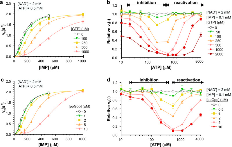
Fig. 1 MsmIMPDH is regulated by both GTP and ppGpp in an ATP-dependent manner. a, c Initial velocity, fitted with the Hill equation, is plotted as a function of IMP concentration in the presence of 500 µM ATP and varied concentrations of (a) GTP and (c) ppGpp, respectively (n = 3). Control kinetics in the presence of 500 µM ATP only are shown in black dashed line. b, d Relative velocity is plotted as a function of the ATP concentration at set concentrations of (b) GTP and (d) ppGpp (n = 2). The substrates IMP and NAD+ were fixed at concentrations of 100 µM and 2 mM, respectively. The relative velocity value was calculated as the ratio of the initial velocity of the reaction in the presence of ATP and GTP or ATP and ppGpp to the control reaction containing only the substrates and the corresponding ATP concentration. Specific ATP concentration ranges above the graphs indicate where inhibition and reactivation occur. All data are presented as mean values with error bars representing the SD.
Fig. 1 MsmIMPDH is regulated by both GTP and ppGpp in an ATP-dependent manner. a, c Initial velocity, fitted with the Hill equation, is plotted as a function of IMP concentration in the presence of 500 µM ATP and varied concentrations of (a) GTP and (c) ppGpp, respectively (n = 3). Control kinetics in the presence of 500 µM ATP only are shown in black dashed line. b, d Relative velocity is plotted as a function of the ATP concentration at set concentrations of (b) GTP and (d) ppGpp (n = 2). The substrates IMP and NAD+ were fixed at concentrations of 100 µM and 2 mM, respectively. The relative velocity value was calculated as the ratio of the initial velocity of the reaction in the presence of ATP and GTP or ATP and ppGpp to the control reaction containing only the substrates and the corresponding ATP concentration. Specific ATP concentration ranges above the graphs indicate where inhibition and reactivation occur. All data are presented as mean values with error bars representing the SD.CC BY · PubMed Central
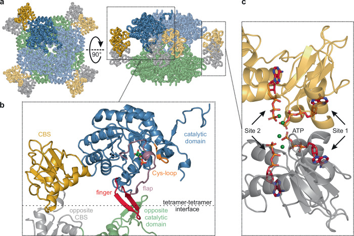
Fig. 2 Architecture of MsmIMPDH. a Side and top views of an MsmIMPDH octamer: the first tetramer is depicted in blue and gold, with its opposite tetramer in green and grey; the MsmIMPDH monomer is highlighted in saturated colours. b Structural details of the highlighted MsmIMPDH monomer. The catalytic domain is depicted in blue and the CBS domain in gold. The flexible loops of the Cys-loop (residues 320–326) are shown in orange, the finger (residues 391–404) in red, the flap (residues 405–450) in purple, and the IMP molecule in green. The two fingers and distal parts of the flaps form a binding interface between the two opposite protomers. c Four ATP molecules (in red) bound within Site 1 and 2 in a complex with four Mg2+ ions (in green) form an interface between two neighbouring CBS domains.
Fig. 2 Architecture of MsmIMPDH. a Side and top views of an MsmIMPDH octamer: the first tetramer is depicted in blue and gold, with its opposite tetramer in green and grey; the MsmIMPDH monomer is highlighted in saturated colours. b Structural details of the highlighted MsmIMPDH monomer. The catalytic domain is depicted in blue and the CBS domain in gold. The flexible loops of the Cys-loop (residues 320–326) are shown in orange, the finger (residues 391–404) in red, the flap (residues 405–450) in purple, and the IMP molecule in green. The two fingers and distal parts of the flaps form a binding interface between the two opposite protomers. c Four ATP molecules (in red) bound within Site 1 and 2 in a complex with four Mg2+ ions (in green) form an interface between two neighbouring CBS domains.CC BY · PubMed Central
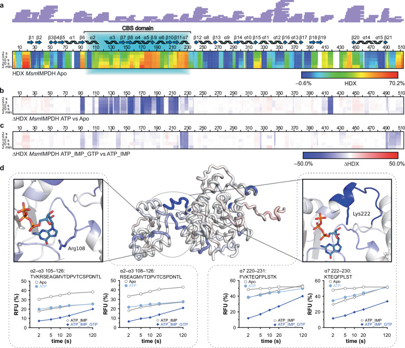
Fig. 3 HDX–MS profiles of MsmIMPDH showing a decrease in the dynamics of the CBS domain upon binding of ATP and GTP. a Heat map of apo MsmIMPDH (constructed from 221 generated peptides) shows the deuteration levels of each amino acid residue in MsmIMPDH obtained at 4 °C over a time course of 2–120 s. Each line represents one time point (2 s, 5 s, 10 s, 20 s, and 2 min). The degree of deuteration is represented by the relative fractional uptake, scaled in rainbow colours from the minimum to the maximum observed uptake. The residues with no sequence coverage are shown as white gaps in the heat map. The sequence coverage is depicted by purple lines above the sequence. Differential heat maps of MsmIMPDH under conditions of (b) apo vs ATP, and (c) ATP–IMP vs ATP–GTP–IMP. Differences in the relative fractional uptake are displayed in a blue–white–red colour scale: residues with decreased accessibility are in blue, and residues with increased HDX are in red. d The ΔHDX changes of ATP–IMP vs ATP–GTP–IMP at 10 s are mapped on the MsmIMPDH monomer structure. Red indicates elevated deuteration, and blue less extensive deuteration of the given region. Hinge regions with residues involved in the binding of GTP (Arg108 and Lys222) are outlined by separate black frames. Deuterium uptake plots show the evolution in the deuteration of four representative peptides in the hinge region. In the graphs, each line represents one protein state, and each point represents one time point over a time course of 2–120 s. For comparison, the protein states are displayed by the same symbols. The drops observed in the HDX rate are displayed in light- and dark-blue colours.
Fig. 3 HDX–MS profiles of MsmIMPDH showing a decrease in the dynamics of the CBS domain upon binding of ATP and GTP. a Heat map of apo MsmIMPDH (constructed from 221 generated peptides) shows the deuteration levels of each amino acid residue in MsmIMPDH obtained at 4 °C over a time course of 2–120 s. Each line represents one time point (2 s, 5 s, 10 s, 20 s, and 2 min). The degree of deuteration is represented by the relative fractional uptake, scaled in rainbow colours from the minimum to the maximum observed uptake. The residues with no sequence coverage are shown as white gaps in the heat map. The sequence coverage is depicted by purple lines above the sequence. Differential heat maps of MsmIMPDH under conditions of (b) apo vs ATP, and (c) ATP–IMP vs ATP–GTP–IMP. Differences in the relative fractional uptake are displayed in a blue–white–red colour scale: residues with decreased accessibility are in blue, and residues with increased HDX are in red. d The ΔHDX changes of ATP–IMP vs ATP–GTP–IMP at 10 s are mapped on the MsmIMPDH monomer structure. Red indicates elevated deuteration, and blue less extensive deuteration of the given region. Hinge regions with residues involved in the binding of GTP (Arg108 and Lys222) are outlined by separate black frames. Deuterium uptake plots show the evolution in the deuteration of four representative peptides in the hinge region. In the graphs, each line represents one protein state, and each point represents one time point over a time course of 2–120 s. For comparison, the protein states are displayed by the same symbols. The drops observed in the HDX rate are displayed in light- and dark-blue colours.CC BY · PubMed Central
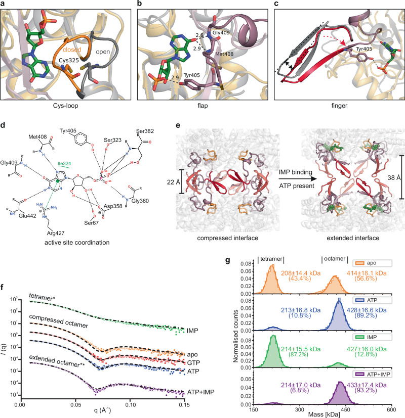
Fig. 4 IMP binding induces reorganization of the MsmIMPDH active site and subsequent dissolution of the tetramer dimerization interface. a The binding of IMP (in green) induces movement of the Cys-loop (residues 320–326, in orange) from an open conformation to a closed conformation. b The initial part of the flap loop (residues 405–415, in purple) interacts with IMP through residues Tyr405, Met408, and Gly409. The contacts are depicted by the grey dotted lines; each number indicates the distance in Å. c IMP binding induces a movement of about 3.5 Å of the entire finger loop (residues 391–404 in red), as indicated by the red arrows. d The two-dimensional representation shows the contacts between IMP and residues forming the MsmIMPDH active site. e Changes to the finger (in red), flap (in purple), and Cys-loop (in orange) after IMP binding (green spheres) lead to extensive rearrangement of the tetramer–tetramer interface, enabling octamer expansion. For consistency, the ATP-bound MsmIMPDH structure was chosen as the model for the compressed conformation across all panels (grey cartoon in a–c). f SAXS profiles of 56 µM MsmIMPDH show the effect of nucleotides on the quaternary structure. For ease of visualization, plots are conveniently displaced along the y axis to represent the three obtained conformations: (green) tetramers induced by 10 mM IMP; (orange) compressed octamers in the apo state, (red) with 10 mM GTP, and (blue) with 10 mM ATP; and (purple) extended octamers induced by 10 mM ATP and IMP. Dashed lines show the theoretical SAXS profiles calculated from the respective cryo-EM structures fitted to the experimental scattering curves. The curve fitted to the IMP dataset (marked by *) represents a 75:25% mixture of tetramers and octamers, while the fit for the ATP + IMP dataset (marked by **) indicates a 38:62% mixture of compressed and extended conformations, as calculated using the OLIGOMER program53. g Mass photometry profiles of 20 nM MsmIMPDH in its apo state and in the presence of 5 mM ATP and/or IMP reveal two distinct peaks. The observed particle masses of the first and second peaks correspond with the MsmIMPDH tetramer (213.2 kDa) and octamer (426.4 kDa).
Fig. 4 IMP binding induces reorganization of the MsmIMPDH active site and subsequent dissolution of the tetramer dimerization interface. a The binding of IMP (in green) induces movement of the Cys-loop (residues 320–326, in orange) from an open conformation to a closed conformation. b The initial part of the flap loop (residues 405–415, in purple) interacts with IMP through residues Tyr405, Met408, and Gly409. The contacts are depicted by the grey dotted lines; each number indicates the distance in Å. c IMP binding induces a movement of about 3.5 Å of the entire finger loop (residues 391–404 in red), as indicated by the red arrows. d The two-dimensional representation shows the contacts between IMP and residues forming the MsmIMPDH active site. e Changes to the finger (in red), flap (in purple), and Cys-loop (in orange) after IMP binding (green spheres) lead to extensive rearrangement of the tetramer–tetramer interface, enabling octamer expansion. For consistency, the ATP-bound MsmIMPDH structure was chosen as the model for the compressed conformation across all panels (grey cartoon in a–c). f SAXS profiles of 56 µM MsmIMPDH show the effect of nucleotides on the quaternary structure. For ease of visualization, plots are conveniently displaced along the y axis to represent the three obtained conformations: (green) tetramers induced by 10 mM IMP; (orange) compressed octamers in the apo state, (red) with 10 mM GTP, and (blue) with 10 mM ATP; and (purple) extended octamers induced by 10 mM ATP and IMP. Dashed lines show the theoretical SAXS profiles calculated from the respective cryo-EM structures fitted to the experimental scattering curves. The curve fitted to the IMP dataset (marked by *) represents a 75:25% mixture of tetramers and octamers, while the fit for the ATP + IMP dataset (marked by **) indicates a 38:62% mixture of compressed and extended conformations, as calculated using the OLIGOMER program53. g Mass photometry profiles of 20 nM MsmIMPDH in its apo state and in the presence of 5 mM ATP and/or IMP reveal two distinct peaks. The observed particle masses of the first and second peaks correspond with the MsmIMPDH tetramer (213.2 kDa) and octamer (426.4 kDa).CC BY · PubMed Central
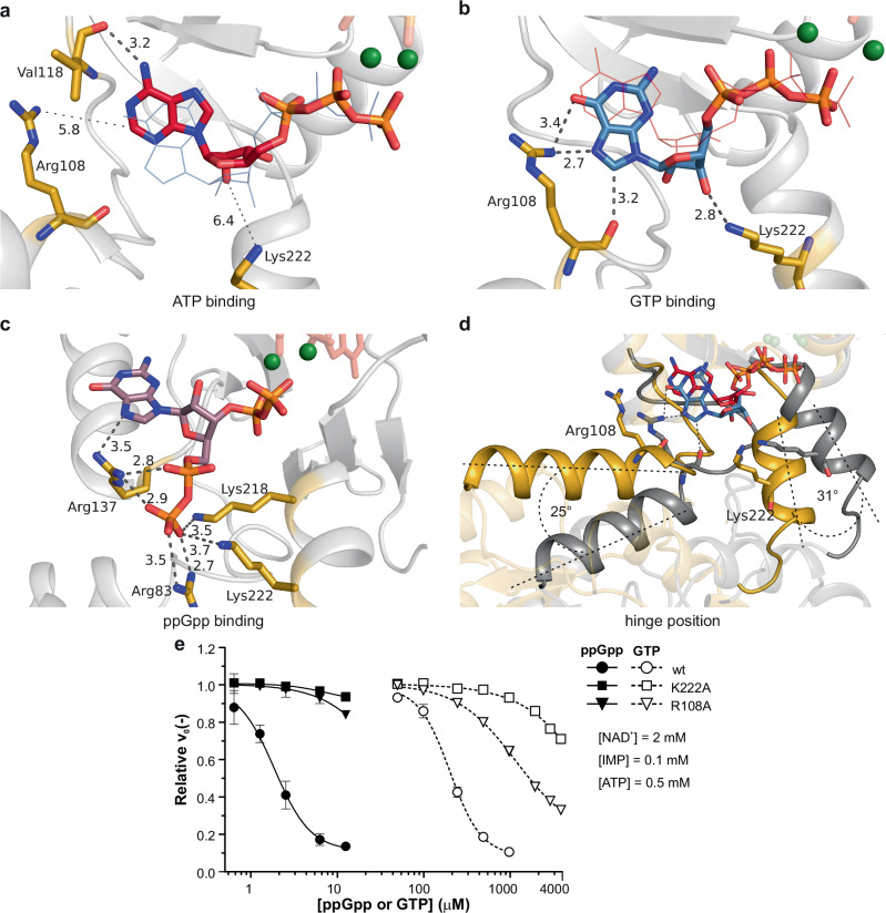
Fig. 5 Differential modes of binding of ATP, GTP, and ppGpp to the CBS domain of MsmIMPDH impact the mobility of the hinge regions. a ATP binds at Site 2 with the base in anti conformation relative to the ribose. b The GTP molecule binds with the base in syn conformation. The other nucleotide is shown as thin lines in the background for comparison of a and b. The contacts with Arg108 and Lys222 (in gold) are depicted by grey dotted lines; each number indicates the distance in Å. c The ppGpp molecule binds at a separate binding site in the CBS domain. d The hinge regions are flexible in the case of ATP binding (in gold), while the GTP locks the hinges in a fixed position (in grey). The side chains of Arg108 and Lys222 are depicted as sticks. e The mutation of Arg108A and Lys222A strongly reduces the inhibition of MsmIMPDH by ppGpp and GTP. NAD+ and IMP substrates were fixed at concentrations of 2 mM and 100 µM, respectively; ATP was fixed at 500 µM. The relative velocity value was calculated as the ratio of the initial velocity of the reaction to the control reaction of the corresponding enzyme (n = 3). Data are presented as mean values with error bars representing the SD.
Fig. 5 Differential modes of binding of ATP, GTP, and ppGpp to the CBS domain of MsmIMPDH impact the mobility of the hinge regions. a ATP binds at Site 2 with the base in anti conformation relative to the ribose. b The GTP molecule binds with the base in syn conformation. The other nucleotide is shown as thin lines in the background for comparison of a and b. The contacts with Arg108 and Lys222 (in gold) are depicted by grey dotted lines; each number indicates the distance in Å. c The ppGpp molecule binds at a separate binding site in the CBS domain. d The hinge regions are flexible in the case of ATP binding (in gold), while the GTP locks the hinges in a fixed position (in grey). The side chains of Arg108 and Lys222 are depicted as sticks. e The mutation of Arg108A and Lys222A strongly reduces the inhibition of MsmIMPDH by ppGpp and GTP. NAD+ and IMP substrates were fixed at concentrations of 2 mM and 100 µM, respectively; ATP was fixed at 500 µM. The relative velocity value was calculated as the ratio of the initial velocity of the reaction to the control reaction of the corresponding enzyme (n = 3). Data are presented as mean values with error bars representing the SD.CC BY · PubMed Central
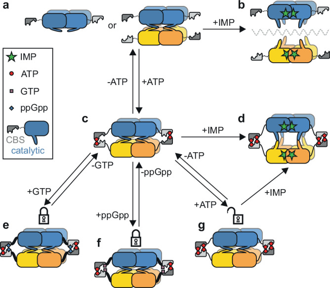
Fig. 6 Schematic model of MsmIMPDH allosteric regulation illustrating the interplay of nucleotide interactions and their impact on the activity and conformation of MsmIMPDH. The top tetramer is depicted in blue, its opposite tetramer in gold, and the CBS domains in shades of grey. a The CBS domains are free in the apo form that exists in both tetrameric and octameric conformations. c The binding of ATP to Site 1 stabilizes the dimers of the CBS domains and thus the octameric conformation. b, d IMP binding drives the formation of the active tetramers (b) or extended octamers stabilized by ATP (d). e, f This extension is blocked by the binding of GTP (e) or ppGpp (f), thus locking the hinge regions that link the CBS and catalytic domains (depicted as thick black linkers). g Binding of ATP to Site 2 competes with GTP and ppGpp, while enabling the IMP-induced extension.
Fig. 6 Schematic model of MsmIMPDH allosteric regulation illustrating the interplay of nucleotide interactions and their impact on the activity and conformation of MsmIMPDH. The top tetramer is depicted in blue, its opposite tetramer in gold, and the CBS domains in shades of grey. a The CBS domains are free in the apo form that exists in both tetrameric and octameric conformations. c The binding of ATP to Site 1 stabilizes the dimers of the CBS domains and thus the octameric conformation. b, d IMP binding drives the formation of the active tetramers (b) or extended octamers stabilized by ATP (d). e, f This extension is blocked by the binding of GTP (e) or ppGpp (f), thus locking the hinge regions that link the CBS and catalytic domains (depicted as thick black linkers). g Binding of ATP to Site 2 competes with GTP and ppGpp, while enabling the IMP-induced extension.CC BY · PubMed Central

Verification

Claims the paper makes

The claims and statistics of this paper haven’t been checked yet.

Run the check — free, no account needed. It reads the open-access text and checks the sources it cites.

A few free checks a day · papers already verified open instantly · results are public

Self-check

Percentages, sums and reported differences re-computed from the paper’s own numbers.

The self-check needs the paper’s full text.

Citations

Cites

Show all 64 references

Metadata

Authors
Bulvas, O., Knejzlík, Z., Sýs, J., Filimoněnko, A., Čížková, M., Clarová, K., Rejman, D., Kouba, T., & Pichová, I.
Affiliations
  • Czech Academy of Sciences, Institute of Organic Chemistry and Biochemistry (CZ)
  • Czech Academy of Sciences (CZ)
Corresponding author
  • Kouba, Tomáš
  • Pichová, Iva
Venue
Nature Communications
Published
2024-08-06
Volume
15
Issue
1
Pages
6673-6673
ISSN
2041-1723 (ISSN-L)
OpenAlex
W4401358016
Cited by
13
References
64
Citations by year
2026: 4 · 2025: 8 · 2024: 1
FWCI
1.77
Type
journal article
Language
en
Open access
Locations
  • Nature Communications (publishedVersion, cc-by)
  • Research Square (acceptedVersion, cc-by)
  • PubMed Central (publishedVersion, cc-by)
  • PubMed (publishedVersion)
  • RePEc: Research Papers in Economics (submittedVersion)
  • DOAJ (DOAJ: Directory of Open Access Journals) (submittedVersion)
Funders
  • European Commission (LX22NPO5103, CZ.02.1.01)
  • Ministerstvo Školství, Mládeže a Tělovýchovy (LM2023050, CZ.02.1.01/0.0/0.0/18_046/0015974, CZ.02.1.01/0.0/0.0/18_046/0016045, CZ.02.1.01, LX22NPO5103, LM2023042)
  • Central European Institute of Technology
  • Masarykova Univerzita
  • Vysoká Škola Chemicko-technologická v Praze
  • European Regional Development Fund (02.1.01/0.0/0.0, CZ.02.1.01/0.0/0.0/18_046/0015974, CZ.02, CZ.02.1.01, CZ.02.01, CZ.02.1.01/0.0/0.0/18_046/0016045, CZ.02.1.01/0.0/0.0, CZ.02.01.01/00/23_015/0008205)
  • NextGenerationEU
Topics
  • Life Sciences › Biochemistry, Genetics and Molecular Biology › Molecular Biology › Biochemical and Molecular Research
  • Life Sciences › Biochemistry, Genetics and Molecular Biology › Structural Biology › Enzyme Structure and Function
  • Life Sciences › Biochemistry, Genetics and Molecular Biology › Molecular Biology › Protein Structure and Dynamics
Keywords
inosine 5'-monophosphate dehydrogenase, allosteric regulation, mycobacteria, purine metabolism, CBS domain, GTP inhibition, (p)ppGpp, nucleotide signaling, enzyme conformational change, active site occlusion, ATP-competitive inhibition, allosteric inhibitors, antibiotic targets
MeSH terms
Adenosine Triphosphate, Allosteric Regulation, Bacterial Proteins, Guanosine Pentaphosphate, Guanosine Triphosphate, IMP Dehydrogenase, Inosine Monophosphate, Models, Molecular, Protein Binding, Mycobacterium smegmatis, Catalytic Domain, Cryoelectron Microscopy

About these checks

Registry, retraction, Cites and statistics results are computed, not judged, with one exception: a printed reference that no index lists is read once by a model before it counts as a citation error. Claims are extracted by a model and kept only when their quote appears verbatim in the text; each cited source is then checked, with a model’s help, for whether it says what the claim relies on. Those verdicts can be wrong — read the claim’s quote and the cited source before relying on one.

Statistics and arithmetic are recomputed, not judged.

Confidence bands are shown only for fields calibrated on our annotated test set. None is calibrated yet, so no band is shown.

What counts as a claim ›How we measure confidence ›How we check this ›

Are you an author, or do you see a problem with this page? Request a review

How to cite

Cite CoChat for the checks on this page, and the paper for its own findings.

This page
CoChat. (2026). Citation check: Deciphering the allosteric regulation of mycobacterial inosine-5′-monophosphate dehydrogenase. https://app.cochat.ai/papers/10.1038/s41467-024-50933-6
The paper
Bulvas, O., Knejzlík, Z., Sýs, J., Filimoněnko, A., Čížková, M., Clarová, K., Rejman, D., Kouba, T., & Pichová, I. (2024). Deciphering the allosteric regulation of mycobacterial inosine-5′-monophosphate dehydrogenase. Nature Communications. https://doi.org/10.1038/s41467-024-50933-6

Data sources

Bibliographic data: Semantic Scholar, OpenAlex, Crossref.

Contains information from the Semantic Scholar Academic Graph Datasets, made available under the ODC Attribution License (ODC-BY 1.0).

Semantic Scholar’s data is described in: Kinney, R., et al. (2023). The Semantic Scholar Open Data Platform. arXiv:2301.10140.