

The DOI matches its Crossref record; 48 of the 48 references it cites were found in a registry or an index.
Metformin hydrolase is a recently evolved nickel-dependent heteromeric ureohydrolase
Open accessPublished version of a preprint
Abstract
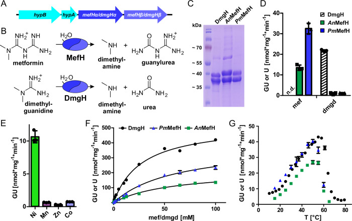
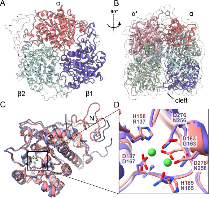
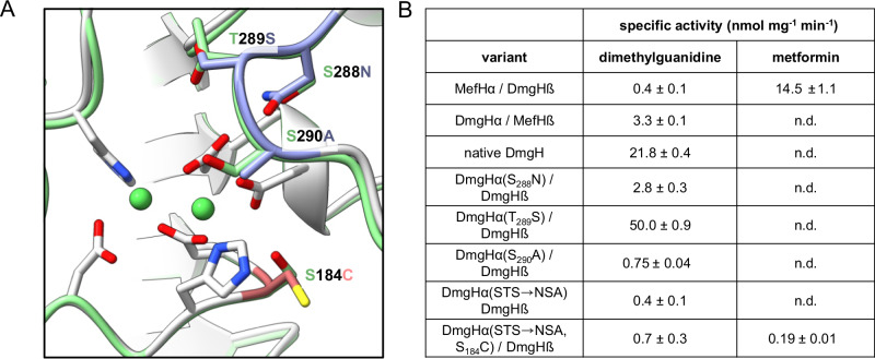
The anti-diabetic drug metformin is one of the most widely prescribed medicines in the world. Together with its degradation product guanylurea, it is a major pharmaceutical pollutant in wastewater treatment plants and surface waters. An operon comprising two genes of the ureohydrolase family in Pseudomonas and Aminobacter species has recently been implicated in metformin degradation. However, the corresponding proteins have not been characterized. Here we show that these genes encode a Ni 2+ -dependent enzyme that efficiently and specifically hydrolyzes metformin to guanylurea and dimethylamine. The active enzyme is a heteromeric complex of α- and β- subunits in which only the α-subunits contain the conserved His and Asp residues for the coordination of two Ni 2+ ions in the active site. A crystal structure of metformin hydrolase reveals an α 2 β 4 stoichiometry of the hexameric complex, which is unprecedented in the ureohydrolase family. By studying a closely related but more widely distributed enzyme, we find that the putative predecessor specifically hydrolyzes dimethylguanidine instead of metformin. Our findings establish the molecular basis for metformin hydrolysis to guanylurea as the primary pathway for metformin biodegradation and provide insight into the recent evolution of ureohydrolase family proteins in response to an anthropogenic compound.
Overview
The claims and statistics of this paper haven’t been checked yet.
Run the check — free, no account needed. It reads the open-access text and checks the sources it cites.
Integrity signals
Registry
Cites
Statistics
Figures
Verification
Claims the paper makes
The claims and statistics of this paper haven’t been checked yet.
Run the check — free, no account needed. It reads the open-access text and checks the sources it cites.
Self-check
Percentages, sums and reported differences re-computed from the paper’s own numbers.
The self-check needs the paper’s full text.
Citations
Cites
Highly accurate protein structure prediction with AlphaFold
Jumper, J., Evans, R., Pritzel, A., Green, T., Figurnov, M., Ronneberger, O., Tunyasuvunakool, K., Bates, R., Žídek, A., Potapenko, A., et al. (2021) · Nature · doi:10.1038/s41586-021-03819-2 · Cited by 43,764
Features and development of Coot
Emsley, P., Lohkamp, B., Scott, W. G., & Cowtan, K. (2010) · Acta Crystallographica Section D Biological Crystallography · doi:10.1107/s0907444910007493 · Cited by 29,915
Phaser crystallographic software
McCoy, A. J., Grosse-Kunstleve, R. W., Adams, P. D., Winn, M. D., Storoni, L. C., & Read, R. J. (2007) · Journal of Applied Crystallography · doi:10.1107/s0021889807021206 · Cited by 20,612
Kabsch, W. (2010) · Acta Crystallographica Section D Biological Crystallography · doi:10.1107/s0907444909047337 · Cited by 16,463
Fast, scalable generation of high‐quality protein multiple sequence alignments using Clustal Omega
Sievers, F., Wilm, A., Dineen, D., Gibson, T. J., Karplus, K., Li, W., Lopez, R., McWilliam, H., Remmert, M., Söding, J., et al. (2011) · Molecular Systems Biology · doi:10.1038/msb.2011.75 · Cited by 15,355
International Diabetes Federation 2017
Carracher, A. M., Marathe, P. H., & Close, K. L. (2018) · Journal of Diabetes · doi:10.1111/1753-0407.12644 · Cited by 13,418
Jalview Version 2—a multiple sequence alignment editor and analysis workbench
Waterhouse, A. M., Procter, J. B., Martin, D. M. A., Clamp, M., & Barton, G. J. (2009) · Bioinformatics · doi:10.1093/bioinformatics/btp033 · Cited by 10,235
UCSF ChimeraX: Structure visualization for researchers, educators, and developers
Pettersen, E. F., Goddard, T. D., Huang, C. C., Meng, E. C., Couch, G. S., Croll, T. I., Morris, J. H., & Ferrin, T. E. (2021) · Protein Science · doi:10.1002/pro.3943 · Cited by 9,514
ColabFold - Making protein folding accessible to all
Mirdita, M., Schütze, K., Moriwaki, Y., Heo, L., Ovchinnikov, S., & Steinegger, M. (2021) · doi:10.1101/2021.08.15.456425 · Cited by 9,391
Liebschner, D., Afonine, P. V., Baker, M. L., Bunkóczi, G., Chen, V. B., Croll, T. I., Hintze, B., Hung, L.-W., Jain, S., McCoy, A. J., et al. (2019) · Acta Crystallographica Section D Structural Biology · doi:10.1107/s2059798319011471 · Cited by 7,953
Research works printed but not linked: 2
- Urakami, T., Araki, H., Oyanagi, H., Suzuki, K.-I. & Komagata, K. Transfer of Pseudomonas aminovorans (den Dooren de Jong 1926) to Aminobacter gen. nov. as Aminobacter aminovorans comb. nov. and Description of Aminobacter aganoensis sp. nov. and Aminobacter niigataensis sp. nov. Int. J. Syst. Evol. Microbiol. 42, 84–92 (1992).
- Hall, T. A. BioEdit: a user-friendly biological sequence alignment editor and analysis program for Windows 95/98/NT. Nucleic Acids Symp. Ser. 41, 95–98 (1999).
Metadata
- Authors
- Sinn, M., Riede, L., Fleming, J. R., Funck, D., Lutz, H., Bachmann, A., Mayans, O., & Hartig, J. S.
- Affiliations
- University of Konstanz (DE)
- Corresponding author
- Sinn, M.
- Hartig, J. S.
- Venue
- Nature Communications
- Published
- 2024-09-14
- Volume
- 15
- Issue
- 1
- Pages
- 8045-8045
- ISSN
- 2041-1723 (ISSN-L)
- PMC
- PMC11399263
- OpenAlex
- W4402503221
- Cited by
- 13
- References
- 48
- Citations by year
- 2026: 6 · 2025: 4 · 2024: 3
- FWCI
- 1.29
- Type
- journal article
- Language
- en
- Open access
- gold
- doi.org
- Licence
- cc-by
- creativecommons.org/licenses/by/4.0 (tdm)
- creativecommons.org/licenses/by/4.0 (vor)
- Locations
- Nature Communications (publishedVersion, cc-by)
- Research Square (acceptedVersion, cc-by)
- PubMed Central (publishedVersion, cc-by)
- PubMed (publishedVersion)
- RePEc: Research Papers in Economics (submittedVersion)
- Desy publication database (The Deutsches Elektronen-Synchrotron) (submittedVersion, other-oa)
- DOAJ (DOAJ: Directory of Open Access Journals) (submittedVersion, cc-by-sa)
- KOPS (University of Konstanz) (submittedVersion, cc-by)
- DESY Publication Database (PUBDB) (Deutsches Elektronen-Synchrotron)
- Funders
- Universität Konstanz
- Topics
- Physical Sciences › Environmental Science › Pollution › Pharmaceutical and Antibiotic Environmental Impacts
- Life Sciences › Biochemistry, Genetics and Molecular Biology › Structural Biology › Enzyme Structure and Function
- Life Sciences › Biochemistry, Genetics and Molecular Biology › Molecular Biology › Enzyme Catalysis and Immobilization
- Keywords
- guanylurea, nickel-dependent enzymes, enzyme evolution, Pseudomonas, Aminobacter, crystal structure, pharmaceutical pollutants, wastewater treatment, enzyme active site, dimethylamine
- MeSH terms
- Bacterial Proteins, Biodegradation, Environmental, Hydrolysis, Metformin, Models, Molecular, Nickel, Pseudomonas, Ureohydrolases, Crystallography, X-Ray, Catalytic Domain
About these checks
Registry, retraction, Cites and statistics results are computed, not judged, with one exception: a printed reference that no index lists is read once by a model before it counts as a citation error. Claims are extracted by a model and kept only when their quote appears verbatim in the text; each cited source is then checked, with a model’s help, for whether it says what the claim relies on. Those verdicts can be wrong — read the claim’s quote and the cited source before relying on one.
Statistics and arithmetic are recomputed, not judged.
Confidence bands are shown only for fields calibrated on our annotated test set. None is calibrated yet, so no band is shown.
What counts as a claim ›How we measure confidence ›How we check this ›
Are you an author, or do you see a problem with this page? Request a review
How to cite
Cite CoChat for the checks on this page, and the paper for its own findings.
- This page
- CoChat. (2026). Citation check: Metformin hydrolase is a recently evolved nickel-dependent heteromeric ureohydrolase. https://app.cochat.ai/papers/10.1038/s41467-024-51752-5
- The paper
- Sinn, M., Riede, L., Fleming, J. R., Funck, D., Lutz, H., Bachmann, A., Mayans, O., & Hartig, J. S. (2024). Metformin hydrolase is a recently evolved nickel-dependent heteromeric ureohydrolase. Nature Communications. https://doi.org/10.1038/s41467-024-51752-5
Data sources
Bibliographic data: Semantic Scholar, OpenAlex, Crossref.
Contains information from the Semantic Scholar Academic Graph Datasets, made available under the ODC Attribution License (ODC-BY 1.0).
Semantic Scholar’s data is described in: Kinney, R., et al. (2023). The Semantic Scholar Open Data Platform. arXiv:2301.10140.
