

The DOI matches its Crossref record; 44 of the 44 references it cites were found in a registry or an index.
Structural insights into epitope-paratope interactions of a monoclonal antibody targeting CEACAM5-expressing tumors
Open accessPublished version of a preprint
Abstract
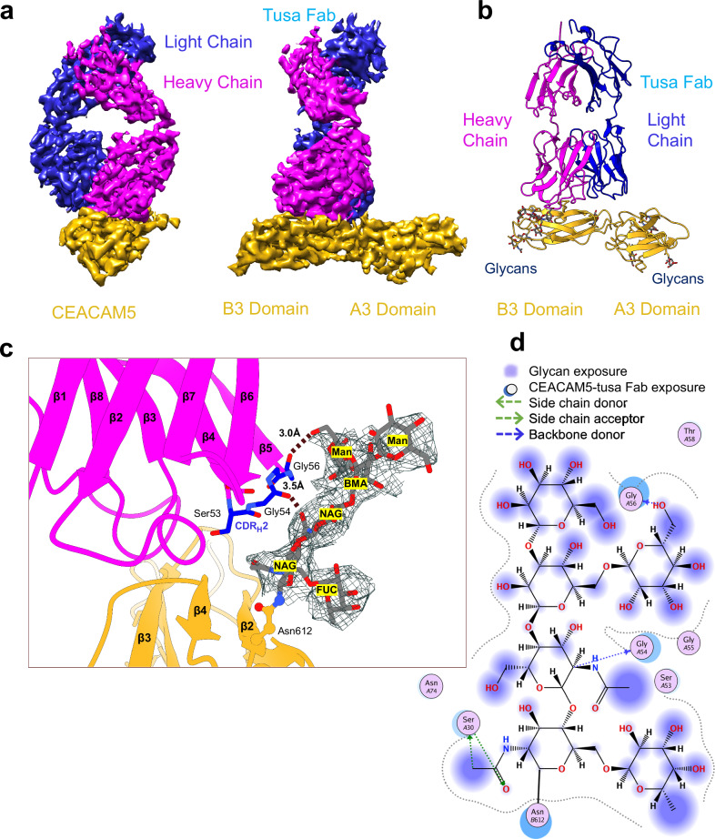
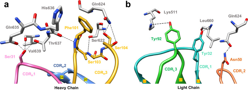
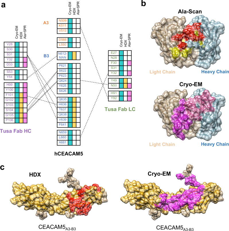
Abstract Carcinoembryonic antigen-related cell adhesion molecules (CEACAMs) are overexpressed in some tumor types. The antibody-drug conjugate, tusamitamab ravtansine, specifically recognizes the A3-B3 domains of human CEACAM5 (hCEACAM5). To understand this specificity, we mapped the epitope-paratope interface between the A3-B3 domains of hCEACAM5 (hCEACAM5A3 − B3) and the antigen-binding fragment of tusamitamab (tusa Fab). Hydrogen/deuterium exchange mass spectrometry identified the tusa Fab paratope, which involves heavy chain (HC) residues 101–109 and light chain residues 48–54 and 88–104. Surface plasmon resonance demonstrated that alanine variants of HC residues 96–108 abolish binding to hCEACAM5, suggesting that these residues are critical for the Fab-antigen complex formation. Cryogenic electron microscopy structure of the tusa Fab-hCEACAM5A3 − B3 complex (3.11 Å overall resolution) revealed a discontinuous epitope involving residues in the A3-B3 domains and an N-linked mannose at residue 612. Conformational constraints on the epitope-paratope interface enable tusamitamab to target hCEACAM5A3 − B3 and differentiate it from other CEACAMs.
Overview
The claims and statistics of this paper haven’t been checked yet.
Run the check — free, no account needed. It reads the open-access text and checks the sources it cites.
Integrity signals
Registry
Cites
Statistics
Figures
Verification
Claims the paper makes
The claims and statistics of this paper haven’t been checked yet.
Run the check — free, no account needed. It reads the open-access text and checks the sources it cites.
Self-check
Percentages, sums and reported differences re-computed from the paper’s own numbers.
The self-check needs the paper’s full text.
Citations
Cites
UCSF Chimera—A visualization system for exploratory research and analysis
Pettersen, E. F., Goddard, T. D., Huang, C. C., Couch, G. S., Greenblatt, D. M., Meng, E. C., & Ferrin, T. E. (2004) · Journal of Computational Chemistry · doi:10.1002/jcc.20084 · Cited by 45,828
Highly accurate protein structure prediction with AlphaFold
Jumper, J., Evans, R., Pritzel, A., Green, T., Figurnov, M., Ronneberger, O., Tunyasuvunakool, K., Bates, R., Žídek, A., Potapenko, A., et al. (2021) · Nature · doi:10.1038/s41586-021-03819-2 · Cited by 43,764
Coot : model-building tools for molecular graphics
Emsley, P., & Cowtan, K. (2004) · Acta Crystallographica Section D Biological Crystallography · doi:10.1107/s0907444904019158 · Cited by 30,571
PHENIX : a comprehensive Python-based system for macromolecular structure solution
Adams, P. D., Afonine, P. V., Bunkóczi, G., Chen, V. B., Davis, I. W., Echols, N., Headd, J. J., Hung, L.-W., Kapral, G. J., Grosse-Kunstleve, R. W., et al. (2010) · Acta Crystallographica Section D Biological Crystallography · doi:10.1107/s0907444909052925 · Cited by 23,716
Fast, scalable generation of high‐quality protein multiple sequence alignments using Clustal Omega
Sievers, F., Wilm, A., Dineen, D., Gibson, T. J., Karplus, K., Li, W., Lopez, R., McWilliam, H., Remmert, M., Söding, J., et al. (2011) · Molecular Systems Biology · doi:10.1038/msb.2011.75 · Cited by 15,355
MolProbity : all-atom structure validation for macromolecular crystallography
Chen, V. B., Arendall, W. B., Headd, J. J., Keedy, D. A., Immormino, R. M., Kapral, G. J., Murray, L. W., Richardson, J. S., & Richardson, D. C. (2010) · Acta Crystallographica Section D Biological Crystallography · doi:10.1107/s0907444909042073 · Cited by 14,115
Inference of Macromolecular Assemblies from Crystalline State
Krissinel, E., & Henrick, K. (2007) · Journal of Molecular Biology · doi:10.1016/j.jmb.2007.05.022 · Cited by 9,984
UniProt: the universal protein knowledgebase in 2021
The UniProt Consortium, Bateman, A., Martin, M.-J., Orchard, S., Magrane, M., Agivetova, R., Ahmad, S., Alpi, E., Bowler-Barnett, E. H., Britto, R., et al. (2021) · Nucleic Acids Research · doi:10.1093/nar/gkaa1100 · Cited by 6,372
The InterPro protein families and domains database: 20 years on
Blum, M., Chang, H.-Y., Chuguransky, S., Grego, T., Kandasaamy, S., Mitchell, A., Nuka, G., Paysan-Lafosse, T., Qureshi, M., Raj, S., et al. (2021) · Nucleic Acids Research · doi:10.1093/nar/gkaa977 · Cited by 2,231
Surface Plasmon Resonance: A Versatile Technique for Biosensor Applications
Nguyen, H., Park, J., Kang, S., & Kim, M. (2015) · Sensors · doi:10.3390/s150510481 · Cited by 1,357
Hammarström, S. (1999) · Seminars in Cancer Biology · doi:10.1006/scbi.1998.0119 · Cited by 1,300
Bork, P., Holm, L., & Sander, C. (1994) · Journal of Molecular Biology · doi:10.1006/jmbi.1994.1582 · Cited by 674
Tate, J. R., & Ward, G. (2004) · PubMed · Cited by 548
Beauchemin, N., & Arabzadeh, A. (2013) · Cancer and Metastasis Reviews · doi:10.1007/s10555-013-9444-6 · Cited by 538
Emerging new therapeutic antibody derivatives for cancer treatment
Jin, S., Sun, Y., Liang, X., Gu, X., Ning, J., Xu, Y., Chen, S., & Pan, L. (2022) · Signal Transduction and Targeted Therapy · doi:10.1038/s41392-021-00868-x · Cited by 459
Hydrogen bonds and salt bridges across protein-protein interfaces
Xu, D., Tsai, C. J., & Nussinov, R. (1997) · Protein Engineering Design and Selection · doi:10.1093/protein/10.9.999 · Cited by 454
The Immunoglobulin Fold Structural Classification, Sequence Patterns and Common Core
BORK, P., HOLM, L., & SANDER, C. (1994) · Journal of Molecular Biology · doi:10.1016/s0022-2836(84)71582-8 · Cited by 412
Kifunensine, a potent inhibitor of the glycoprotein processing mannosidase I.
Elbein, A. D., Tropea, J. E., Mitchell, M., & Kaushal, G. P. (1990) · Journal of Biological Chemistry · doi:10.1016/s0021-9258(18)55439-9 · Cited by 332
Lee, H. S., Qi, Y., & Im, W. (2015) · Scientific Reports · doi:10.1038/srep08926 · Cited by 306
Stock-based detection of protein oligomeric states in jsPISA
Krissinel, E. (2015) · Nucleic Acids Research · doi:10.1093/nar/gkv314 · Cited by 242
Origins of specificity and affinity in antibody–protein interactions
Peng, H., Lee, K. H., Jian, J.-W., & Yang, A. (2014) · Proceedings of the National Academy of Sciences · doi:10.1073/pnas.1401131111 · Cited by 205
Beauchemin, N., Benchimol, S., Cournoyer, D., Fuks, A., & Stanners, C. P. (1987) · Molecular and Cellular Biology · doi:10.1128/mcb.7.9.3221-3230.1987 · Cited by 190
Zhou, Q., & Qiu, H. (2018) · Journal of Pharmaceutical Sciences · doi:10.1016/j.xphs.2018.11.029 · Cited by 169
Protein–protein binding affinity prediction from amino acid sequence
Yugandhar, K., & Gromiha, M. M. (2014) · Bioinformatics · doi:10.1093/bioinformatics/btu580 · Cited by 130
Sinha, N., Mohan, S., Lipschultz, C. A., & Smith‐Gill, S. J. (2002) · Biophysical Journal · doi:10.1016/s0006-3495(02)75302-2 · Cited by 121
Many Routes to an Antibody Heavy-Chain CDR3: Necessary, Yet Insufficient, for Specific Binding
D’Angelo, S., Ferrara, F., Naranjo, L. A., Erasmus, M. F., Hraber, P., & Bradbury, A. M. (2018) · Frontiers in Immunology · doi:10.3389/fimmu.2018.00395 · Cited by 98
Gazzah, A., Bedard, P. L., Hierro, C., Kang, Y., Razak, A. A., Ryu, M.-H., Demers, B., Fagniez, N., Henry, C., Hospitel, M., et al. (2022) · Annals of Oncology · doi:10.1016/j.annonc.2021.12.012 · Cited by 89
Kuroda, D., & Gray, J. J. (2016) · Bioinformatics · doi:10.1093/bioinformatics/btw197 · Cited by 86
Decary, S., Berne, P., Nicolazzi, C., Lefebvre, A., Dabdoubi, T., Cameron, B., Rival, P., Devaud, C., Prades, C., Bouchard, H., et al. (2020) · Clinical Cancer Research · doi:10.1158/1078-0432.ccr-19-4051 · Cited by 84
Rapid Development of Affinity Matured Monoclonal Antibodies Using RIMMS
Kilpatrick, K. E., Wring, S. A., Walker, D. H., et al. (1997) · Hybridoma · doi:10.1089/hyb.1997.16.381 · Cited by 81
The interfacial character of antibody paratopes: analysis of antibody–antigen structures
Nguyen, M. N., Pradhan, M. R., Verma, C. S., & Zhong, P. (2017) · Bioinformatics · doi:10.1093/bioinformatics/btx389 · Cited by 71
Boehm, M. K., Mayans, O. M., Thornton, J. D., Beǵent, R. H. J., Keep, P. A., & Perkins, S. J. (1996) · Journal of Molecular Biology · doi:10.1006/jmbi.1996.0353 · Cited by 66
Zhao, Q., Zhan, T., Deng, Z., Li, Q., Liu, Y., Yang, S., Ji, D., & Li, Y. (2018) · Clinical Proteomics · doi:10.1186/s12014-018-9182-4 · Cited by 65
A predicted three‐dimensional structure for the carcinoembryonic antigen (CEA)
Bates, P. A., Luo, J., & Sternberg, M. J. E. (1992) · FEBS Letters · doi:10.1016/0014-5793(92)81249-l · Cited by 61
Role of Salt Bridge Formation in Antigen-Antibody Interaction
Tsumoto, K., Ogasahara, K., Ueda, Y., Watanabe, K., Yutani, K., & Kumagai, I. (1996) · Journal of Biological Chemistry · doi:10.1074/jbc.271.51.32612 · Cited by 59
Pont, L., Kuzyk, V., Benavente, F., Sanz‐Nebot, V., Mayboroda, O. A., Wuhrer, M., & Lageveen‐Kammeijer, G. S. M. (2021) · Journal of Proteome Research · doi:10.1021/acs.jproteome.0c00875 · Cited by 52
Diverse oligomeric states of CEACAM IgV domains
Bonsor, D. A., Günther, S., Beadenkopf, R., Beckett, D., & Sundberg, E. J. (2015) · Proceedings of the National Academy of Sciences · doi:10.1073/pnas.1509511112 · Cited by 51
Chiang, W., Cheng, T.-M., Chang, C.-C., Pan, S.-H., Changou, C. A., Chang, T.-H., Lee, K.-H., Wu, S., Chen, Y.-F., Chuang, K.-H., et al. (2017) · Oncogene · doi:10.1038/onc.2017.303 · Cited by 50
Asymmetric Preorganization of Inverted Pair Residues in the Sodium-Calcium Exchanger
Giladi, M., Almagor, L., van Dijk, L., Hiller, R., Man, P., Forest, É., & Khananshvili, D. (2016) · Scientific Reports · doi:10.1038/srep20753 · Cited by 41
Gazzah, A., Ricordel, C., Cousin, S., et al. (2020) · Journal of Clinical Oncology · doi:10.1200/jco.2020.38.15_suppl.9505 · Cited by 38
Baek, D.-S., Kim, Y., Vergara, S. V., Conard, A., Adams, C., Calero, G. A., Ishima, R., Mellors, J. W., & Dimitrov, D. S. (2021) · Cancer Letters · doi:10.1016/j.canlet.2021.10.041 · Cited by 33
Characterizing CEACAM5 interaction with CD8α and CD1d in intestinal homeostasis
Roda, G., Jianyu, X., Park, M. S., DeMarte, L., Hovhannisyan, Z., Couri, R., Stanners, C. P., Yeretssian, G., & Mayer, L. (2013) · Mucosal Immunology · doi:10.1038/mi.2013.80 · Cited by 32
Giladi, M., van Dijk, L., Refaeli, B., Almagor, L., Hiller, R., Man, P., Forest, É., & Khananshvili, D. (2017) · Journal of Biological Chemistry · doi:10.1074/jbc.m117.787168 · Cited by 32
Ricordel, C., Barlési, F., Cousin, S., et al. (2022) · Journal of Clinical Oncology · doi:10.1200/jco.2022.40.16_suppl.9039 · Cited by 27
Metadata
- Authors
- Kumar, A., Duffieux, F., Gagnaire, M., Rapisarda, C., Bertrand, T., & Rak, A.
- Affiliations
- Sanofi (France) (FR)
- Corresponding author
- Rak, Alexey
- Venue
- Nature Communications
- Published
- 2024-10-30
- Volume
- 15
- Issue
- 1
- Pages
- 9377-9377
- ISSN
- 2041-1723 (ISSN-L)
- PMC
- PMC11525548
- OpenAlex
- W4403890164
- Cited by
- 11
- References
- 44
- Citations by year
- 2026: 7 · 2025: 4
- FWCI
- 2.80
- Type
- journal article
- Language
- en
- Open access
- gold
- doi.org
- Licence
- cc-by-nc-nd
- creativecommons.org/licenses/by-nc-nd/4.0 (tdm)
- creativecommons.org/licenses/by-nc-nd/4.0 (vor)
- Locations
- Nature Communications (publishedVersion, cc-by-nc-nd)
- Research Square (acceptedVersion, cc-by)
- PubMed Central (publishedVersion, cc-by-nc-nd)
- PubMed (publishedVersion)
- RePEc: Research Papers in Economics (submittedVersion)
- DOAJ (DOAJ: Directory of Open Access Journals) (submittedVersion, cc-by-sa)
- Funders
- Sanofi
- Topics
- Life Sciences › Immunology and Microbiology › Immunology › Monoclonal and Polyclonal Antibodies Research
- Life Sciences › Biochemistry, Genetics and Molecular Biology › Structural Biology › Enzyme Structure and Function
- Life Sciences › Biochemistry, Genetics and Molecular Biology › Molecular Biology › vaccines and immunoinformatics approaches
- Keywords
- CEACAM5, monoclonal antibodies, antibody-drug conjugates, antigen-binding fragments, hydrogen-deuterium exchange, alanine scanning, cryo-EM structure, tumor targeting
- MeSH terms
- Protein Domains, Antibodies, Monoclonal, Epitopes, Carcinoembryonic Antigen, Humans, Immunoglobulin Fab Fragments, Models, Molecular, Neoplasms, Protein Binding, Cell Adhesion Molecules, Immunoconjugates, Cryoelectron Microscopy, Surface Plasmon Resonance, GPI-Linked Proteins
About these checks
Registry, retraction, Cites and statistics results are computed, not judged, with one exception: a printed reference that no index lists is read once by a model before it counts as a citation error. Claims are extracted by a model and kept only when their quote appears verbatim in the text; each cited source is then checked, with a model’s help, for whether it says what the claim relies on. Those verdicts can be wrong — read the claim’s quote and the cited source before relying on one.
Statistics and arithmetic are recomputed, not judged.
Confidence bands are shown only for fields calibrated on our annotated test set. None is calibrated yet, so no band is shown.
What counts as a claim ›How we measure confidence ›How we check this ›
Are you an author, or do you see a problem with this page? Request a review
Data sources
Bibliographic data: Semantic Scholar, OpenAlex, Crossref.
Contains information from the Semantic Scholar Academic Graph Datasets, made available under the ODC Attribution License (ODC-BY 1.0).
Semantic Scholar’s data is described in: Kinney, R., et al. (2023). The Semantic Scholar Open Data Platform. arXiv:2301.10140.
