

The DOI matches its Crossref record; 1 of the 1 references it cites were found in a registry or an index.
Structure and function of the geldanamycin amide synthase from Streptomyces hygroscopicus
Open accessPublished version of a preprint
Abstract
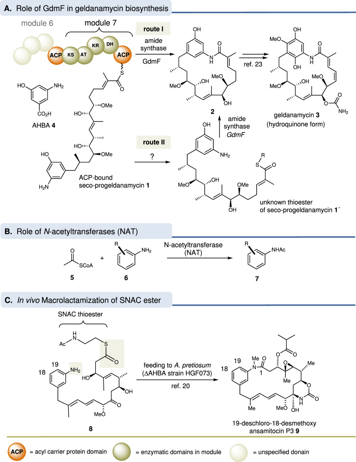
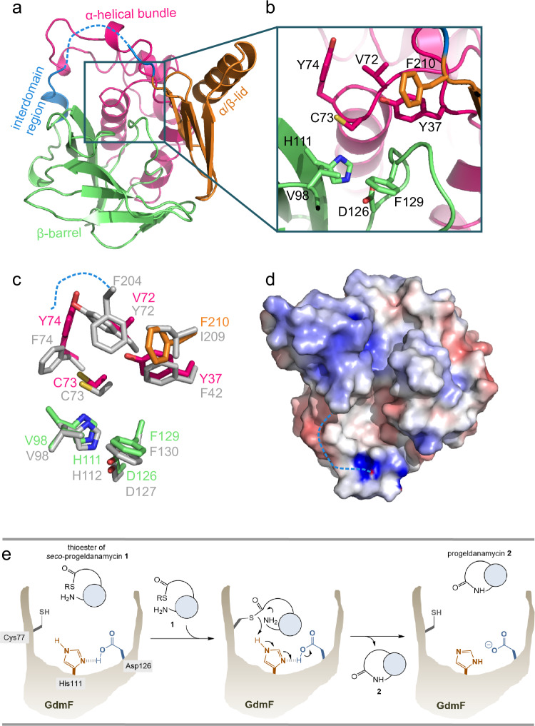
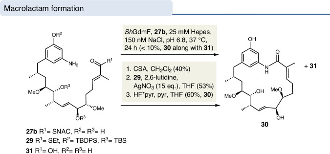
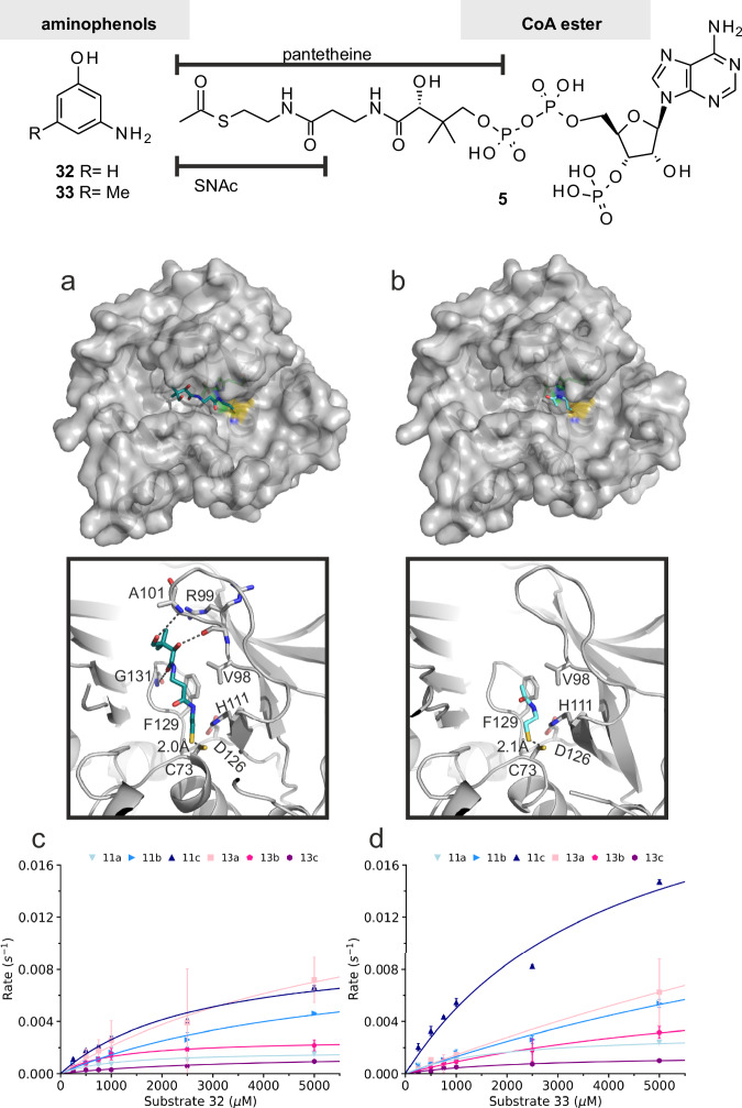
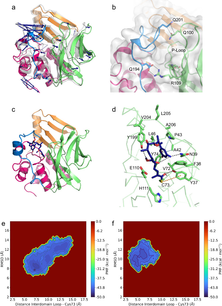
Amide synthases catalyze the formation of macrolactam rings from aniline-containing polyketide-derived seco-acids as found in the important class of ansamycin antibiotics. One of these amide synthases is the geldanamycin amide synthase GdmF, which we recombinantly expressed, purified and studied in detail both functionally as well as structurally. Here we show that purified GdmF catalyzes the amide formation using synthetically derived substrates. The atomic structures of the ligand-free enzyme and in complex with simplified substrates reveal distinct structural features of the substrate binding site and a putative role of the flexible interdomain region for the catalysis reaction.
Overview
The claims and statistics of this paper haven’t been checked yet.
Run the check — free, no account needed. It reads the open-access text and checks the sources it cites.
Integrity signals
Registry
Cites
Statistics
Figures
Verification
Claims the paper makes
The claims and statistics of this paper haven’t been checked yet.
Run the check — free, no account needed. It reads the open-access text and checks the sources it cites.
Self-check
Percentages, sums and reported differences re-computed from the paper’s own numbers.
The self-check needs the paper’s full text.
Citations
Cites
Highly accurate protein structure prediction with AlphaFold
Jumper, J., Evans, R., Pritzel, A., Green, T., Figurnov, M., Ronneberger, O., Tunyasuvunakool, K., Bates, R., Žídek, A., Potapenko, A., et al. (2021) · Nature · doi:10.1038/s41586-021-03819-2 · Cited by 43,764
Research works printed but not linked: 67
- Franke, J., Eichner, S., Zeilinger, C. & Kirschning, A. Targeting Heat-shock-protein 90 (Hsp90) by natural products: Geldanamycin, a show case in cancer therapy. Nat. Prod. Rep. 30, 1299–1323 (2013). · DOI
- Russell, R. A., Kitson, R. R. A. & Moody, C. J. Learning from Nature: Advances in Geldanamycin- and Radicicol-Based Inhibitors of Hsp90. J. Org. Chem. 78, 5117–5141 (2013). · DOI
- Cassady, J. M., Chan, K. K., Floss, H. G. & Leistner, E. Recent developments in the maytansinoid antitumor agents. Chem. Pharm. Bull. 52, 1–26 (2004). · DOI
- Kirschning, A., Harmrolfs, K. & Knobloch, T. The chemistry and biology of the maytansinoid antitumor Agents. C. R. Chim. 11, 1523–1543 (2008). · DOI
- Workman, P. Overview: translating Hsp90 biology into Hsp90 drugs. Curr. Cancer Drug Targets 3, 297–300 (2003). · DOI
- Neckers, L. & Neckers, K. Heat-shock protein 90 inhibitors as novel cancer chemotherapeutics – an update. Expert Opin. Emerg. Drugs. 10, 137–149 (2005). · DOI
- Whitesell, L. & Lindquist, S. L. HSP90 and the chaperoning of cancer. Nat. Rev. Cancer 5, 761–772 (2005). · DOI · Paper page
- Kitson, R. R. A. & Moody, C. J. Learning from nature: advances in geldanamycin-and radicicol-based inhibitors of Hsp90. J. Org. Chem. 78, 5117–5141 (2013). · DOI
- Kang, Q., Shen, Y. & Bai, L. Biosynthesis of 3,5-AHBA-derived natural products. Nat. Prod. Rep. 29, 243–263 (2012). · DOI
- Carroll, B. J. et al. Identification of a set of genes involved in the formation of the substrate for the incorporation of the unusual “glycolate” chain extension unit in ansamitocin biosynthesis. J. Am. Chem. Soc. 124, 4176–4177 (2002). · DOI
- Kato, Y. et al. Floss, Functional expression of genes involved in the biosynthesis of the novel polyketide chain extension unit, methoxymalonyl-acyl carrier protein, and engineered biosynthesis of 2-desmethyl-2-methoxy-6-deoxyerythronolide B. J. Am. Chem. Soc. 124, 5268–5269 (2002). · DOI
- Qin, H.-L. & Panek, J. S. Total synthesis of the Hsp90 inhibitor geldanamycin. Org. Lett. 12, 2477–2479 (2008). · DOI
- Holton, S. J. et al. Structure of mesorhizobium loti arylamine N-acetyltransferase 1. Acta Crystallogr. Sect. F Struct. Biol. Cryst. Commun. 61, 14–16 (2005). · DOI
- Evans, D. A. N-Acetyltransferase. Pharmacol. Therap. 42, 157–234 (1989). · DOI
- Ma, Y. et al. Cell-free expression of human glucosamine 6-phosphate N-acetyltransferase (HsGNA1) for inhibitor screening. Protein Expr. Purif. 86, 120–126 (2012). · DOI
- Sim, E., Lack, N., Wang, C.-J., llam, E. & Kawamura, A. Arylamine N-acetyltransferases: Structural and functional implications of polymorphisms. Toxicology 254, 170–183 (2008). · DOI
- Sinclair, J. C., Sandy, J., Delgoda, R., Sim, E. & Noble, M. E. M. Structure of arylamine N-acetyltransferase reveals a catalytic triad. Nat. Struct. Biol. 7, 560–564 (2000). · DOI
- Riddle, B. & Jencks, W. P. Acetyl-coenzyme A: arylamine N-acetyltransferase. Role of the acetyl-enzyme intermediate and the effects of substituents on the rate. J. Biol. Chem. 246, 3250–3258 (1971). · DOI
- Pompeo, F., Mushtaq, A. & Sim, E. Expression and purification of the rifamycin amide synthase, RifF, an enzyme homologous to the prokaryoticarylamine N-acetyltransferases. Protein Expr. Purif. 24, 138–151 (2002). · DOI
- Harmrolfs, K. et al. Cyclization of synthetic seco-Proansamitocins to ansamitocin macrolactams by actinosynnema pretiosum as biocatalyst. ChemBioChem. 11, 2517–2520 (2010). · DOI
- Fullam, E. et al. Divergence of cofactor recognition across evolution: coenzyme A binding in a prokaryotic arylamine N-acetyltransferase. J. Mol. Biol. 375, 178–191 (2008). · DOI
- Kirschning, A. & Hahn, F. Merging chemical synthesis and biosynthesis: A new chapter in the total synthesis of natural products and natural product libraries. Angew. Chem. 124, 4086–4096 (2012). · DOI
- Eichner, S. et al. Broad substrate specificity of the amide synthase in S. hygroscopicus - new 20-membered macrolactones derived from geldanamycin. J. Am. Chem. Soc. 134, 1673–1679 (2012). · DOI
- Wesemann, F., Heutling, A., Wienecke, P. & Kirschning, A. First ring-expanded maytansin lactone accessed by a new mutasynthetic variant. ChemBioChem 21, 2927–2930 (2020). · DOI
- Wu, H. et al. Structural basis of substrate-binding specificity of human arylamine N-acetyltransferases. J. Biol. Chem. 282, 30189–30197 (2007). · DOI
- Watanabe, M., Sofuni, T. & Nohmi, T. Involvement of Cys69 residue in the catalytic mechanism of N-hydroxyarylamine O-acetyltransferase of Salmonella typhimurium. Sequence similarity at the amino acid level suggests a common catalytic mechanism of acetyltransferase for S. typhimurium and higher organisms. J. Biol. Chem. 267, 8429–8436 (1992). · DOI
- Payton, M. et al. Eubacterial arylamine N-acetyltransferases – identification and comparison of 18 members of the protein family with conserved active site cysteine, histidine and aspartate residues. Microbiology 147, 1137–1147 (2001). · DOI
- Gaudelli, N. M. & Townsend, C. A. Stereocontrolled syntheses of peptide thioesters containing modified seryl residues as probes of antibiotic biosynthesis. J. Org. Chem. 78, 6412–6426 (2013). · DOI
- Pfrengle, F., Dekaris, V., Schefzig, L., Zimmer, R., Reissig, H. U. Indium trichloride mediated cleavage of acetonides in the presence of acid-labile functional groups – enhancing the synthetic utility of 1,3-dioxolanyl-substituted 1,2-oxazines. Synlett 2965–2968 https://doi.org/10.1055/s-0028-1083628 (2008). · DOI
- Frenzel, T., Brünjes, M., Quitschalle, M. & Kirschning, A. Synthesis of the N-acetylcysteamine thioester of seco-proansamitocin. Org. Lett. 8, 135–138 (2006). · DOI
- Hoye, T. R., Jeffrey, C. S. & Shao, F. Mosher ester analysis for the determination of absolute configuration of stereogenic (chiral) carbinol carbons. Nat. Protoc. 2, 2451–2458 (2007). · DOI
- Keck, G. E. & Murry, J. A. Total synthesis of (-)-colletol. J. Org. Chem. 56, 6606–6611 (1991). · DOI
- Sim, E., Walters, K. & Boukouvala, S. Arylamine N-acetyltransferases: From Structure to Function. Drug Metab. Rev. 40, 479–510 (2008). · DOI
- Xu, X. et al. Insight into cofactor recognition in arylamine N-acetyltransferase enzymes: structure of Mesorhizobium loti arylamine N-acetyltransferase in complex with coenzyme A. Acta Crystallogr. D Biol. Crystallogr. 71, 266–273 (2015). · DOI
- Liebschner, D. et al. Polder maps: improving OMIT maps by excluding bulk solvent. Acta Crystallogr. D Biol. Crystallogr. 73, 148–157 (2017). · DOI
- Andres, H. H., Kolb, H. J., Schreiber, R. J. & Weiss, L. Characterization of the active site, substrate specificity and kinetic properties of acetyl-CoA: Arylamine N-acetyltransferase from pigeon liver. Biochim. Biophys. Acta Protein Struct. Mol. Enzymol. 746, 193–201 (1983). · DOI
- Andres, H. H., Klem, A. J., Schopfer, L. M., Harrison, J. K. & Weber, W. W. On the active site of liver acetyl-CoA. Arylamine N-acetyltransferase from rapid acetylator rabbits (III/J). J. Biol. Chem. 263, 7521–7527 (1988). · DOI
- Sinclair, J. & Sim, E. A fragment consisting of the first 204 amino-terminal amino acids of human arylamine N-acetyltransferase one (NAT1) and the first transacetylation step of catalysis. Biochem. Pharmacol. 53, 11–16 (1997). · DOI
- Sandy, J., Holton, S., Fullam, E., Sim, E. & Noble, M. Binding of the anti-tubercular drug isoniazid to the arylamine N-acetyltransferase protein from Mycobacterium smegmatis. Protein Sci. 14, 775–782 (2005). · DOI
- Abuhammad, A. et al. Structure of arylamine N-acetyltransferase from Mycobacterium tuberculosis determined by cross-seeding with the homologous protein from M. marinum: Triumph over adversity. Acta Crystallogr. Sect. D Biol. Crystallogr. 69, 1433–1446 (2013). · DOI
- Hermane, J. et al. New geldanamycin derivatives with anti Hsp properties by mutasynthesis. Org. Biomol. Chem. 17, 5269–5278 (2019). · DOI
- Riddles, P. W., Blakeley, R. L. & Zerner, B. Reassessment of Ellman’s reagent. Methods Enzymol. 91, 49–60 (1983). · DOI
- Eyer, P. et al. Molar absorption coefficients for the reduced Ellman reagent: Reassessment. Anal. Biochem. 312, 224–227 (2003). · DOI
- Šali, A. & Blundell, T. L. Comparative Protein Modelling by Satisfaction of Spatial Restraints. J. Mol. Biol. 234, 779–815 (1993). · DOI · Paper page
- Land, H. & Humble, M. S. YASARA: A tool to obtain structural guidance in biocatalytic investigations. Methods Mol. Biol. 1685, 43–67 (2018). · DOI
- Kabsch, W. Xds. Acta Crystallogr. D Biol. Crystallogr. 66, 125–132 (2010). · DOI · Paper page
- Evans, P. R. & Murshudov, G. N. How good are my data and what is the resolution? Acta Crystallogr. D Biol. Crystallogr. 69, 1204–1214 (2013). · DOI
- Emsley, P., Lohkamp, B., Scott, W. G. & Cowtan, K. Features and development of coot. Acta Crystallogr. D Biol. Crystallogr. 66, 486–501 (2010). · DOI · Paper page
- Winn, M. D. et al. Overview of the CCP4 suite and current developments. Acta Crystallogr. D Biol. Crystallogr. 67, 235–242 (2011). · DOI · Paper page
- Langer, G., Cohen, S. X., Lamzin, V. S. & Perrakis, A. Automated macromolecular model building for X-ray crystallography using ARP/wARP version 7. Nat. Protoc. 3, 1171–1179 (2008). · DOI · Paper page
- McCoy, A. J. et al. Phaser crystallographic software. J. Appl. Crystallogr. 40, 658–674 (2007). · DOI · Paper page
- Afonine, P. V. et al. Real-space refinement in PHENIX for cryo-EM and crystallography. Acta Crystallogr. D Struct. Biol. 74, 531–544 (2018). · DOI
- Moriarty, N. W., Grosse-Kunstleve, R. W. & Adams, P. D. Electronic ligand builder and optimization workbench (eLBOW): A tool for ligand coordinate and restraint generation. Acta Crystallogr. D Biol. Crystallogr. 65, 1074–1080 (2009). · DOI
- Phillips, J. C. et al. Scalable molecular dynamics with NAMD. J. Comput. Chem. 26, 1781–1802 (2005). · DOI · Paper page
- Best, R. B. et al. Optimization of the additive CHARMM all-atom protein force field targeting improved sampling of the backbone ϕ, ψ and side-chain χ 1 and χ 2 dihedral angles. J. Chem. Theory Comput. 8, 3257–3273 (2012). · DOI
- Hart, K. et al. Optimization of the CHARMM additive force field for DNA: Improved treatment of the BI/BII conformational equilibrium. J. Chem. Theory Comput. 8, 348–362 (2012). · DOI
- Vanommeslaeghe, K. & MacKerell, A. D. Automation of the CHARMM general force field (CGenFF) I: bond perception and atom typing. J. Chem. Inf. Model. 52, 3144–3154 (2012). · DOI
- Jorgensen, W. L. et al. Comparison of simple potential functions for simulating liquid water. J. Chem. Phys. 79, 926–935 (1983). · DOI
- Darden, T. A. & Pedersen, L. G. Molecular modeling: an experimental tool. Environ. Health Perspect. 101, 410–412 (1993). · DOI
- Hamelberg, D., Mongan, J. & McCammon, J. A. Accelerated molecular dynamics: A promising and efficient simulation method for biomolecules. J. Chem. Phys. 120, 11919–11929 (2004). · DOI
- Wang, Y. et al. Implementation of accelerated molecular dynamics in NAMD. Comput. Sci. Discov. 4, 015002 (2011). · DOI
- Michaud-Agrawal, N. et al. MDAnalysis: A toolkit for the analysis of molecular dynamics simulations. J. Comput. Chem. 32, 2319–2327 (2011). · DOI
- Gowers, R. et al. MDAnalysis: A Python package for the rapid analysis of molecular dynamics simulations. In Proceedings of the 15th Python in Science Conference, 98–105 (2016). · DOI
- Humphrey, W., Dalke, A. & Schulten, K. VMD: Visual molecular dynamics. J. Mol. Graphics 14, 33–38 (1996). · DOI · Paper page
- Miao, Y. et al. Improved reweighting of accelerated molecular dynamics simulations for free energy calculation. J. Chem. Theory Comput. 10, 2677–2689 (2014). · DOI
- Wienken, C. J., Baaske, P., Rothbauer, U., Braun, D. & Duhr, S. Protein-binding assays in biological liquids using microscale thermophoresis. Nat. Commun. 1, 1–7 (2010). · DOI
- Duhr, S. & Braun, D. Why molecules move along a temperature gradient. Proc. Natl. Acad. Sci. USA 103, 19678–19682 (2006). · DOI
Metadata
- Authors
- Ewert, W., Bartens, C., Ongouta, J., Holmes, M., Heutling, A., Kishore, A., Urbansky, T., Zeilinger, C., Preller, M., & Kirschning, A.
- Affiliations
- Medizinische Hochschule Hannover (DE)
- Leibniz University Hannover (DE)
- Hochschule Bonn-Rhein-Sieg (DE)
- Corresponding author
- Zeilinger, Carsten
- Preller, Matthias
- Kirschning, Andreas
- ORCID
- Venue
- Nature Communications
- Published
- 2025-03-12
- Volume
- 16
- Issue
- 1
- Pages
- 2464-2464
- ISSN
- 2041-1723 (ISSN-L)
- PMC
- PMC11903869
- OpenAlex
- W4408348578
- Cited by
- 5
- References
- 1
- Citations by year
- 2026: 3 · 2025: 2
- FWCI
- 2.83
- Type
- journal article
- Language
- en
- Open access
- gold
- doi.org
- Locations
- Nature Communications (publishedVersion, cc-by)
- Research Square (acceptedVersion, cc-by)
- Europe PMC (PubMed Central) (publishedVersion, cc-by)
- PubMed Central (publishedVersion, cc-by)
- PubMed (publishedVersion)
- Publication Server of Bonn-Rhein-Sieg University of Applied Sciences (Bonn-Rhein-Sieg University of Applied Sciences) (publishedVersion, cc-by)
- Uppsala University Publications (Uppsala University) (submittedVersion, cc-by)
- Institutional Repository of Leibniz Universität Hannover (Leibniz Universität Hannover) (submittedVersion, cc-by-nc-nd)
- RePEc: Research Papers in Economics (submittedVersion)
- DOAJ (DOAJ: Directory of Open Access Journals) (submittedVersion, cc-by-sa)
- Leibniz Universität Hannover (cc-by)
- Funders
- Deutsche Forschungsgemeinschaft (Ki397/13-1)
- Topics
- Life Sciences › Immunology and Microbiology › Applied Microbiology and Biotechnology › Microbial Natural Products and Biosynthesis
- Life Sciences › Biochemistry, Genetics and Molecular Biology › Structural Biology › Enzyme Structure and Function
- Life Sciences › Biochemistry, Genetics and Molecular Biology › Molecular Biology › Biochemical and Structural Characterization
- Keywords
- Streptomyces hygroscopicus, ansamycin antibiotics, amide synthesis, enzyme structure, substrate binding site, interdomain flexibility, enzyme catalysis
- MeSH terms
- Bacterial Proteins, Binding Sites, Models, Molecular, Recombinant Proteins, Streptomyces, Substrate Specificity, Benzoquinones, Crystallography, X-Ray, Amide Synthases, Lactams, Macrocyclic
About these checks
Registry, retraction, Cites and statistics results are computed, not judged, with one exception: a printed reference that no index lists is read once by a model before it counts as a citation error. Claims are extracted by a model and kept only when their quote appears verbatim in the text; each cited source is then checked, with a model’s help, for whether it says what the claim relies on. Those verdicts can be wrong — read the claim’s quote and the cited source before relying on one.
Statistics and arithmetic are recomputed, not judged.
Confidence bands are shown only for fields calibrated on our annotated test set. None is calibrated yet, so no band is shown.
What counts as a claim ›How we measure confidence ›How we check this ›
Are you an author, or do you see a problem with this page? Request a review
