Student or educator? Sign up with your school email and get 6 months of Pro free.
Cited by 10Enzyme Structure and FunctionAdvanced Electron Microscopy Techniques and ApplicationsMetalloenzymes and iron-sulfur proteins

The DOI matches its Crossref record; 66 of the 66 references it cites were found in a registry or an index.

Structure of dimerized assimilatory NADPH-dependent sulfite reductase reveals the minimal interface for diflavin reductase binding

Open access

Published version of a preprint

Abstract

Escherichia coli NADPH-dependent assimilatory sulfite reductase (SiR) reduces sulfite by six electrons to make sulfide for incorporation into sulfur-containing biomolecules. SiR has two subunits: an NADPH, FMN, and FAD-binding diflavin flavoprotein and a siroheme/Fe4S4 cluster-containing hemoprotein. The molecular interactions that govern subunit binding have been unknown since the discovery of SiR over 50 years ago because SiR is flexible, thus has been intransigent for traditional high-resolution structural analysis. We use a combination of the chameleon® plunging system with a fluorinated lipid to overcome the challenges of preserving a flexible molecule to determine a 2.78 Å-resolution cryo-EM structure of a minimal heterodimer complex. Chameleon®, combined with the fluorinated lipid, overcomes persistent denaturation at the air-water interface. Using a previously characterized minimal heterodimer reduces the heterogeneity of a structurally heterogeneous complex to a level that we analyze using multi-conformer cryo-EM image analysis algorithms. Here, we report the near-atomic resolution structure of the flavoprotein/hemoprotein complex, revealing how they interact in a minimal interface. Further, we determine the structural elements that discriminate between pairing a hemoprotein with a diflavin reductase, as in the E. coli homolog, or a ferredoxin partner, as in maize (Zea mays). Here, the authors probe E. coli NADPH-dependent assimilatory sulfite reductase (SiR) by determining the cryo-EM structure of the SiRFP/SiRHP dimer, revealing the minimal binding interface for diflavin reductase and electron transfer partner binding.

Overview

The claims and statistics of this paper haven’t been checked yet.

Run the check — free, no account needed. It reads the open-access text and checks the sources it cites.

A few free checks a day · papers already verified open instantly · results are public

Integrity signals

Registry

Crossref · 10.1038/s41467-025-58037-5

Cites

66 of 66 references verified0 citation errors · 0 not checkable

Statistics

Figures

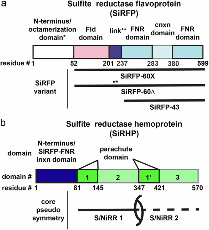
Fig. 1 Sulfite Reductase (SiR) components are highly modular. a Sulfite Reductase Flavoprotein (SiRFP) is composed of an N-terminal octamerization domain (not present in the constructs studied here), a flavin mononucleotide (FMN)-binding flavodoxin (Fld) domain (pink) connected through a linker (purple) to a flavin adenine dinucleotide (FAD)-binding ferredoxin-NADP+ reductase (FNR) domain (blue) interrupted by a connection (cnxn) domain (light blue). The variants and their names are denoted below: SiRFP-60X has a truncated octamerization domain (*) to make a 60 kDa monomer with engineered crosslinks between the Fld and FNR domain and a full-length linker. SiRFP-60Δ has a truncated octamerization domain (*) to make a 60 kDa monomer with a shortened linker (**, Δ212-217). SiRFP-43 only contains the FNR and connection domains to make a 43 kDa monomer. b Sulfite Reductase Hemoprotein’s (SiRHP) N-terminal 80 residues (dark blue) are solely responsible for forming a stable interaction with the FNR domain of SiRFP. The pseudosymmetric core is composed of sequential Sulfite and Nitrite Reductase Repeats (S/NiRRs) (green and light green, domains 1/2 and 1’/3) that include a parachute domain (green, domains 1/1’).
Fig. 1 Sulfite Reductase (SiR) components are highly modular. a Sulfite Reductase Flavoprotein (SiRFP) is composed of an N-terminal octamerization domain (not present in the constructs studied here), a flavin mononucleotide (FMN)-binding flavodoxin (Fld) domain (pink) connected through a linker (purple) to a flavin adenine dinucleotide (FAD)-binding ferredoxin-NADP+ reductase (FNR) domain (blue) interrupted by a connection (cnxn) domain (light blue). The variants and their names are denoted below: SiRFP-60X has a truncated octamerization domain (*) to make a 60 kDa monomer with engineered crosslinks between the Fld and FNR domain and a full-length linker. SiRFP-60Δ has a truncated octamerization domain (*) to make a 60 kDa monomer with a shortened linker (**, Δ212-217). SiRFP-43 only contains the FNR and connection domains to make a 43 kDa monomer. b Sulfite Reductase Hemoprotein’s (SiRHP) N-terminal 80 residues (dark blue) are solely responsible for forming a stable interaction with the FNR domain of SiRFP. The pseudosymmetric core is composed of sequential Sulfite and Nitrite Reductase Repeats (S/NiRRs) (green and light green, domains 1/2 and 1’/3) that include a parachute domain (green, domains 1/1’).CC BY-NC-ND · PubMed Central
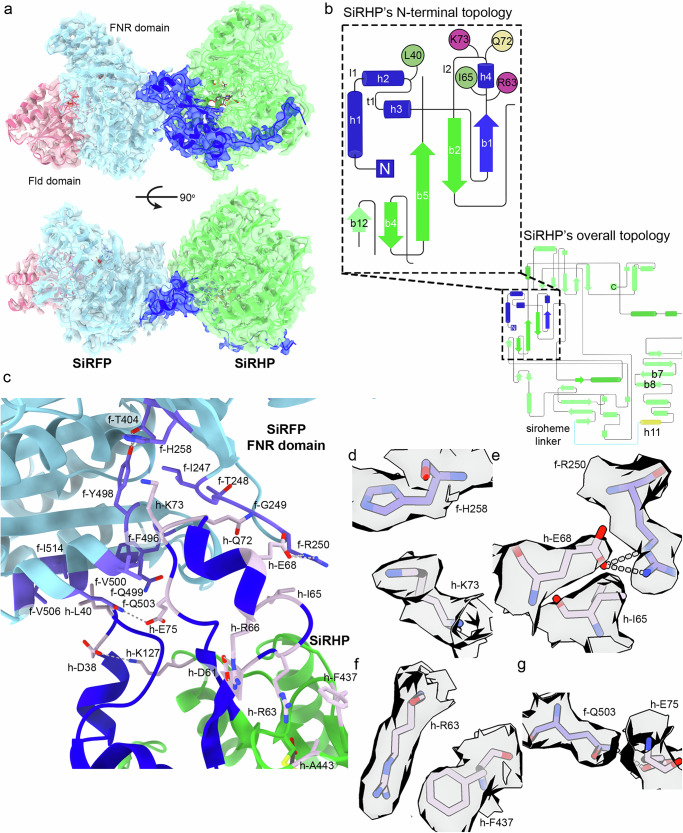
Fig. 2 Sulfite Reductase Flavoprotein (SiRFP)and hemoprotein (SiRHP) interact through the SiRFP ferredoxin-NADP+ reductase (FNR) domain and the SiRHP N-terminus. a The SiRFP/SiRHP interface is minimal, governed by residues from three loops in SiRHP that fit into pockets on SiRFP’s FNR domain. SiRFP’s Fld domain is pink, SiRFP’s FNR domain is light blue. SiRHP’s N-terminal 80 residues are dark blue. SiRHP’s core two Sulfite and Nitrite Reductase Repeats (S/NiRRs) are green. The map is shown at contour level 0.3. b The topology diagram of SiRHP highlights the N-terminal 80 residues whose structure was previously unknown but that govern the interactions with SiRFP. The residues that interact with SiRFP are localized to three loops. Hydrophobic residues are green, cationic residues are purple, and polar residues are yellow. The first S/NiRR is dark green, whereas the second S/NiRR is light green, with the connecting h11 and linker in yellow and cyan, respectively (“Created in BioRender. Ghazi Esfahani, B. (2025) https://BioRender.com/ q86t823.”). c The interface between SiRFP and SiRHP is minimal. The SiRFP-FNR domain is light blue with light purple highlighted residues that are important for forming the SiRHP binding site. SiRHP’S N-terminal 80 residues are blue, and the S/NiRR repeats are green with mauve highlighted residues that mediate the interaction with SiRFP. d–g Select densities for important side-chain interactions that mediate the interface between the SiRFP and SiRHP interface are rendered at contour level 0.4 and colored as in (c).
Fig. 2 Sulfite Reductase Flavoprotein (SiRFP)and hemoprotein (SiRHP) interact through the SiRFP ferredoxin-NADP+ reductase (FNR) domain and the SiRHP N-terminus. a The SiRFP/SiRHP interface is minimal, governed by residues from three loops in SiRHP that fit into pockets on SiRFP’s FNR domain. SiRFP’s Fld domain is pink, SiRFP’s FNR domain is light blue. SiRHP’s N-terminal 80 residues are dark blue. SiRHP’s core two Sulfite and Nitrite Reductase Repeats (S/NiRRs) are green. The map is shown at contour level 0.3. b The topology diagram of SiRHP highlights the N-terminal 80 residues whose structure was previously unknown but that govern the interactions with SiRFP. The residues that interact with SiRFP are localized to three loops. Hydrophobic residues are green, cationic residues are purple, and polar residues are yellow. The first S/NiRR is dark green, whereas the second S/NiRR is light green, with the connecting h11 and linker in yellow and cyan, respectively (“Created in BioRender. Ghazi Esfahani, B. (2025) https://BioRender.com/ q86t823.”). c The interface between SiRFP and SiRHP is minimal. The SiRFP-FNR domain is light blue with light purple highlighted residues that are important for forming the SiRHP binding site. SiRHP’S N-terminal 80 residues are blue, and the S/NiRR repeats are green with mauve highlighted residues that mediate the interaction with SiRFP. d–g Select densities for important side-chain interactions that mediate the interface between the SiRFP and SiRHP interface are rendered at contour level 0.4 and colored as in (c).CC BY-NC-ND · PubMed Central
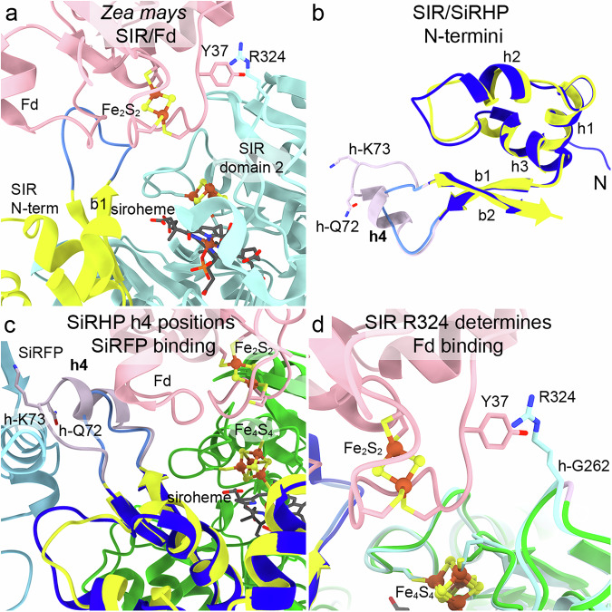
Fig. 3 Zea mays Sulfite Reductase (SIR) and Escherichia coli Sulfite Reductase Hemoprotein (SiRHP) interact with their reductase partners in different ways. a SIR binds a ferredoxin (Fd, pink) such that its Fe2S2 cluster is close to SIR’s Fe4S4 cluster, positioned between SIR’s N-terminus (yellow) and domain 2 (cadet blue) (PDB 5H9236). b SiRHP h4 (mauve) is the sole secondary structural feature that discriminates between SiRFP binding and Fd binding in Z. mays. SiRHP’s N-terminal 80 residues are dark blue. SIR’s N-terminal residues are yellow, with the element equivalent to SiRHP h4 in medium blue. c SiRFP (light blue) and Fd (pink) bind on different faces of SiRHP (dark blue) or SIR (yellow). Only the core Sulfite and Nitrite Reductase Repeats (S/NiRRs) from SiRHP (green) are shown for clarity. Other elements are colored as in (b). d SIR R324 (cadet blue) interacts with Fd Y37 (pink). That interaction is prevented in SiRHP (green) because the position equivalent to SIR R324 is h-G262 (mauve). Z. mays Fd Fe2S2 and SIR Fe4S4 are shown as in panel (a).
Fig. 3 Zea mays Sulfite Reductase (SIR) and Escherichia coli Sulfite Reductase Hemoprotein (SiRHP) interact with their reductase partners in different ways. a SIR binds a ferredoxin (Fd, pink) such that its Fe2S2 cluster is close to SIR’s Fe4S4 cluster, positioned between SIR’s N-terminus (yellow) and domain 2 (cadet blue) (PDB 5H9236). b SiRHP h4 (mauve) is the sole secondary structural feature that discriminates between SiRFP binding and Fd binding in Z. mays. SiRHP’s N-terminal 80 residues are dark blue. SIR’s N-terminal residues are yellow, with the element equivalent to SiRHP h4 in medium blue. c SiRFP (light blue) and Fd (pink) bind on different faces of SiRHP (dark blue) or SIR (yellow). Only the core Sulfite and Nitrite Reductase Repeats (S/NiRRs) from SiRHP (green) are shown for clarity. Other elements are colored as in (b). d SIR R324 (cadet blue) interacts with Fd Y37 (pink). That interaction is prevented in SiRHP (green) because the position equivalent to SIR R324 is h-G262 (mauve). Z. mays Fd Fe2S2 and SIR Fe4S4 are shown as in panel (a).CC BY-NC-ND · PubMed Central
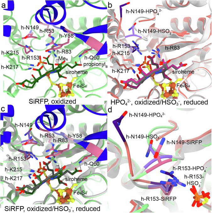
Fig. 4 Sulfite Reductase Hemoprotein’s (SiRHP) active site loop that forms the complete substrate binding site is in its closed conformation with the intact N-terminus, bound to Sulfite Reductase Flavoprotein (SiRFP). a When SiRFP binds oxidized SiRHP with the bound, inhibitory phosphate (orange and red), the anion-binding loop containing h-N149 is ordered but with h-Arg153 in a similar orientation as in free SiRHP. Panels are labeled by their binding partner and oxidation state. Residues important for anion binding or loop position are dark pink. The siroheme methyl group from ring A (MeA) is a gray sphere, and the interaction between h-Q60 and the ring B propionyl group is marked by a gray dash. b In the absence of its N-terminal 80 residues, SiRFP, or the sulfite substrate (yellow and red), SiRHP’s anion binding loop is disordered (missing salmon ribbons marked by salmon *s). The residues that are important for anion binding in response to oxidation state in this structure are dark purple, those that are unchanged are salmon (PDB 1AOP23). Upon reduction and with bound sulfite, the active site loop becomes ordered, and h-R153 flips over to bind the substrate (light gray ribbons). The residues that are important for anion binding in response to oxidation state in this structure are light purple, and those that are unchanged are light gray (PDB 2GEP24,25). c Superimposition of the phosphate (orange and red) and SiRFP-bound SiRHP (green) with the reduced, sulfite-bound (yellow and red), SiRHP (light gray) shows the anion-binding loop to be in similar position as in reduced/SO32--bound SiRHP, primed for sulfite binding. d Superimposition of the three SiRHP structures bound to different anions or binding partners highlights the intermediate conformation induced by SiRFP binding. This intermediate conformation shows the ordering of SiRHP’s N-terminus and the active site loop in a phosphate-bound, oxidized state. Models are colored as in (a–c).
Fig. 4 Sulfite Reductase Hemoprotein’s (SiRHP) active site loop that forms the complete substrate binding site is in its closed conformation with the intact N-terminus, bound to Sulfite Reductase Flavoprotein (SiRFP). a When SiRFP binds oxidized SiRHP with the bound, inhibitory phosphate (orange and red), the anion-binding loop containing h-N149 is ordered but with h-Arg153 in a similar orientation as in free SiRHP. Panels are labeled by their binding partner and oxidation state. Residues important for anion binding or loop position are dark pink. The siroheme methyl group from ring A (MeA) is a gray sphere, and the interaction between h-Q60 and the ring B propionyl group is marked by a gray dash. b In the absence of its N-terminal 80 residues, SiRFP, or the sulfite substrate (yellow and red), SiRHP’s anion binding loop is disordered (missing salmon ribbons marked by salmon *s). The residues that are important for anion binding in response to oxidation state in this structure are dark purple, those that are unchanged are salmon (PDB 1AOP23). Upon reduction and with bound sulfite, the active site loop becomes ordered, and h-R153 flips over to bind the substrate (light gray ribbons). The residues that are important for anion binding in response to oxidation state in this structure are light purple, and those that are unchanged are light gray (PDB 2GEP24,25). c Superimposition of the phosphate (orange and red) and SiRFP-bound SiRHP (green) with the reduced, sulfite-bound (yellow and red), SiRHP (light gray) shows the anion-binding loop to be in similar position as in reduced/SO32--bound SiRHP, primed for sulfite binding. d Superimposition of the three SiRHP structures bound to different anions or binding partners highlights the intermediate conformation induced by SiRFP binding. This intermediate conformation shows the ordering of SiRHP’s N-terminus and the active site loop in a phosphate-bound, oxidized state. Models are colored as in (a–c).CC BY-NC-ND · PubMed Central
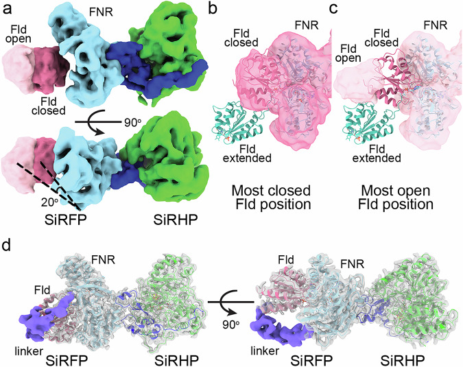
Fig. 5 The Sulfite Reductase Flavoprotein (SiRFP) flavodoxin domain (Fld) domain is highly mobile relative to its ferredoxin-NAD+ reductase (FNR) domain. a One of the latent variable dimensions of motion demonstrates an opening of the Fld domain, even in SiRFP-60Δ, with the motion parallel to the midline of the FNR domain (light blue). The most-closed position is dark pink, the most open is light pink. SiRHP is green, with the N-terminus in dark blue. The angle is measured between the lines connecting the first traceable residues in the FNR domain with the open and closed Fld volume center of mass. b The map of the most closed conformation of the Fld domain (dark pink) corresponds to the position of the Fld domain in the dimer formed with SiRFP-60X, far from where it is in the extended conformation of SiRFP-60Δ in crystals (teal, PDB 6EFV31). c The map of the most opened conformation (light pink) shows that the Fld domain lands in an intermediary position between a highly extended position seen in the X-ray crystal structure (teal, PDB 6EFV31) and the closed conformation seen in (b). d Even with the Fld and FNR domains constrained by a crosslink, the 36 residue-long linker between the domains is largely disordered, visible only in a small (~20,000) subset of particles (purple volume), shown at contour level 0.06.
Fig. 5 The Sulfite Reductase Flavoprotein (SiRFP) flavodoxin domain (Fld) domain is highly mobile relative to its ferredoxin-NAD+ reductase (FNR) domain. a One of the latent variable dimensions of motion demonstrates an opening of the Fld domain, even in SiRFP-60Δ, with the motion parallel to the midline of the FNR domain (light blue). The most-closed position is dark pink, the most open is light pink. SiRHP is green, with the N-terminus in dark blue. The angle is measured between the lines connecting the first traceable residues in the FNR domain with the open and closed Fld volume center of mass. b The map of the most closed conformation of the Fld domain (dark pink) corresponds to the position of the Fld domain in the dimer formed with SiRFP-60X, far from where it is in the extended conformation of SiRFP-60Δ in crystals (teal, PDB 6EFV31). c The map of the most opened conformation (light pink) shows that the Fld domain lands in an intermediary position between a highly extended position seen in the X-ray crystal structure (teal, PDB 6EFV31) and the closed conformation seen in (b). d Even with the Fld and FNR domains constrained by a crosslink, the 36 residue-long linker between the domains is largely disordered, visible only in a small (~20,000) subset of particles (purple volume), shown at contour level 0.06.CC BY-NC-ND · PubMed Central

Verification

Claims the paper makes

The claims and statistics of this paper haven’t been checked yet.

Run the check — free, no account needed. It reads the open-access text and checks the sources it cites.

A few free checks a day · papers already verified open instantly · results are public

Self-check

Percentages, sums and reported differences re-computed from the paper’s own numbers.

The self-check needs the paper’s full text.

Citations

Cites

Show all 66 references

Metadata

Authors
Ghazi Esfahani, B., Walia, N., Neselu, K., Garg, Y., Aragon, M., Askenasy, I., Wei, H. A., Mendez, J. H., & Stroupe, M. E.
Affiliations
  • Florida State University (US)
  • New York Structural Biology Center (US)
Corresponding author
Stroupe, M. Elizabeth
Venue
Nature Communications
Published
2025-03-26
Volume
16
Issue
1
Pages
2955-2955
ISSN
2041-1723 (ISSN-L)
OpenAlex
W4408866252
Cited by
10
References
66
Citations by year
2026: 8 · 2025: 2
FWCI
2.11
Type
journal article
Language
en
Open access
Licence
Locations
  • Nature Communications (publishedVersion, cc-by-nc-nd)
  • Europe PMC (PubMed Central) (publishedVersion, cc-by-nc-nd)
  • PubMed Central (publishedVersion, cc-by-nc-nd)
  • PubMed (publishedVersion)
  • DOAJ (DOAJ: Directory of Open Access Journals) (submittedVersion, cc-by-sa)
Funders
  • National Science Foundation (MCB1856502, CHE1904612, MRI2017869)
  • U.S. Department of Health & Human Services | National Institutes of Health (S10 RR024564, R24 GM145964)
  • Simons Foundation (SF349247)
Topics
  • Life Sciences › Biochemistry, Genetics and Molecular Biology › Structural Biology › Enzyme Structure and Function
  • Life Sciences › Biochemistry, Genetics and Molecular Biology › Structural Biology › Advanced Electron Microscopy Techniques and Applications
  • Life Sciences › Biochemistry, Genetics and Molecular Biology › Biochemistry › Metalloenzymes and iron-sulfur proteins
Keywords
Escherichia coli, siroheme, Fe4S4 cluster, ferredoxin, cryo-EM structure, sulfite reduction
MeSH terms
Zea mays, Escherichia coli, Flavoproteins, Models, Molecular, NADP, Oxidoreductases, Protein Binding, Protein Structure, Tertiary, Catalytic Domain, Cryoelectron Microscopy, Protein Structure, Quaternary, Escherichia coli Proteins, Sulfite Reductase (NADPH), Protein Multimerization

About these checks

Registry, retraction, Cites and statistics results are computed, not judged, with one exception: a printed reference that no index lists is read once by a model before it counts as a citation error. Claims are extracted by a model and kept only when their quote appears verbatim in the text; each cited source is then checked, with a model’s help, for whether it says what the claim relies on. Those verdicts can be wrong — read the claim’s quote and the cited source before relying on one.

Statistics and arithmetic are recomputed, not judged.

Confidence bands are shown only for fields calibrated on our annotated test set. None is calibrated yet, so no band is shown.

What counts as a claim ›How we measure confidence ›How we check this ›

Are you an author, or do you see a problem with this page? Request a review

How to cite

Cite CoChat for the checks on this page, and the paper for its own findings.

This page
CoChat. (2026). Citation check: Structure of dimerized assimilatory NADPH-dependent sulfite reductase reveals the minimal interface for diflavin reductase binding. https://app.cochat.ai/papers/10.1038/s41467-025-58037-5
The paper
Ghazi Esfahani, B., Walia, N., Neselu, K., Garg, Y., Aragon, M., Askenasy, I., Wei, H. A., Mendez, J. H., & Stroupe, M. E. (2025). Structure of dimerized assimilatory NADPH-dependent sulfite reductase reveals the minimal interface for diflavin reductase binding. Nature Communications. https://doi.org/10.1038/s41467-025-58037-5

Data sources

Bibliographic data: Semantic Scholar, OpenAlex, Crossref.

Contains information from the Semantic Scholar Academic Graph Datasets, made available under the ODC Attribution License (ODC-BY 1.0).

Semantic Scholar’s data is described in: Kinney, R., et al. (2023). The Semantic Scholar Open Data Platform. arXiv:2301.10140.